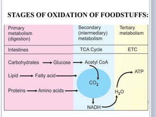
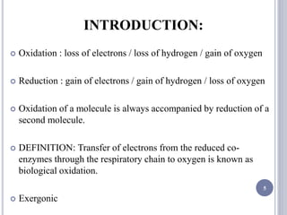
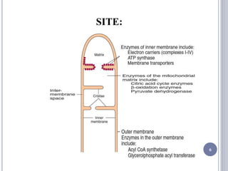
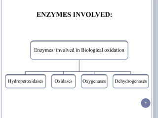
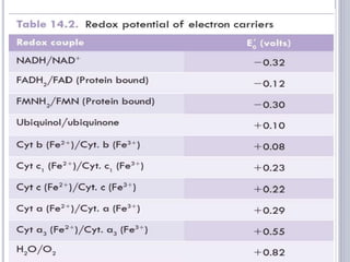
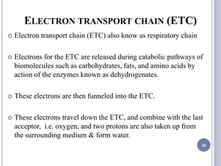
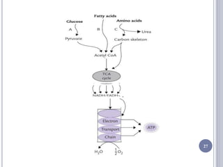
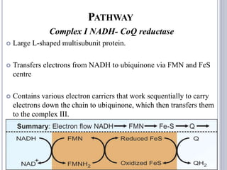
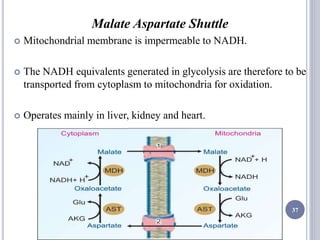
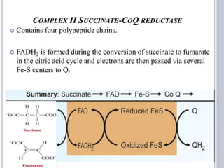
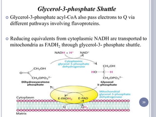
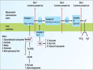
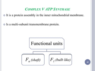
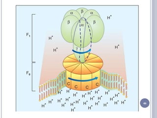
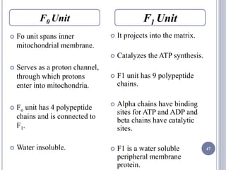
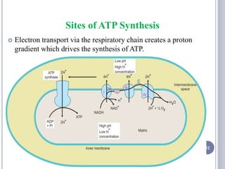
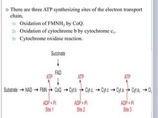
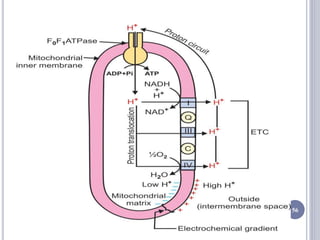
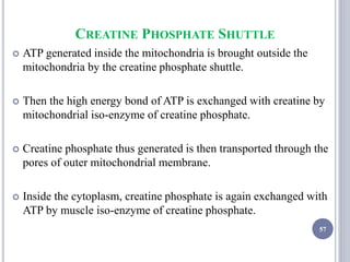
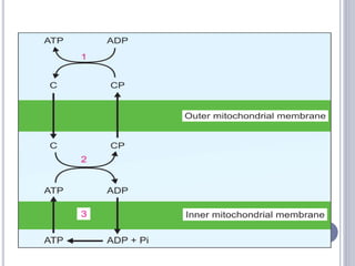
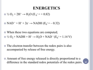
BIOLOGICAL OXIDATION
&
ELECTRON TRANSPORT
CHAIN
By: Nidhi Argade
1
LEARNING OBJECTIVES :
 BIOLOGICAL OXIDATION
 OXIDATIVE PHOSPHORYLATION
2
BIOLOGICAL OXIDATION
 STAGES OF OXIDATION OF FOODSTUFFS
 INTRODUCTION
 SITE
 ENZYMES INVOLVED
 OTHER SPECIAL TERMS
3
STAGES OF OXIDATION OF FOODSTUFFS:
4
INTRODUCTION:
 Oxidation : loss of electrons / loss of hydrogen / gain of oxygen
 Reduction : gain of electrons / gain of hydrogen / loss of oxygen
 Oxidation of a molecule is always accompanied by reduction of a
second molecule.
 DEFINITION: Transfer of electrons from the reduced co-
enzymes through the respiratory chain to oxygen is known as
biological oxidation.
 Exergonic
5
SITE:
6
ENZYMES INVOLVED:
7
Enzymes involved in Biological oxidation
Hydroperoxidases Oxidases Oxygenases Dehydrogenases
HYDROPEROXIDASES
 Use hydrogen peroxide / an organic peroxide as substrate.
 Important role in protecting the body against the harmful
effects of reactive oxygen species (ROS).
 Oxidation in which H2O2 acts as hydrogen acceptor and is
reduced to water.
 H2O2 H2O + O2
8
9
 Reduce peroxides using
various electron acceptors.
 Prosthetic group is
protoheme.
 Examples;
Glutathione peroxidase
Leukocyte peroxidase
Horse radish peroxidase
 H2O2 + AH2
2H2O + A
 Uses hydrogen peroxide as
electron donor & electron
acceptor.
 Catalase is a hemoprotein
containing four heme groups.
 Catalase can react directly
with H2O2.
 Km of catalase for H 2 O 2 is
much greater.
 2H2O 2
2H 2 O + O 2
Peroxidase Catalase
peroxidase
catalase
OXIDASES
 Use oxygen as hydrogen acceptor.
 Catalyze the removal of hydrogen from substrates
 Only oxygen can act as acceptor of hydrogen, and form water or
hydrogen peroxide as a reaction product.
 Includes; Cytochrome oxidase,
Tyrosinase,
Polyphenol oxidase,
Catechol oxidase
Monoamine oxidase.
10
OXYGENASES
 Catalyze the direct transfer and incorporation of oxygen into
a substrate molecule in two steps:
1. Oxygen is bound to the enzyme at the active site
2. The bound oxygen is reduced or transferred to the
substrate.
 Concerned with the synthesis or degradation of many
different types of metabolites.
11
Subgroups
Mono-oxygenases Di-oxygenases
12
 Incorporate O2 into their
substrates and the other
oxygen atom is reduced to
water.
 An additional electron donor
or co-substrate necessary.
 Also called hydroxylases
because OH group is
incorporated into the
substrate.
 A-H+ O2+ BH2
A-OH+ H2O+ B
 Incorporate both atoms of
molecular oxygen into the
substrate.
 E.g.
 Tryptophan pyrolase (utilizes
heme)
 Homogentisate dioxygenase
 3-Hydroxyanthranilate
dioxygenase
 A+O2 AO2
Mono-oxygenases Di-oxygenases
Hydroxylase
DEHYDROGENASES
 Most common of all oxidoreductases.
 Cannot use oxygen as a hydrogen accceptor.
 Catalyze transfer of hydrogen between a substrate and a
coenzyme, most commonly NAD, FAD, NADP or FMN.
13
Subgroups
Aerobic
Dehydrogenases
Anaerobic
Dehydrogenases
14
 Catalyze the removal of
hydrogen from a substrate,
but oxygen can act as the
acceptor.
 Enzymes are flavoproteins
and the product is usually
hydrogen peroxide.
 Flavoproteins contain either
FMN or FAD as prosthetic
group.
 Examples;
 L-amino acid oxidase
 Xanthine oxidase
 AH2 + O2 A + H2 O2
 Catalyze the removal of
hydrogen from a substrate but
oxygen cannot act as hydrogen
acceptor.
 Require co-enzymes as
acceptors of the hydrogen
atoms.
 When the substrate is oxidized,
the coenzyme is reduced.
 Examples;
 NAD+ linked
dehydrogenases
 NADP+ linked
dehydrogenases
Aerobic
Dehydrogenases
Anaerobic
Dehydrogenases
NAD+ LINKED DEHYDROGENASES
 When a substrate is oxidized, it loses 2 hydrogen atoms and 2
electrons.
 One H+ and both electrons are accepted by NAD+ to form NADH
and the other H+ is released.
 E.g;
 Glyceraldehyde-3-phosphate dehydrogenase
 Pyruvate dehydrogenase
 Isocitrate dehydrogenase
 Malate dehydrogenase
 Glutamate dehydrogenase
 Alpha ketoglutarate dehydrogenase
 Beta hydroxyacyl CoA dehydrogenase
15
NADP+ LINKED DEHYDROGENASES
 NADPH cannot be oxidized with concomitant production of
energy.
 Used in reductive biosynthetic reactions
FAD LINKED DEHYDROGENASES
 When FAD is the coenzyme, both the hydrogen atoms are
attached to the flavin ring.
 Examples; Succinate dehydrogenase
Fatty acyl CoA dehydrogenase
Glycerolphosphate dehydrogenase
16
CYTOCHROMES
 Iron-containing hemoproteins
 All the cytochromes hemoproteins having iron atom, except
cytochrome oxidase.
17
Location of Cytochromes
Mitochondria Endoplasmic Reticulum
Cytochrome b,
Cytochrome c1,
Cytochrome c
Cytochrome P-450
Cytochrome b5
SUBSTRATE LEVEL PHOSPHORYLATION:
 Energy from a high energy compound is directly transferred to
nucleoside diphosphate to form a triphosphate without the help
of electron transport chain.
 E.g.
 Bisphosphoglycerate kinase
18
 Pyruvate kinase
 Succinate thiokinase
19
OXIDATIVE PHOSPHORYLATION
 INTRODUCTION
 SITE
 REDOX COUPLE & POTENTIAL
 ELECTRON TRANSPORT CHAIN
20
INTRODUCTION:
 DEFINITION: Coupling of oxidation with phosphorylation is
called oxidative phosphorylation.
 Respiration is coupled to the generation of the high-energy
intermediate by oxidative phosphorylation.
 Involves the reduction of O2 to H2O with electrons donated by
NADH and FADH2 .
Site:
 Inner Mitochondrial Matrix (IMM) 21
REDOX COUPLE AND REDOX POTENTIAL:
 DEFINITION: When a substance exists both in the reduced
state and in the oxidized state, the pair is called a redox couple.
 The tendency of a redox couple to donate or accept electrons is
determined by standard redox potential (E0
’).
 Potential of this couple is estimated by measuring the
electromotive force (EMF) of a sample half cell connected to a
standard half cell.
22
REDOX POTENTIAL
23
 When a substance has
lower affinity for electrons
than hydrogen.
 NADH, a strong reducing
agent
 Redox potential –0.32 V
 When a substance has a
higher affinity for electrons
than hydrogen.
 Oxygen strong oxidant
 Redox potential +0.82 V
Positive Redox Potential
 Electrons always flow from the lower to the higher redox
potential, so that redox systems with negative E0 tend to donate
electrons to redox systems with positive E0.
Negative Redox Potential
24
ELECTRON TRANSPORT CHAIN:
 COMPONENTS OF ETC
 PATHWAY
 MECHANISM OF ATP SYNTHESIS
 ENERGETICS
 REGULATION
 INHIBITORS
 CLINICAL SIGNIFICANCE
25
ELECTRON TRANSPORT CHAIN (ETC)
 Electron transport chain (ETC) also know as respiratory chain
 Electrons for the ETC are released during catabolic pathways of
biomolecules such as carbohydrates, fats, and amino acids by
action of the enzymes known as dehydrogenates.
 These electrons are then funneled into the ETC.
 These electrons travel down the ETC, and combine with the last
acceptor, i.e. oxygen, and two protons are also taken up from
the surrounding medium & form water.
26
27
COMPONENTS OF ETC
 Nicotinamide Nucleotides
 Flavoproteins
 Iron–sulphur centres
 Ubiquinone
 Cytochromes:-
b,
c1,
c,
a
a3
28
1) NICOTINAMIDE NUCLEOTIDES
 NAD+ involved in the ETC.
 Large protein complex embedded in IMM.
 Reduced to NADH by transfer of a pair of electrons from the substrate
by action of various dehydrogenases.
 NAD+ + H + + 2e- NADH + H +
 NADH subsequently loses its electrons to the initial component of
ETC.
 NADP is similarly produced by action of NADP dependent
dehydrogenases, but it is mostly involved in reductive biosynthesis of
biomolecules.
29
2) FLAVOPROTEINS
 Important hydrogen carriers
 FAD and FMN react with two protons plus two electron, in
alternating between the reduced and the oxidized state.
 FAD + 2H + 2e- FADH2
 FMN is prosthetic group of NADH dehydrogenase, bound
firmly to the enzyme protein and does not function as a
diffusible co-substrate.
 FAD linked to another flavoprotein, succinate dehydrogenase,
and functions as a diffusible co-substrate.
30
3) IRON–SULPHUR CENTRES
 Iron atoms are coordinated to inorganic sulphur atoms.
 Within an FeS cluster, an electron is carried by the iron atom,
which accepts the electron, changes from the Fe3 state to the
Fe2 state.
 The electron is passed to another electron carrier, the iron
atom of the FeS cluster changes back again to the ferric state.
31
4) UBIQUINONE
 Mobile diffusible hydrogen carrier, which can move from donor to
acceptor molecules during electron transport.
 Benzoquinone with a hydrocarbon tail of 10 isoprene units which
makes it strongly hydrophobic and confines it to the lipid bilayer
of the inner mitochondrial membrane.
32
 It can accept electrons from FMNH2 or from FADH2 and
transfers them to cytochromes.
 Ubiquinone carries two hydrogen atoms, but can also act in
one-electron transfers by forming a free radical intermediate,
called semiquinone intermediate.
 Reduced form is called ubiquinol.
33
5) CYTOCHROMES
 Integral membrane proteins.
 Final class of components that participate in electron
transport.
 Conjugated proteins, containing an iron-porphyrin as a
prosthetic group.
 Electrons are transported from co-enzyme Q to cytochromes
in the order: b, c1, c, a and a3.
34
STRUCTURAL ORGANIZATION OF COMPONENTS
35
PATHWAY
Complex I NADH- CoQ reductase
 Large L-shaped multisubunit protein.
 Transfers electrons from NADH to ubiquinone via FMN and FeS
centre
 Contains various electron carriers that work sequentially to carry
electrons down the chain to ubiquinone, which then transfers them
to the complex III.
36
Malate Aspartate Shuttle
 Mitochondrial membrane is impermeable to NADH.
 The NADH equivalents generated in glycolysis are therefore to be
transported from cytoplasm to mitochondria for oxidation.
 Operates mainly in liver, kidney and heart.
37
COMPLEX II SUCCINATE-COQ REDUCTASE
 Contains four polypeptide chains.
 FADH2 is formed during the conversion of succinate to fumarate
in the citric acid cycle and electrons are then passed via several
Fe-S centers to Q.
38
Glycerol-3-phosphate Shuttle
 Glycerol-3-phosphate acyl-CoA also pass electrons to Q via
different pathways involving flavoproteins.
 Reducing equivalents from cytoplasmic NADH are transported to
mitochondria as FADH2 through glycerol-3- phosphate shuttle.
39
Co -Enzyme Q
 The ubiquinone (Q) is reduced successively to semiquinone (QH)
and finally to quinol (QH2).
 It accepts a pair of electrons from NADH or FADH2.
 The Q cycle thus facilitates the switching from the two electron
carrier ubiquinol to the single electron carrier cytochrome c.
40
41
Complex III CoQ- Cytochrome c reductase
 Cluster of iron-sulfur proteins.
 Contain cytochrome b and cytochrome c1.
 During this process of transfer of electron, the iron in heme
group shuttles between Fe3+ and Fe2+ forms.
Cytochrome C
 Peripheral membrane protein containing one heme prosthetic
group.
 Cytochrome c shuttles electrons from Complex III to Complex
IV.
42
Complex IV Cyctochrome oxidase
 Transfers electrons from cytochrome c to O2 to form two
molecules of water.
 Transfer of four electrons from cytochrome c to O2 involves two
heme groups, a and a3, and Cu.
 Electrons are passed initially to a Cu center which contains 2Cu
atoms linked to two protein cysteine-SH groups then in sequence
to heme a, heme a3, a second Cu center, which is linked to heme
a3, and finally to O2.
43
44
COMPLEX V ATP SYNTHASE
 It is a protein assembly in the inner mitochondrial membrane.
 Is a multi-subunit transmembrane protein.
45
Functional units
Fo (shaft) F1 (bulb like)
46
47
 Fo unit spans inner
mitochondrial membrane.
 Serves as a proton channel,
through which protons
enter into mitochondria.
 Fo unit has 4 polypeptide
chains and is connected to
F1.
 Water insoluble.
 It projects into the matrix.
 Catalyzes the ATP synthesis.
 F1 unit has 9 polypeptide
chains.
 Alpha chains have binding
sites for ATP and ADP and
beta chains have catalytic
sites.
 F1 is a water soluble
peripheral membrane
protein.
F0 Unit F1 Unit
MECHANISM OF ATP SYNTHESIS
 Translocation of protons carried out by the Fo catalyzes the
formation of phospho-anhydride bond of ATP by F1.
 Coupling of the dissipation of proton gradient with ATP synthesis
is through the interaction of F1 and Fo.
Binding Change Mechanism;
 Fo is the wheel.
 Flow of protons is the waterfall and the structural changes in F1
lead to ATP coin being minted for each turn of the wheel.
48
 F1 has 3 conformation states for the alpha-beta functional unit:
1) O state—Does not bind substrate or products
2) L state—Loose binding of substrate and products
3) T state—Tight binding of substrate and products
 O form is open and has no affinity for substrates.
 L form binds substrate with sluggish affinity.
 T form binds substrate tightly and catalyzes ATP synthesis.
49
50
1. ADP and Pi bind to L binding site.
2. L to T conversion is by energy driven conformational change
that catalyzes the formation of ATP.
3. T state reverts to O state when ATP is released.
4. L state is regenerated for further ADP binding.
 For the complete rotation of F1 head through the 3 states, 10
protons are translocated.
FORMATION OF ATP
 Energy of electron transfer is used to drive protons out of the
matrix by the complexes I, III and IV that are proton pumps.
 The proton gradient thus created is maintained across the inner
mitochondrial membrane till electrons are transferred to oxygen
to form water.
 The electrochemical potential of this gradient is used to
synthesize ATP.
 According to the estimated free energy of synthesis, it was
presumed that around 3 protons are required per ATP
synthesized.
51
Sites of ATP Synthesis
 Electron transport via the respiratory chain creates a proton
gradient which drives the synthesis of ATP.
52
 There are three ATP synthesizing sites of the electron transport
chain,
1) Oxidation of FMNH2 by CoQ.
2) Oxidation of cytochrome b by cytochrome c1.
3) Cytochrome oxidase reaction.
53
P : O RATIO
 Number of inorganic phosphate molecules incorporated into ATP
for every atom of oxygen consumed.
 When a pair of electrons from NADH reduces an atom of oxygen
(½ O2), 2.5 mol of ATP are formed per 0.5 mol of O2 consumed.
 The P:O ratio of NADH oxidation is 2.5.
 The P:O value of FADH2 is 1.5.
54
CHEMIOSMOTIC THEORY
 The coupling of oxidation with phosphorylation is termed
oxidative phosphorylation.
 Peter Mitchell
 The transport of protons from inside to outside of inner
mitochondrial membrane is accompanied by the generation of a
proton gradient across the membrane.
 Protons accumulate outside the membrane, creating an
electrochemical potential difference.
 Proton motive force drives the synthesis of ATP by ATP synthase
complex
55
56
CREATINE PHOSPHATE SHUTTLE
 ATP generated inside the mitochondria is brought outside the
mitochondria by the creatine phosphate shuttle.
 Then the high energy bond of ATP is exchanged with creatine by
mitochondrial iso-enzyme of creatine phosphate.
 Creatine phosphate thus generated is then transported through the
pores of outer mitochondrial membrane.
 Inside the cytoplasm, creatine phosphate is again exchanged with
ATP by muscle iso-enzyme of creatine phosphate.
57
58
59
ENERGETICS
 ½ O2 + 2H+ → H2O (E 0‫׳‬ = + 0.82)
 NAD + + H+ + 2e- → NADH (E0‫׳‬ = – 0.32)
 When these two equations are computed;
 ½ O2 + NADH + H+ → H2O + NAD + (E0‫׳‬ = 1.14 V)
 The electron transfer between the redox pairs is also
accompanied by release of free energy.
 Amount of free energy released is directly proportional to a
difference in the standard redox potentials of the redox pairs. 60
 Δ G0‫׳‬ = -nF E 0‫׳‬
= –2 × 23.06 × 1.14
= –52.6 kcal/mol
 Δ G0‫=׳‬ Standard free energy change in kcal/mole.
 n = number of reducing equivalents transferred
 F = Faraday’s constant (23.062 kcal V–1 mol–1)
 E 0‫׳‬ = difference between the standard redox potentials of
the electron-donor and the electron-acceptor redox systems
 Used to calculate the energy changes for individual segments of
the electron transport chain.
61
 The free energy change between NAD+ and water is equal to 53
kcal/mol.
 Hence, with the help of ETC assembly, the total energy change
is released in small increments so that energy can be trapped as
chemical bond energy.
62
REGULATION
 Availability of ADP regulates the process.
 When ATP level is low and ADP level is high, oxidative
phosphorylation proceeds at a rapid rate. This is called
respiratory control or acceptor control.
 The major source of NADH and FADH2 is the citric acid cycle,
the rate of which is regulated by the energy charge of the cell.
 Electron transport system is regulated by the magnitude of the
PMF.
 Electron transport is directly coupled to proton translocation,
the flow of electrons through the higher the PMF, the lower the
rate of electron transport, and vice versa.
63
 Under resting conditions, with a high cell energy charge, the
demand for new synthesis of ATP is limited and, although the
PMF is high, flow of protons back into the mitochondria through
ATP synthase is minimal.
 When energy demands are increased, cytosolic ADP rises and is
exchanged with intramitochondrial ATP via the transmembrane
adenine nucleotide carrier ADP/ATP translocase.
 Increased intramitochondrial concentrations of ADP cause the
PMF to become discharged as protons pour through ATP
synthase, regenerating the ATP pool.
 While the rate of electron transport is dependent on the PMF, the
magnitude of the PMF at any moment simply reflects the
energy charge of the cell. 64
Inhibitors of ETC
Electron
transport chain
proper
Oxidative
phosphorylation
Uncouplers of
oxidative
phosphorylation
65
INHIBITORS OF ELECTRON TRANSPORT
CHAIN PROPER
 Inhibitors that inhibit the flow of electrons through the
respiratory chain.
Complex l (NADH to CoQ)
 These inhibitors prevent the oxidation of substrates by
blocking the transfer of reducing equivalents from Fe-S
protein to CoQ.
1. Alkylguanides , hypotensive drug
2. Rotenone, insecticide and fish poison
3. Barbiturates (amobarbital), sedative
4. Chlorpromazine, tranquilizer
5. Piericidin, antibiotic
66
Complex II to Co-Q
1. Carboxin
Site between succinate dehydrogenase and Co-Q
1. Carboxin, inhibits transfer of ions from FADH2
2. Malonate, competitive inhibitor of succinate Dehydrogenase
Complex III
 Prevent the transfer of electrons from cytochrome b to
cytochrome c1.
1. Dimercaprol
2. Antimycin A (antibiotics)
3. British antilewisite (BAL),
67
Complex IV (cytochrome oxidase)
 Prevent transfer of electrons from cyt a a3 to molecular oxygen
by inhibiting cytochrome oxidase and can therefore totally arrest
respiration.
1. Carbon monoxide, inhibits cellular respiration
2. Cyanide (CN–)
3. Azide (N3–)
4. Hydrogen sulfide (H2S)
68
INHIBITORS OF OXIDATIVE
PHOSPHORYLATION (F0 F1 ATPASE)
 These compounds block phosphorylation directly by
inhibiting F0 F1 ATPase enzyme.
1. Atractyloside, inhibits translocase
2. Oligomycin, inhibits flow of protons through Fo
3. Ionophores, e.g. Valinomycin
69
70
U
N
C
O
U
P
L
E
R
S
Inhibitors of Oxidative
Phosphorylation
Uncouplers of Oxidative Phosphorylation
 Uncouplers are chemical substances that allow electron transport
chain in mitochondria but prevent the phosphorylation of ADP to
ATP by uncoupling the essential linkage between electron transport
and phosphorylation for the synthesis of ATP.
 Lipophilic
 Readily diffuse through the mitochondrial membrane and are
capable of binding H+ ion.
 Allow transport of H+ ion across the membrane towards the side
with the lower H+ ion concentration, thus preventing the formation
of proton gradient which is required for the formation of ATP.
71
 These compounds make the inner mitochondrial membrane
abnormally permeable to protons.
 The energy produced by the transport of electrons is released as
heat rather than being used for synthesis of ATP.
1. 2,4-dinitrophenol (2,4-DNP)
2. 2,4-dinitrocresol (2,4-DNC)
3. Chloro Carbonyl Cyanide Phenyl hydrazone (CCCP)
72
Physiological Uncouplers
 Sometimes, the uncoupling of oxidative phosphorylation is
useful biologically.
 In hibernating animals and in newborn human infants, the
liberation of heat energy is required to maintain body
temperature.
 In hibernating animals Thermogenin, a uncoupling protein
present in the inner mitochondrial membrane of adipocytes,
provides an alternate pathway for protons.
 Thyroxine act as a physiological uncoupler. 73
 In newborn human infants Newborn babies contain Brown
adipose tissue in their neck and upper back that serves the
function of non-shivering thermogenesis.
 Uncoupling of proton flow releases the energy of the
electrochemical proton gradient as heat.
 Non-shivering thermogenesis is a hormonal stimulus for heat
generation without the associate muscle contractions of
shivering.
 The process of thermogenesis in brown fat is initiated by the
release of free fatty acids from the triglycerides stored in the
adipose cells.
 Phosphorylation is uncoupled from oxidation leading to release
of the energy of the gradient as heat. 74
DISEASES ASSOCIATED WITH MITOCHONDRIA
 Mitochondrial DNA is inherited cytoplasmically and is therefore
transmitted maternally.
 Mutations in mitochondrial DNA are responsible for;
1. Lethal infantile mitochondrial ophthalmoplegia
2. Leber’s hereditary optic neuropathy (LHON)
3. Myoclonic epilepsy
4. Mitochondrial encephalomyopathy lactic acidosis stroke like
episodes (MELAS)
75
REFERENCES:
 Textbook of Biochemistry for Medical Students 7th Edition, DM
Vasudevan, Biological Oxidation and Electron Transport Chain;
pg; 56-269.
 Textbook of Medical Biochemistry, 3rd Edition, Dinesh Puri,
Electron Transport , Oxidative Phosphorylation &
Mitochondrial Membrane Transporters; pg; 301-314.
 Essentials of Biochemistry Pankaja Naik, Biological Oxidation
pg; 135-144.
 Harper’s Illustrated Biochemistry 30th Edition, Section III
Bioenergetics, Biologic Oxidation; pg; 119-125, The
Respiratory Chain & Oxidative Phosphorylation; pg; 126-136.
 Textbook of Medical Biochemistry 8th Edition Dr MN
Chatterjea & Rana Shinde, Section II Chemistry Of
Biomolecules; Biological Oxidation pg; 135-149.
76
77

Biological oxidation and etc

  • 1.
  • 2.
    LEARNING OBJECTIVES : BIOLOGICAL OXIDATION  OXIDATIVE PHOSPHORYLATION 2
  • 3.
    BIOLOGICAL OXIDATION  STAGESOF OXIDATION OF FOODSTUFFS  INTRODUCTION  SITE  ENZYMES INVOLVED  OTHER SPECIAL TERMS 3
  • 4.
    STAGES OF OXIDATIONOF FOODSTUFFS: 4
  • 5.
    INTRODUCTION:  Oxidation :loss of electrons / loss of hydrogen / gain of oxygen  Reduction : gain of electrons / gain of hydrogen / loss of oxygen  Oxidation of a molecule is always accompanied by reduction of a second molecule.  DEFINITION: Transfer of electrons from the reduced co- enzymes through the respiratory chain to oxygen is known as biological oxidation.  Exergonic 5
  • 6.
  • 7.
    ENZYMES INVOLVED: 7 Enzymes involvedin Biological oxidation Hydroperoxidases Oxidases Oxygenases Dehydrogenases
  • 8.
    HYDROPEROXIDASES  Use hydrogenperoxide / an organic peroxide as substrate.  Important role in protecting the body against the harmful effects of reactive oxygen species (ROS).  Oxidation in which H2O2 acts as hydrogen acceptor and is reduced to water.  H2O2 H2O + O2 8
  • 9.
    9  Reduce peroxidesusing various electron acceptors.  Prosthetic group is protoheme.  Examples; Glutathione peroxidase Leukocyte peroxidase Horse radish peroxidase  H2O2 + AH2 2H2O + A  Uses hydrogen peroxide as electron donor & electron acceptor.  Catalase is a hemoprotein containing four heme groups.  Catalase can react directly with H2O2.  Km of catalase for H 2 O 2 is much greater.  2H2O 2 2H 2 O + O 2 Peroxidase Catalase peroxidase catalase
  • 10.
    OXIDASES  Use oxygenas hydrogen acceptor.  Catalyze the removal of hydrogen from substrates  Only oxygen can act as acceptor of hydrogen, and form water or hydrogen peroxide as a reaction product.  Includes; Cytochrome oxidase, Tyrosinase, Polyphenol oxidase, Catechol oxidase Monoamine oxidase. 10
  • 11.
    OXYGENASES  Catalyze thedirect transfer and incorporation of oxygen into a substrate molecule in two steps: 1. Oxygen is bound to the enzyme at the active site 2. The bound oxygen is reduced or transferred to the substrate.  Concerned with the synthesis or degradation of many different types of metabolites. 11 Subgroups Mono-oxygenases Di-oxygenases
  • 12.
    12  Incorporate O2into their substrates and the other oxygen atom is reduced to water.  An additional electron donor or co-substrate necessary.  Also called hydroxylases because OH group is incorporated into the substrate.  A-H+ O2+ BH2 A-OH+ H2O+ B  Incorporate both atoms of molecular oxygen into the substrate.  E.g.  Tryptophan pyrolase (utilizes heme)  Homogentisate dioxygenase  3-Hydroxyanthranilate dioxygenase  A+O2 AO2 Mono-oxygenases Di-oxygenases Hydroxylase
  • 13.
    DEHYDROGENASES  Most commonof all oxidoreductases.  Cannot use oxygen as a hydrogen accceptor.  Catalyze transfer of hydrogen between a substrate and a coenzyme, most commonly NAD, FAD, NADP or FMN. 13 Subgroups Aerobic Dehydrogenases Anaerobic Dehydrogenases
  • 14.
    14  Catalyze theremoval of hydrogen from a substrate, but oxygen can act as the acceptor.  Enzymes are flavoproteins and the product is usually hydrogen peroxide.  Flavoproteins contain either FMN or FAD as prosthetic group.  Examples;  L-amino acid oxidase  Xanthine oxidase  AH2 + O2 A + H2 O2  Catalyze the removal of hydrogen from a substrate but oxygen cannot act as hydrogen acceptor.  Require co-enzymes as acceptors of the hydrogen atoms.  When the substrate is oxidized, the coenzyme is reduced.  Examples;  NAD+ linked dehydrogenases  NADP+ linked dehydrogenases Aerobic Dehydrogenases Anaerobic Dehydrogenases
  • 15.
    NAD+ LINKED DEHYDROGENASES When a substrate is oxidized, it loses 2 hydrogen atoms and 2 electrons.  One H+ and both electrons are accepted by NAD+ to form NADH and the other H+ is released.  E.g;  Glyceraldehyde-3-phosphate dehydrogenase  Pyruvate dehydrogenase  Isocitrate dehydrogenase  Malate dehydrogenase  Glutamate dehydrogenase  Alpha ketoglutarate dehydrogenase  Beta hydroxyacyl CoA dehydrogenase 15
  • 16.
    NADP+ LINKED DEHYDROGENASES NADPH cannot be oxidized with concomitant production of energy.  Used in reductive biosynthetic reactions FAD LINKED DEHYDROGENASES  When FAD is the coenzyme, both the hydrogen atoms are attached to the flavin ring.  Examples; Succinate dehydrogenase Fatty acyl CoA dehydrogenase Glycerolphosphate dehydrogenase 16
  • 17.
    CYTOCHROMES  Iron-containing hemoproteins All the cytochromes hemoproteins having iron atom, except cytochrome oxidase. 17 Location of Cytochromes Mitochondria Endoplasmic Reticulum Cytochrome b, Cytochrome c1, Cytochrome c Cytochrome P-450 Cytochrome b5
  • 18.
    SUBSTRATE LEVEL PHOSPHORYLATION: Energy from a high energy compound is directly transferred to nucleoside diphosphate to form a triphosphate without the help of electron transport chain.  E.g.  Bisphosphoglycerate kinase 18
  • 19.
     Pyruvate kinase Succinate thiokinase 19
  • 20.
    OXIDATIVE PHOSPHORYLATION  INTRODUCTION SITE  REDOX COUPLE & POTENTIAL  ELECTRON TRANSPORT CHAIN 20
  • 21.
    INTRODUCTION:  DEFINITION: Couplingof oxidation with phosphorylation is called oxidative phosphorylation.  Respiration is coupled to the generation of the high-energy intermediate by oxidative phosphorylation.  Involves the reduction of O2 to H2O with electrons donated by NADH and FADH2 . Site:  Inner Mitochondrial Matrix (IMM) 21
  • 22.
    REDOX COUPLE ANDREDOX POTENTIAL:  DEFINITION: When a substance exists both in the reduced state and in the oxidized state, the pair is called a redox couple.  The tendency of a redox couple to donate or accept electrons is determined by standard redox potential (E0 ’).  Potential of this couple is estimated by measuring the electromotive force (EMF) of a sample half cell connected to a standard half cell. 22
  • 23.
    REDOX POTENTIAL 23  Whena substance has lower affinity for electrons than hydrogen.  NADH, a strong reducing agent  Redox potential –0.32 V  When a substance has a higher affinity for electrons than hydrogen.  Oxygen strong oxidant  Redox potential +0.82 V Positive Redox Potential  Electrons always flow from the lower to the higher redox potential, so that redox systems with negative E0 tend to donate electrons to redox systems with positive E0. Negative Redox Potential
  • 24.
  • 25.
    ELECTRON TRANSPORT CHAIN: COMPONENTS OF ETC  PATHWAY  MECHANISM OF ATP SYNTHESIS  ENERGETICS  REGULATION  INHIBITORS  CLINICAL SIGNIFICANCE 25
  • 26.
    ELECTRON TRANSPORT CHAIN(ETC)  Electron transport chain (ETC) also know as respiratory chain  Electrons for the ETC are released during catabolic pathways of biomolecules such as carbohydrates, fats, and amino acids by action of the enzymes known as dehydrogenates.  These electrons are then funneled into the ETC.  These electrons travel down the ETC, and combine with the last acceptor, i.e. oxygen, and two protons are also taken up from the surrounding medium & form water. 26
  • 27.
  • 28.
    COMPONENTS OF ETC Nicotinamide Nucleotides  Flavoproteins  Iron–sulphur centres  Ubiquinone  Cytochromes:- b, c1, c, a a3 28
  • 29.
    1) NICOTINAMIDE NUCLEOTIDES NAD+ involved in the ETC.  Large protein complex embedded in IMM.  Reduced to NADH by transfer of a pair of electrons from the substrate by action of various dehydrogenases.  NAD+ + H + + 2e- NADH + H +  NADH subsequently loses its electrons to the initial component of ETC.  NADP is similarly produced by action of NADP dependent dehydrogenases, but it is mostly involved in reductive biosynthesis of biomolecules. 29
  • 30.
    2) FLAVOPROTEINS  Importanthydrogen carriers  FAD and FMN react with two protons plus two electron, in alternating between the reduced and the oxidized state.  FAD + 2H + 2e- FADH2  FMN is prosthetic group of NADH dehydrogenase, bound firmly to the enzyme protein and does not function as a diffusible co-substrate.  FAD linked to another flavoprotein, succinate dehydrogenase, and functions as a diffusible co-substrate. 30
  • 31.
    3) IRON–SULPHUR CENTRES Iron atoms are coordinated to inorganic sulphur atoms.  Within an FeS cluster, an electron is carried by the iron atom, which accepts the electron, changes from the Fe3 state to the Fe2 state.  The electron is passed to another electron carrier, the iron atom of the FeS cluster changes back again to the ferric state. 31
  • 32.
    4) UBIQUINONE  Mobilediffusible hydrogen carrier, which can move from donor to acceptor molecules during electron transport.  Benzoquinone with a hydrocarbon tail of 10 isoprene units which makes it strongly hydrophobic and confines it to the lipid bilayer of the inner mitochondrial membrane. 32
  • 33.
     It canaccept electrons from FMNH2 or from FADH2 and transfers them to cytochromes.  Ubiquinone carries two hydrogen atoms, but can also act in one-electron transfers by forming a free radical intermediate, called semiquinone intermediate.  Reduced form is called ubiquinol. 33
  • 34.
    5) CYTOCHROMES  Integralmembrane proteins.  Final class of components that participate in electron transport.  Conjugated proteins, containing an iron-porphyrin as a prosthetic group.  Electrons are transported from co-enzyme Q to cytochromes in the order: b, c1, c, a and a3. 34
  • 35.
  • 36.
    PATHWAY Complex I NADH-CoQ reductase  Large L-shaped multisubunit protein.  Transfers electrons from NADH to ubiquinone via FMN and FeS centre  Contains various electron carriers that work sequentially to carry electrons down the chain to ubiquinone, which then transfers them to the complex III. 36
  • 37.
    Malate Aspartate Shuttle Mitochondrial membrane is impermeable to NADH.  The NADH equivalents generated in glycolysis are therefore to be transported from cytoplasm to mitochondria for oxidation.  Operates mainly in liver, kidney and heart. 37
  • 38.
    COMPLEX II SUCCINATE-COQREDUCTASE  Contains four polypeptide chains.  FADH2 is formed during the conversion of succinate to fumarate in the citric acid cycle and electrons are then passed via several Fe-S centers to Q. 38
  • 39.
    Glycerol-3-phosphate Shuttle  Glycerol-3-phosphateacyl-CoA also pass electrons to Q via different pathways involving flavoproteins.  Reducing equivalents from cytoplasmic NADH are transported to mitochondria as FADH2 through glycerol-3- phosphate shuttle. 39
  • 40.
    Co -Enzyme Q The ubiquinone (Q) is reduced successively to semiquinone (QH) and finally to quinol (QH2).  It accepts a pair of electrons from NADH or FADH2.  The Q cycle thus facilitates the switching from the two electron carrier ubiquinol to the single electron carrier cytochrome c. 40
  • 41.
    41 Complex III CoQ-Cytochrome c reductase  Cluster of iron-sulfur proteins.  Contain cytochrome b and cytochrome c1.  During this process of transfer of electron, the iron in heme group shuttles between Fe3+ and Fe2+ forms.
  • 42.
    Cytochrome C  Peripheralmembrane protein containing one heme prosthetic group.  Cytochrome c shuttles electrons from Complex III to Complex IV. 42
  • 43.
    Complex IV Cyctochromeoxidase  Transfers electrons from cytochrome c to O2 to form two molecules of water.  Transfer of four electrons from cytochrome c to O2 involves two heme groups, a and a3, and Cu.  Electrons are passed initially to a Cu center which contains 2Cu atoms linked to two protein cysteine-SH groups then in sequence to heme a, heme a3, a second Cu center, which is linked to heme a3, and finally to O2. 43
  • 44.
  • 45.
    COMPLEX V ATPSYNTHASE  It is a protein assembly in the inner mitochondrial membrane.  Is a multi-subunit transmembrane protein. 45 Functional units Fo (shaft) F1 (bulb like)
  • 46.
  • 47.
    47  Fo unitspans inner mitochondrial membrane.  Serves as a proton channel, through which protons enter into mitochondria.  Fo unit has 4 polypeptide chains and is connected to F1.  Water insoluble.  It projects into the matrix.  Catalyzes the ATP synthesis.  F1 unit has 9 polypeptide chains.  Alpha chains have binding sites for ATP and ADP and beta chains have catalytic sites.  F1 is a water soluble peripheral membrane protein. F0 Unit F1 Unit
  • 48.
    MECHANISM OF ATPSYNTHESIS  Translocation of protons carried out by the Fo catalyzes the formation of phospho-anhydride bond of ATP by F1.  Coupling of the dissipation of proton gradient with ATP synthesis is through the interaction of F1 and Fo. Binding Change Mechanism;  Fo is the wheel.  Flow of protons is the waterfall and the structural changes in F1 lead to ATP coin being minted for each turn of the wheel. 48
  • 49.
     F1 has3 conformation states for the alpha-beta functional unit: 1) O state—Does not bind substrate or products 2) L state—Loose binding of substrate and products 3) T state—Tight binding of substrate and products  O form is open and has no affinity for substrates.  L form binds substrate with sluggish affinity.  T form binds substrate tightly and catalyzes ATP synthesis. 49
  • 50.
    50 1. ADP andPi bind to L binding site. 2. L to T conversion is by energy driven conformational change that catalyzes the formation of ATP. 3. T state reverts to O state when ATP is released. 4. L state is regenerated for further ADP binding.  For the complete rotation of F1 head through the 3 states, 10 protons are translocated.
  • 51.
    FORMATION OF ATP Energy of electron transfer is used to drive protons out of the matrix by the complexes I, III and IV that are proton pumps.  The proton gradient thus created is maintained across the inner mitochondrial membrane till electrons are transferred to oxygen to form water.  The electrochemical potential of this gradient is used to synthesize ATP.  According to the estimated free energy of synthesis, it was presumed that around 3 protons are required per ATP synthesized. 51
  • 52.
    Sites of ATPSynthesis  Electron transport via the respiratory chain creates a proton gradient which drives the synthesis of ATP. 52
  • 53.
     There arethree ATP synthesizing sites of the electron transport chain, 1) Oxidation of FMNH2 by CoQ. 2) Oxidation of cytochrome b by cytochrome c1. 3) Cytochrome oxidase reaction. 53
  • 54.
    P : ORATIO  Number of inorganic phosphate molecules incorporated into ATP for every atom of oxygen consumed.  When a pair of electrons from NADH reduces an atom of oxygen (½ O2), 2.5 mol of ATP are formed per 0.5 mol of O2 consumed.  The P:O ratio of NADH oxidation is 2.5.  The P:O value of FADH2 is 1.5. 54
  • 55.
    CHEMIOSMOTIC THEORY  Thecoupling of oxidation with phosphorylation is termed oxidative phosphorylation.  Peter Mitchell  The transport of protons from inside to outside of inner mitochondrial membrane is accompanied by the generation of a proton gradient across the membrane.  Protons accumulate outside the membrane, creating an electrochemical potential difference.  Proton motive force drives the synthesis of ATP by ATP synthase complex 55
  • 56.
  • 57.
    CREATINE PHOSPHATE SHUTTLE ATP generated inside the mitochondria is brought outside the mitochondria by the creatine phosphate shuttle.  Then the high energy bond of ATP is exchanged with creatine by mitochondrial iso-enzyme of creatine phosphate.  Creatine phosphate thus generated is then transported through the pores of outer mitochondrial membrane.  Inside the cytoplasm, creatine phosphate is again exchanged with ATP by muscle iso-enzyme of creatine phosphate. 57
  • 58.
  • 59.
  • 60.
    ENERGETICS  ½ O2+ 2H+ → H2O (E 0‫׳‬ = + 0.82)  NAD + + H+ + 2e- → NADH (E0‫׳‬ = – 0.32)  When these two equations are computed;  ½ O2 + NADH + H+ → H2O + NAD + (E0‫׳‬ = 1.14 V)  The electron transfer between the redox pairs is also accompanied by release of free energy.  Amount of free energy released is directly proportional to a difference in the standard redox potentials of the redox pairs. 60
  • 61.
     Δ G0‫׳‬= -nF E 0‫׳‬ = –2 × 23.06 × 1.14 = –52.6 kcal/mol  Δ G0‫=׳‬ Standard free energy change in kcal/mole.  n = number of reducing equivalents transferred  F = Faraday’s constant (23.062 kcal V–1 mol–1)  E 0‫׳‬ = difference between the standard redox potentials of the electron-donor and the electron-acceptor redox systems  Used to calculate the energy changes for individual segments of the electron transport chain. 61
  • 62.
     The freeenergy change between NAD+ and water is equal to 53 kcal/mol.  Hence, with the help of ETC assembly, the total energy change is released in small increments so that energy can be trapped as chemical bond energy. 62
  • 63.
    REGULATION  Availability ofADP regulates the process.  When ATP level is low and ADP level is high, oxidative phosphorylation proceeds at a rapid rate. This is called respiratory control or acceptor control.  The major source of NADH and FADH2 is the citric acid cycle, the rate of which is regulated by the energy charge of the cell.  Electron transport system is regulated by the magnitude of the PMF.  Electron transport is directly coupled to proton translocation, the flow of electrons through the higher the PMF, the lower the rate of electron transport, and vice versa. 63
  • 64.
     Under restingconditions, with a high cell energy charge, the demand for new synthesis of ATP is limited and, although the PMF is high, flow of protons back into the mitochondria through ATP synthase is minimal.  When energy demands are increased, cytosolic ADP rises and is exchanged with intramitochondrial ATP via the transmembrane adenine nucleotide carrier ADP/ATP translocase.  Increased intramitochondrial concentrations of ADP cause the PMF to become discharged as protons pour through ATP synthase, regenerating the ATP pool.  While the rate of electron transport is dependent on the PMF, the magnitude of the PMF at any moment simply reflects the energy charge of the cell. 64
  • 65.
    Inhibitors of ETC Electron transportchain proper Oxidative phosphorylation Uncouplers of oxidative phosphorylation 65
  • 66.
    INHIBITORS OF ELECTRONTRANSPORT CHAIN PROPER  Inhibitors that inhibit the flow of electrons through the respiratory chain. Complex l (NADH to CoQ)  These inhibitors prevent the oxidation of substrates by blocking the transfer of reducing equivalents from Fe-S protein to CoQ. 1. Alkylguanides , hypotensive drug 2. Rotenone, insecticide and fish poison 3. Barbiturates (amobarbital), sedative 4. Chlorpromazine, tranquilizer 5. Piericidin, antibiotic 66
  • 67.
    Complex II toCo-Q 1. Carboxin Site between succinate dehydrogenase and Co-Q 1. Carboxin, inhibits transfer of ions from FADH2 2. Malonate, competitive inhibitor of succinate Dehydrogenase Complex III  Prevent the transfer of electrons from cytochrome b to cytochrome c1. 1. Dimercaprol 2. Antimycin A (antibiotics) 3. British antilewisite (BAL), 67
  • 68.
    Complex IV (cytochromeoxidase)  Prevent transfer of electrons from cyt a a3 to molecular oxygen by inhibiting cytochrome oxidase and can therefore totally arrest respiration. 1. Carbon monoxide, inhibits cellular respiration 2. Cyanide (CN–) 3. Azide (N3–) 4. Hydrogen sulfide (H2S) 68
  • 69.
    INHIBITORS OF OXIDATIVE PHOSPHORYLATION(F0 F1 ATPASE)  These compounds block phosphorylation directly by inhibiting F0 F1 ATPase enzyme. 1. Atractyloside, inhibits translocase 2. Oligomycin, inhibits flow of protons through Fo 3. Ionophores, e.g. Valinomycin 69
  • 70.
  • 71.
    Uncouplers of OxidativePhosphorylation  Uncouplers are chemical substances that allow electron transport chain in mitochondria but prevent the phosphorylation of ADP to ATP by uncoupling the essential linkage between electron transport and phosphorylation for the synthesis of ATP.  Lipophilic  Readily diffuse through the mitochondrial membrane and are capable of binding H+ ion.  Allow transport of H+ ion across the membrane towards the side with the lower H+ ion concentration, thus preventing the formation of proton gradient which is required for the formation of ATP. 71
  • 72.
     These compoundsmake the inner mitochondrial membrane abnormally permeable to protons.  The energy produced by the transport of electrons is released as heat rather than being used for synthesis of ATP. 1. 2,4-dinitrophenol (2,4-DNP) 2. 2,4-dinitrocresol (2,4-DNC) 3. Chloro Carbonyl Cyanide Phenyl hydrazone (CCCP) 72
  • 73.
    Physiological Uncouplers  Sometimes,the uncoupling of oxidative phosphorylation is useful biologically.  In hibernating animals and in newborn human infants, the liberation of heat energy is required to maintain body temperature.  In hibernating animals Thermogenin, a uncoupling protein present in the inner mitochondrial membrane of adipocytes, provides an alternate pathway for protons.  Thyroxine act as a physiological uncoupler. 73
  • 74.
     In newbornhuman infants Newborn babies contain Brown adipose tissue in their neck and upper back that serves the function of non-shivering thermogenesis.  Uncoupling of proton flow releases the energy of the electrochemical proton gradient as heat.  Non-shivering thermogenesis is a hormonal stimulus for heat generation without the associate muscle contractions of shivering.  The process of thermogenesis in brown fat is initiated by the release of free fatty acids from the triglycerides stored in the adipose cells.  Phosphorylation is uncoupled from oxidation leading to release of the energy of the gradient as heat. 74
  • 75.
    DISEASES ASSOCIATED WITHMITOCHONDRIA  Mitochondrial DNA is inherited cytoplasmically and is therefore transmitted maternally.  Mutations in mitochondrial DNA are responsible for; 1. Lethal infantile mitochondrial ophthalmoplegia 2. Leber’s hereditary optic neuropathy (LHON) 3. Myoclonic epilepsy 4. Mitochondrial encephalomyopathy lactic acidosis stroke like episodes (MELAS) 75
  • 76.
    REFERENCES:  Textbook ofBiochemistry for Medical Students 7th Edition, DM Vasudevan, Biological Oxidation and Electron Transport Chain; pg; 56-269.  Textbook of Medical Biochemistry, 3rd Edition, Dinesh Puri, Electron Transport , Oxidative Phosphorylation & Mitochondrial Membrane Transporters; pg; 301-314.  Essentials of Biochemistry Pankaja Naik, Biological Oxidation pg; 135-144.  Harper’s Illustrated Biochemistry 30th Edition, Section III Bioenergetics, Biologic Oxidation; pg; 119-125, The Respiratory Chain & Oxidative Phosphorylation; pg; 126-136.  Textbook of Medical Biochemistry 8th Edition Dr MN Chatterjea & Rana Shinde, Section II Chemistry Of Biomolecules; Biological Oxidation pg; 135-149. 76
  • 77.

Editor's Notes

  • #5 One molecule of glucose is 2850 kJ/mol and that of palmitate is 9781 kJ/mol. This energy is then used for synthetic purpose
  • #6 Energy released during this process is trapped as ATP.
  • #7 inner membrane is highly selective in its permeability, containing specific transport proteins. The knob like protrusions represent the ATP synthase system Inner membrane is 22% cardiolipin and contains no cholesterol, whereas outer membrane is similar to cell membrane, with less than 3% cardiolipin and 45% cholesterol.
  • #8 they belong to the class I, i.e. oxidoreductases
  • #9 ROS are highly reactive oxygen-containing molecules such as peroxides which are formed during normal metabolism, but can be damaging if they accumulate. They are believed to contribute to the causation of diseases such as cancer and atherosclerosis, as well as the aging process in general
  • #10 P (Low Km compared to catalase) Found in milk and in leukocytes, platelets, and other tissues involved in eicosanoid metabolism.
  • #11 (terminal component of ETC),
  • #13 M: Not concerned with energy production. Cytochromes P450 are monooxygenases important in steroid metabolism & for the detoxification of many drugs. Examples; Microsomal cyt-D5 monooxygenase, Mitochondrial cyt P450 mono-oxygenase located mainly in the endoplasmic reticulum in the liver and intestine, but are also found in the mitochondria in some tissues.
  • #16 Derived from nicotinic acid, a member of the vitamin B complex
  • #17 fatty acid synthesis and cholesterol synthesis
  • #18 cytochrome oxidase. peripheral membrane protein containing one heme prosthetic group.
  • #22 Inner mt membrane bears the component of the respiratory chain and the ATP synthase
  • #27 The final steps in the overall oxidation of food stuffs (carbohydrate, fat and amino acids) result in formation of NADH and FADH2. The electron transport chain (ETC) oxidizes NADH and FADH2 by transferring electrons (reducing equivalents) by a series of oxidation reduction reactions to O2, the terminal electron acceptor. In the presence of O2, the ETC converts reducing equivalents into energy, (ATP) by oxidative phosphorylation.
  • #30 coenzymes derived from the vitamin niacin in form of hydride ion
  • #31 Prosthetic groups are flavins (FMN or FAD), which are derivatives of the vitamin riboflavin FAD has an additional sugar group and an adenine base, which completes its structure
  • #32 also known as non-haem iron proteins or the iron-sulphur clusters. (ferric) state to the (ferrous) state one group of ironsulphur protein participates in the transfer of electrons from FMN to ubiquinone (CoQ), and the other from cytochrome b to cytochrome c1
  • #33 coenzyme Q CH3O group, CH3 group, 10 isoprene units.
  • #35 exception of cytochrome c In cytochromes, the iron group of the haem is anchored on both sides, on one side by histidine and on the other by methionine. In haemoglobin, it is only anchored on one side by histidine, enabling oxygen to interact with the free side of Fe.
  • #36 four components of the respiratory chain are freely diffusible (NADH, ubiquinone, cytochrome c and oxygen), and the rest are organized as constituents of large protein complexes that a complex contains many polypeptide or protein subunits and several iron centres, (ii) that these components can be readily reduced or oxidized, and (iii) that they transfer electrons as they fl ip between the reduced state and the oxidized state.
  • #37 comprises 28–41 protein subunits FMN as a prosthetic group and about 7-iron-sulphur (FeS) centres There is a large negative free energy change; the energy released is 12 kcal/mol. This is utilized to drive 4 protons out of the mitochondria. Ubiquinone has a long, flexible, lipid soluble arm, and so it can readily move throughthe inner membrane to transfer the electrons to the next enzyme in the sequence, i.e. the Complex III As the electron pair fl ows from NADH to complex I, it is accepted together with a hydrogen ions, H, such that two electrons and two H are accepted in total. As a result FMN is converted into FMNH2. The electrons are then transferred within the complex I to iron-sulphur clusters, and then passed onto ubiquinone, which is thereby converted to ubiquinol
  • #38 MDH = malate dehydrogenase; AST = aspartate aminotransferase; Glu= glutamic acid; AKG = alpha ketoglutaric acid; 1= malate transporter;2=glutamate aspartate transporter
  • #39 substrates oxidized by FAD-linked enzymes bypass complex-I. three major enzyme systems that transfer their electrons directly to ubiquinone from the FAD prosthetic group are: i. Succinate dehydrogenase, ii. Fatty acyl CoA dehydrogenase iii. Mitochondrial glycerol phosphate dehydrogenase
  • #41 an unusual Fe-S in which one of the Fe atoms is linked to two histidine residues rather than two cysteine residues Q may exist in three forms: the oxidized quinone, the reduced quinol, or the semiquinone. The semiquinone is formed transiently during the cycle, one turn of which results in the oxidation of 2QH2 to Q, releasing 4H+ into the intermembrane space, and the reduction of one Q to QH2, causing 2H+ to be taken up from the matrix. Note that while Q carries two electrons, the cytochromes carry only one, thus the oxidation of one QH2 is coupled to the reduction of two molecules of cytochrome c via the Q cycle.
  • #42 free energy change is—10 kcal/mol; and 4 protons are pumped out.
  • #44 Molecular Oxygen Is Reduced to Water via Complex IV The O2 remains tightly bound to Complex IV until it is fully reduced, and this minimizes the release of potentially damaging intermediates such as superoxide anions or peroxide which are formed when O2 accepts one or two electrons respectively
  • #45 Of the eight H+ removed from the matrix, four are used to form two water molecules and four are pumped into the intermembrane space
  • #48 Fo is inhibited by oligomycin ATP synthesis requires Mg++ ions
  • #49 (oxidative phosphorylation) Paul Boyer
  • #52 The free energy released by electron transport through complex I to IV must be conserved in a form that ATP synthase can perform energy coupling Peter Hinkle when one NADH transfers its electrons to oxygen, 10 protons are pumped out. This would account for the synthesis of approximately 3 ATP. Similarly the oxidation of 1 FADH2 is accompanied by the pumping of 6 protons, accounting for 2 molecules of ATP.
  • #54 Hydrolysis of ATP to ADP (under standard conditions) releases –30.5 kJ/mol or –7.3 kcal/mol
  • #59 1 = CKm (muscle creatine kinase); 2 = CKmt (mitochondrial creatine phosphate); 3 = ANT (adenine nucleotide transporter) C = creatine; CP = creatine phosphate
  • #61 E0’ and G0 ׳ of biological oxidation may be calculated as follows:
  • #64 electron transport is directly coupled to proton translocation, the flow of electrons through the
  • #67 Rotenone: Blocks transfer of electrons from NADH to ubiquinone.
  • #77 V C20: 256-269 DP C14: 301-314 PN C10:135-144 H C12: 119-125 C13: 126-136 C C10:135-149