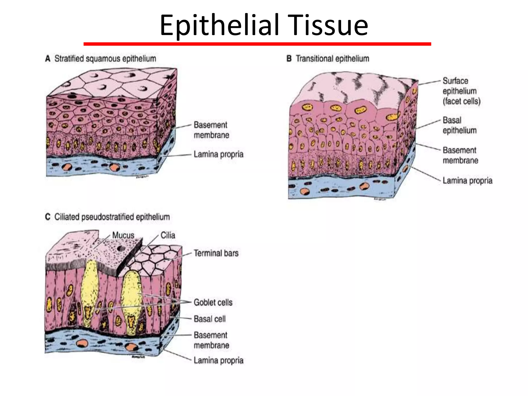
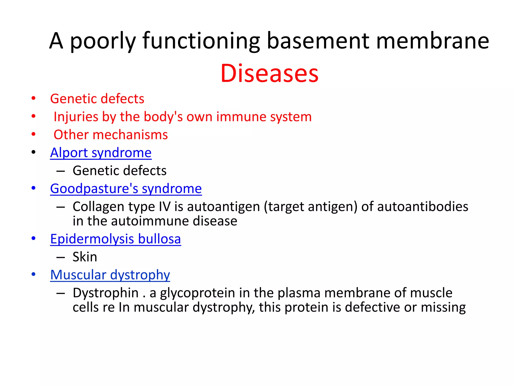
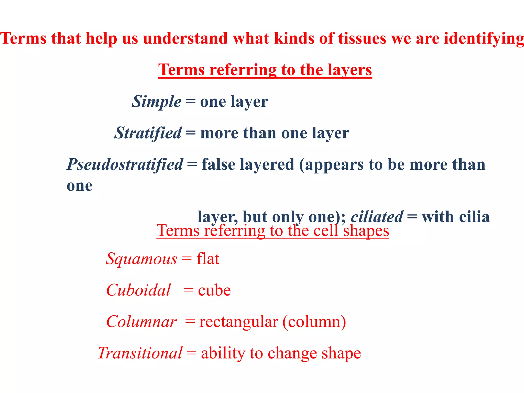
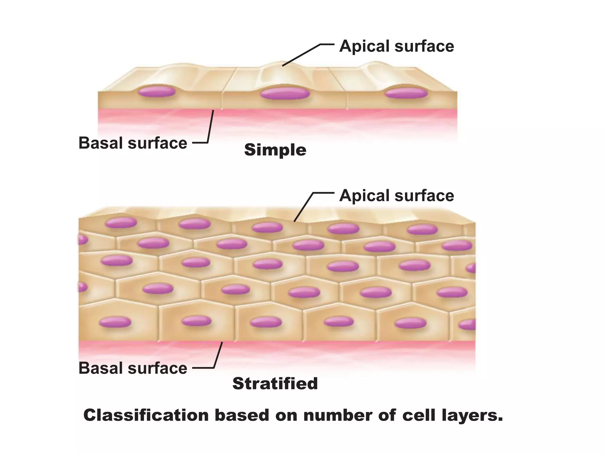
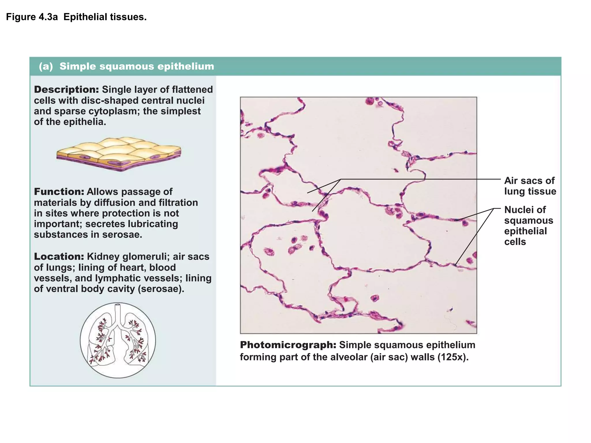
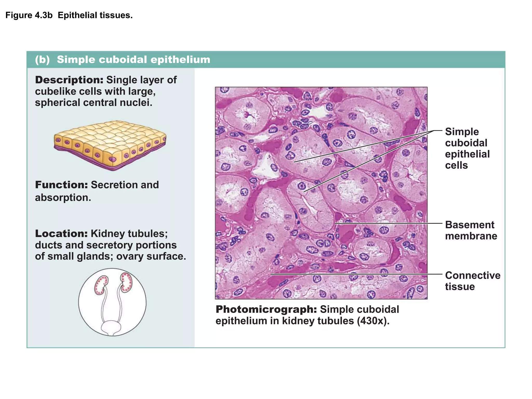
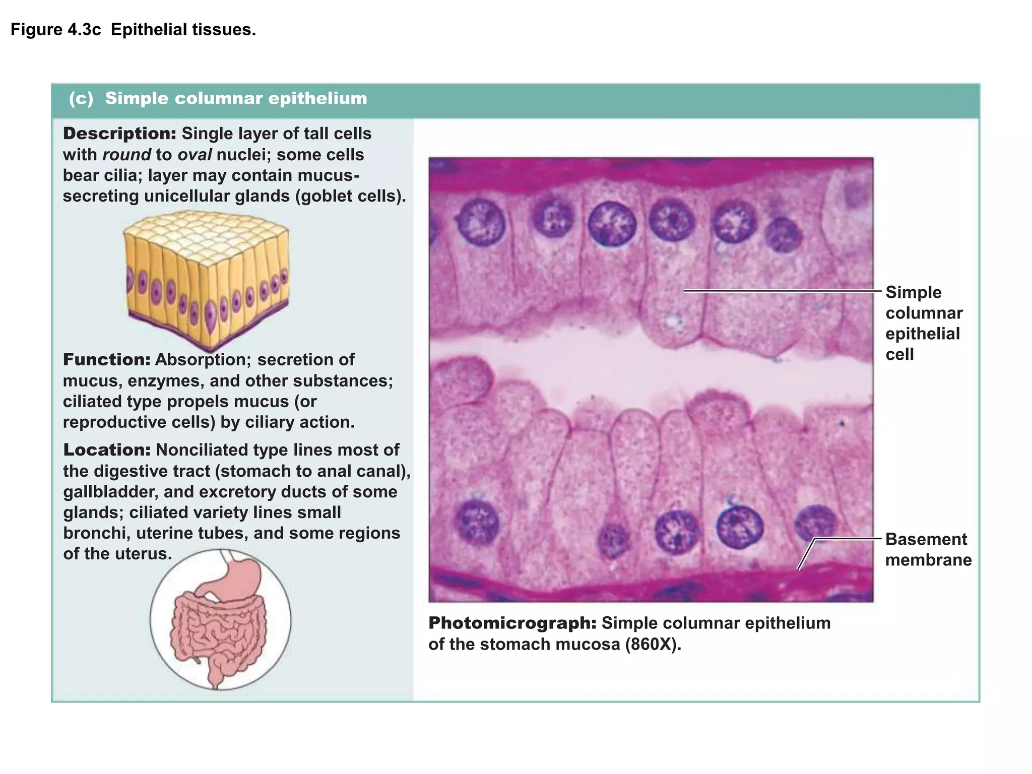
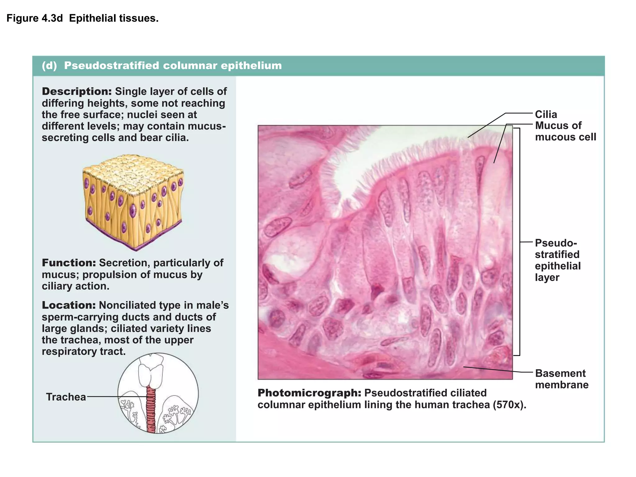
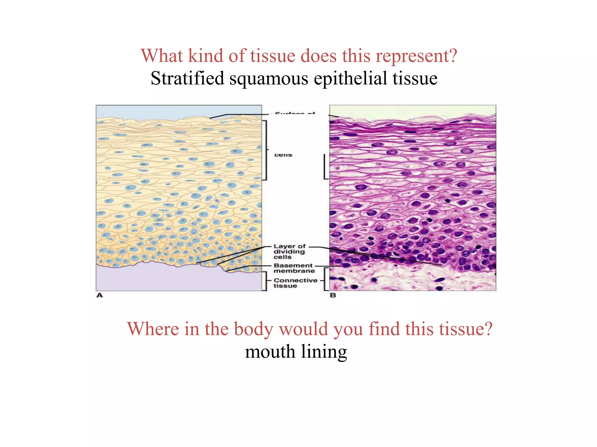
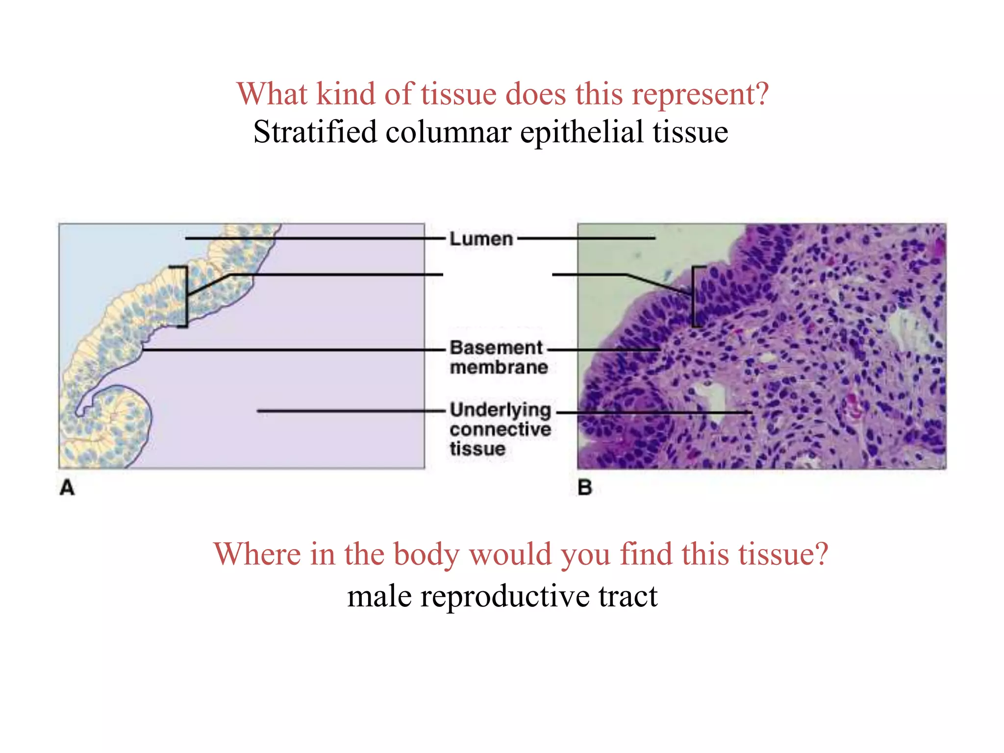
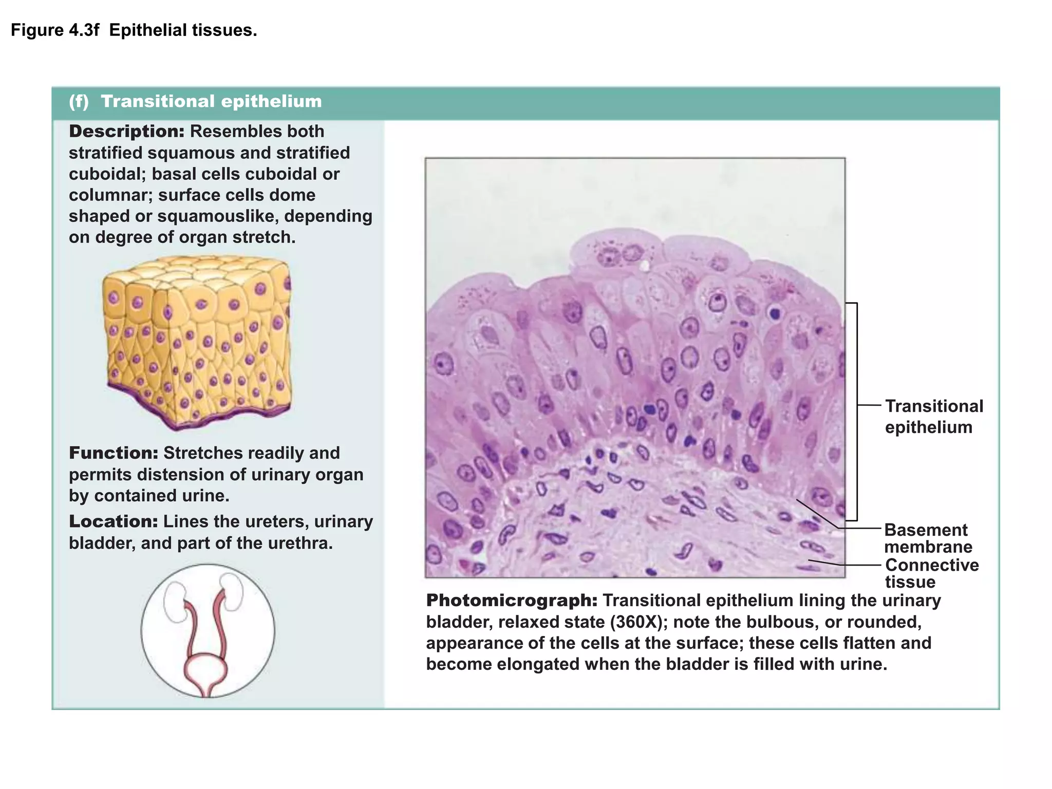
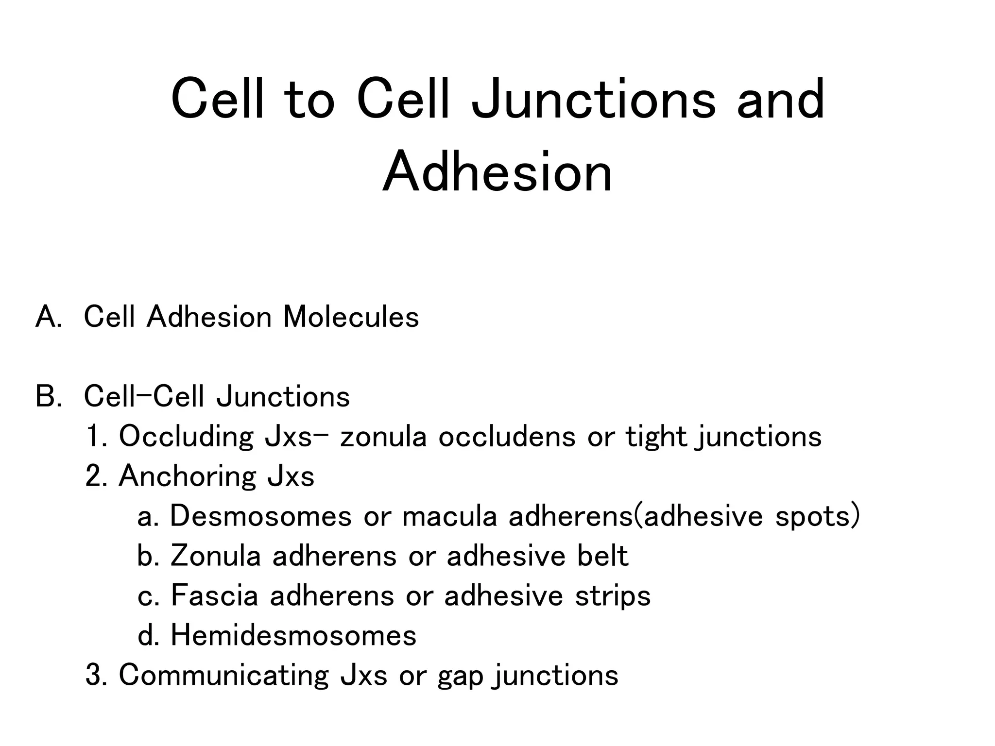
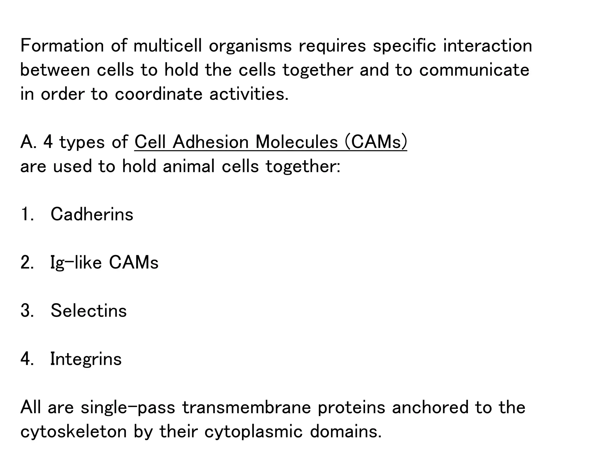
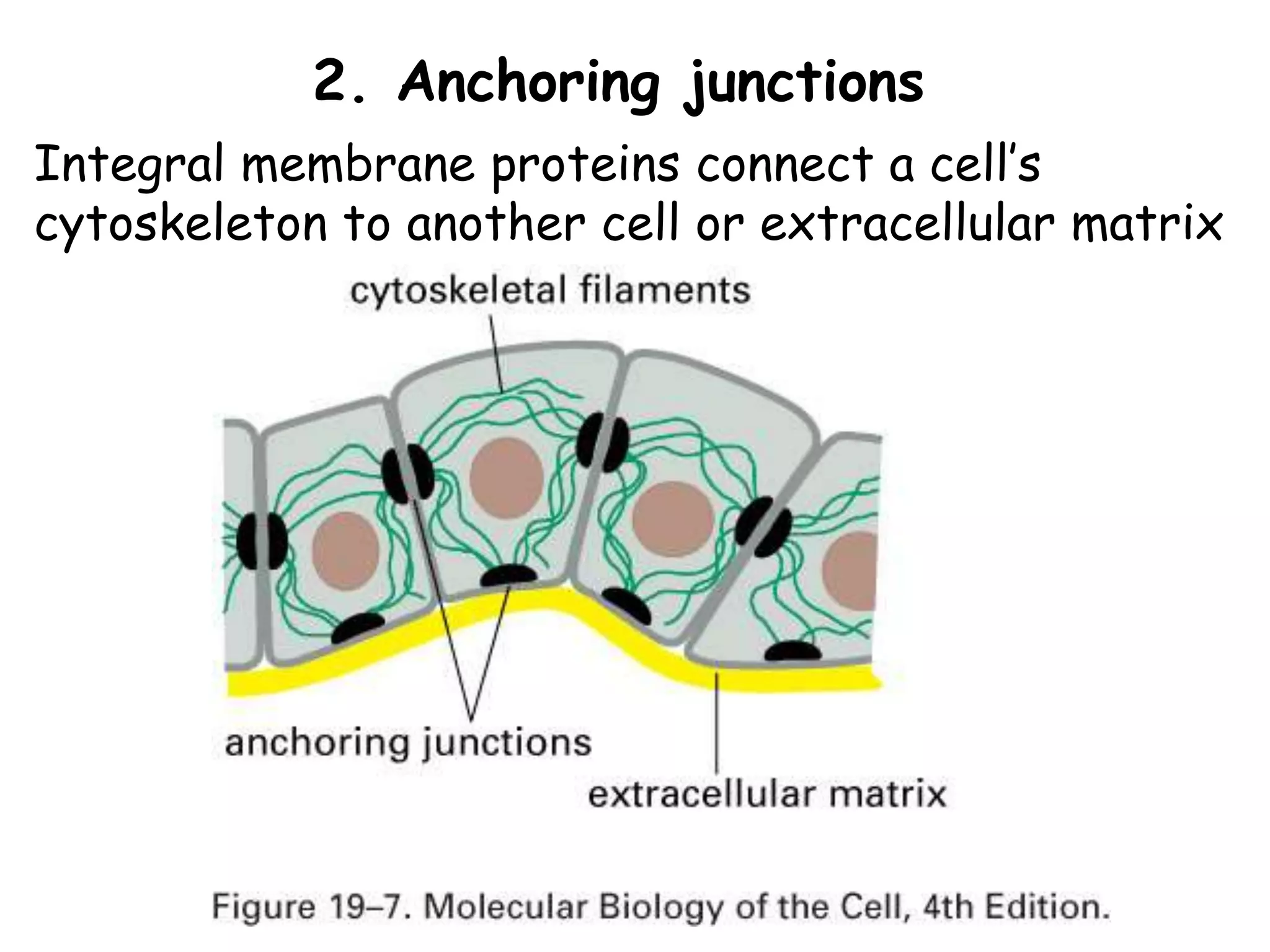
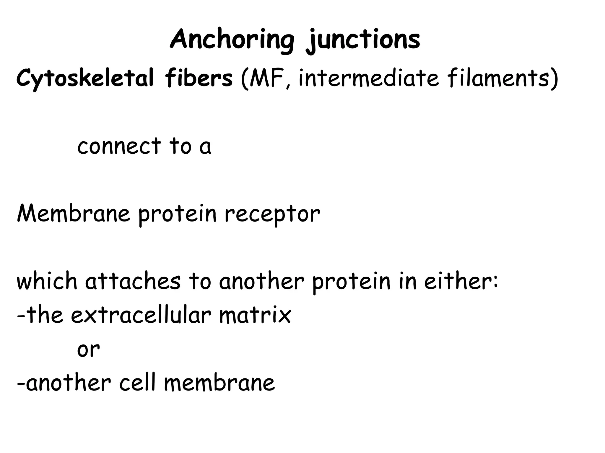
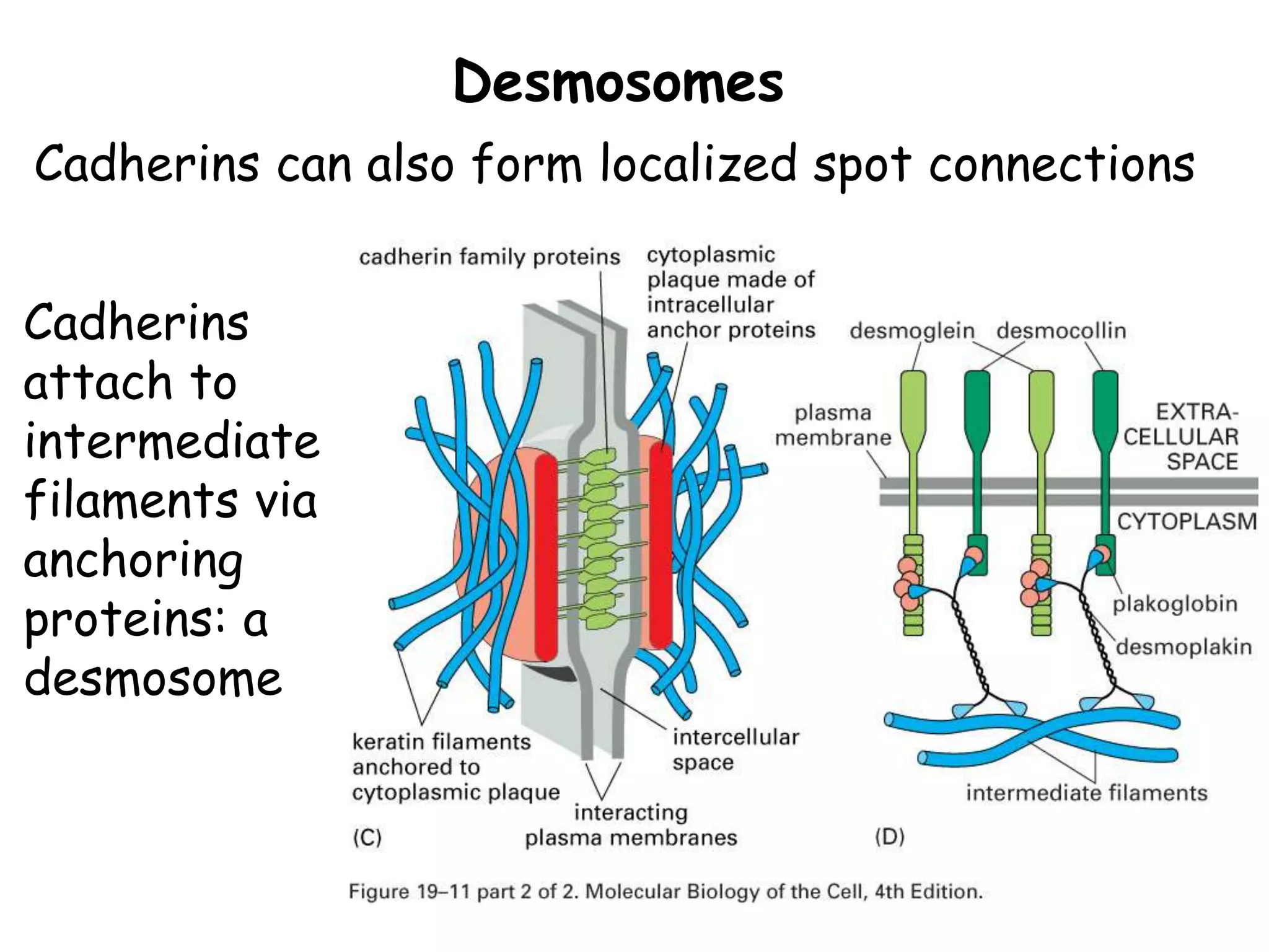
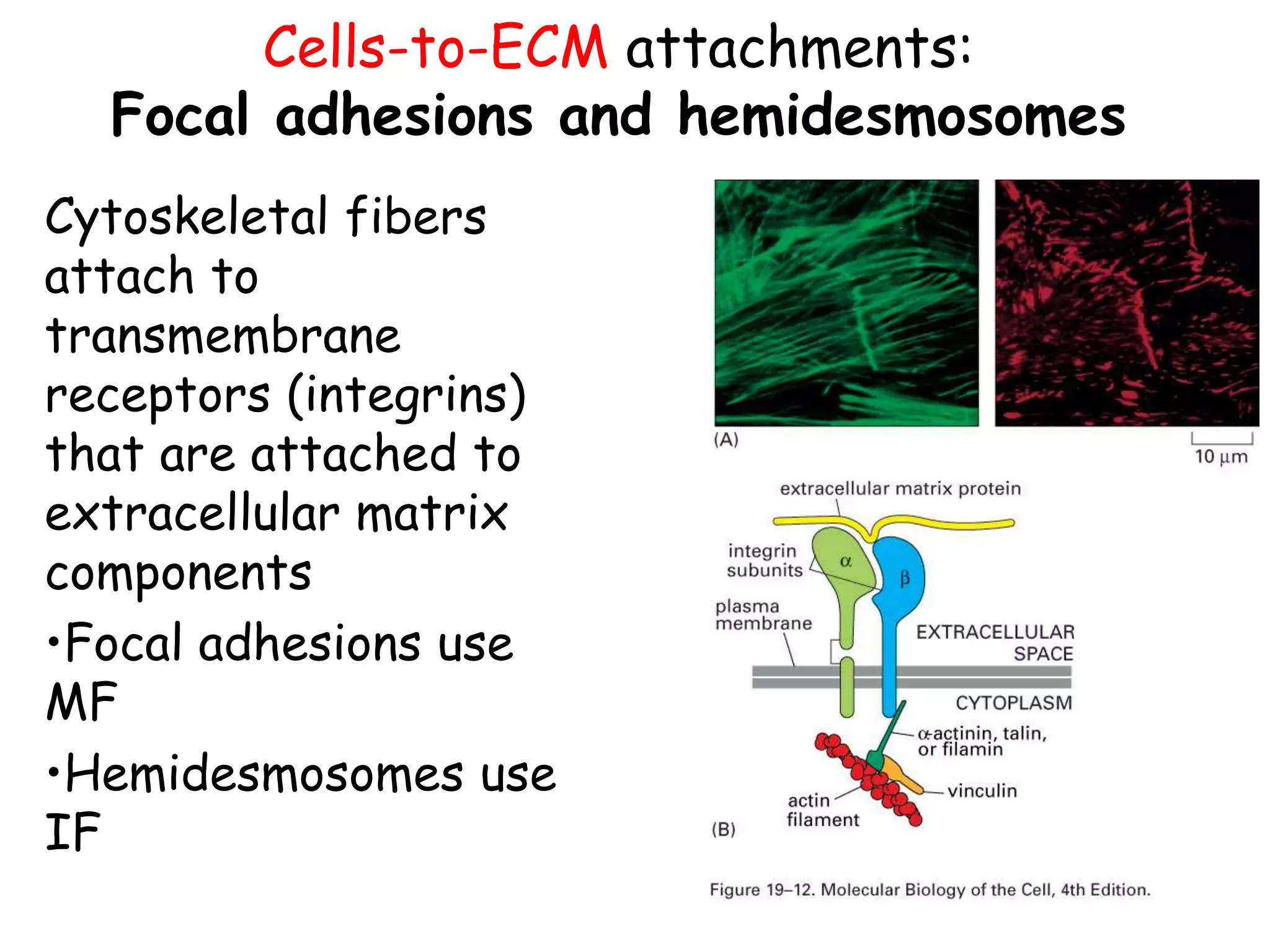
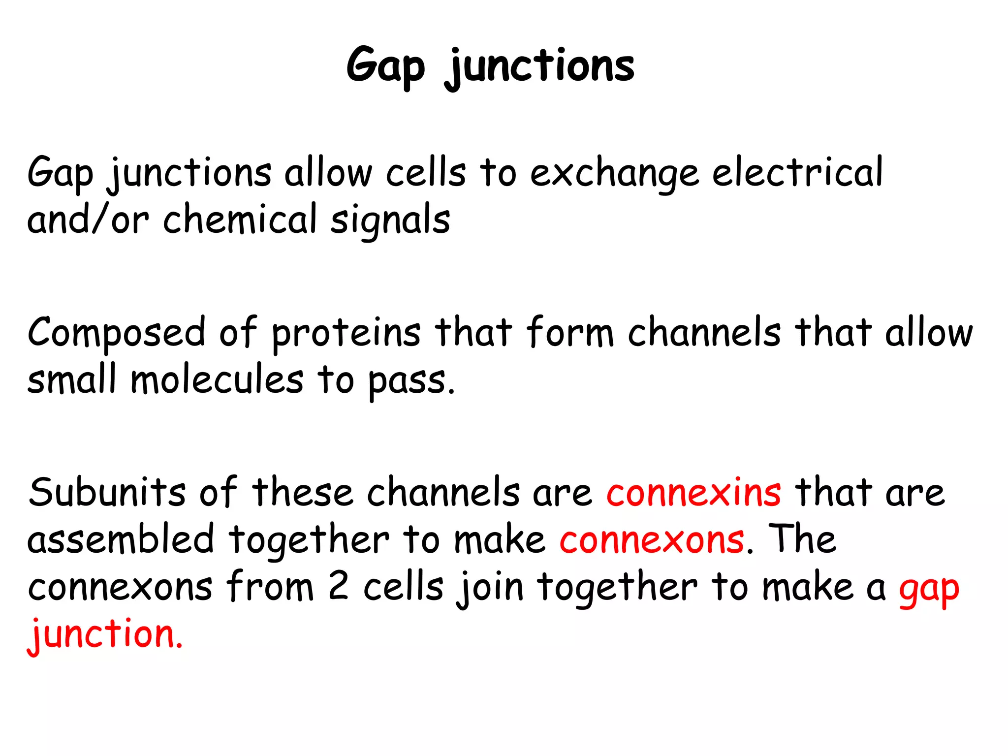
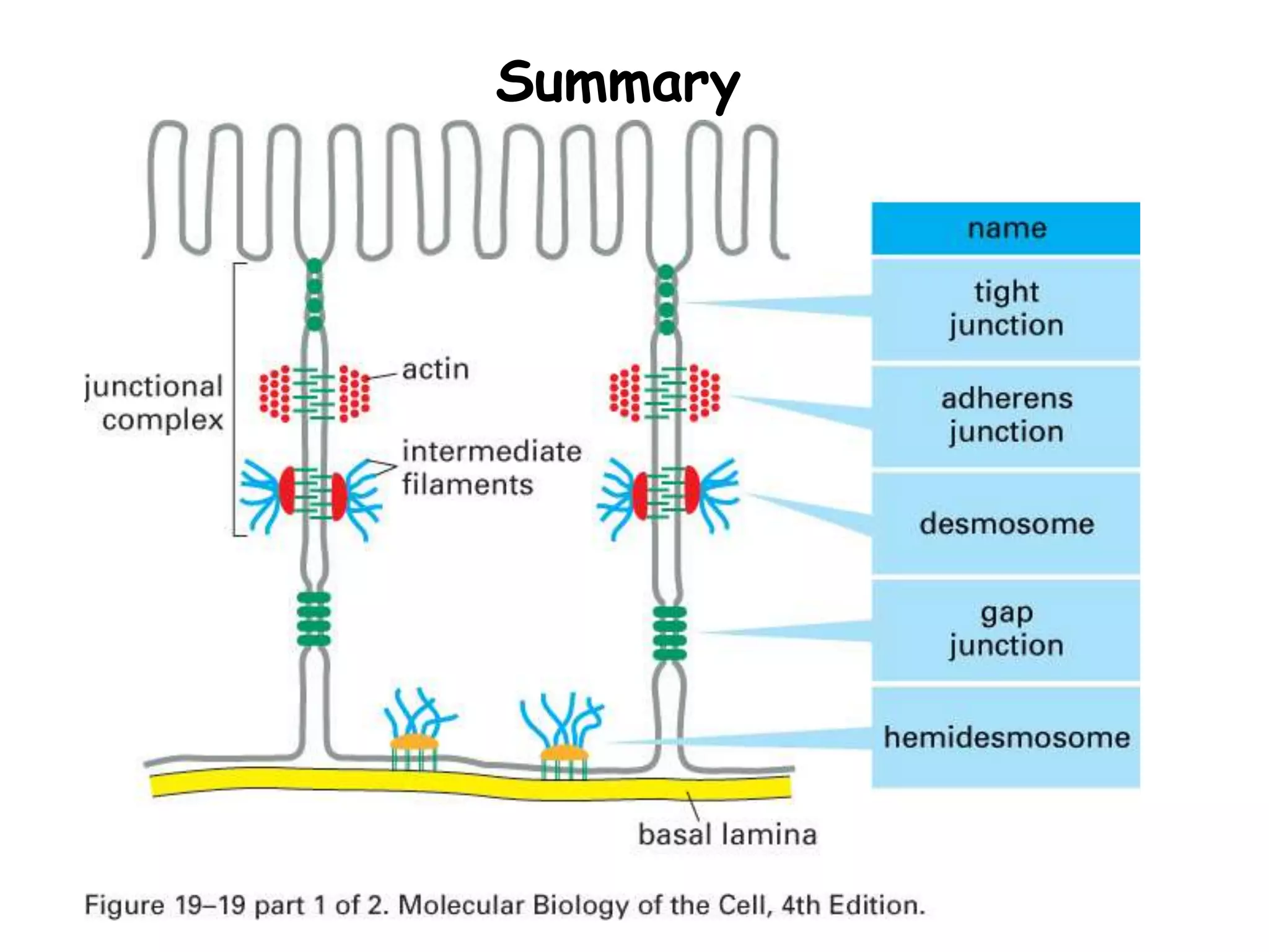
This document provides information on epithelial tissue and cell junctions. It discusses the general features of epithelial tissue, including that epithelial cells are closely packed with many cell junctions. It also describes the different types of epithelial tissue (simple vs stratified), the cell shapes (squamous, cuboidal, columnar), and locations in the body. The document further explains the structure and functions of the basement membrane and cell junctions, including occluding junctions, anchoring junctions, and communicating junctions. Key cellular adhesion molecules and proteins involved in different junction types are also outlined.
Epithelial tissue is a type of tissue that covers the surfaces of organs, lines body cavities and forms glands. This PDF provides a comprehensive overview of epithelial tissue, including its types, structure, function, and location in the body. The document covers the different types of epithelial cells, their shapes, and layers, as well as their functions in different organs and tissues. The PDF also delves into the unique features of epithelial tissue, such as its polarity, cell junctions, and the basement membrane. Additionally, Whether you are a student, medical professional, or someone interested in learning about the human body, this PDF provides a detailed understanding of epithelial tissue and its importance in the body.
Palestine last event orientationfvgnh .pptxRaedMohamed3
An EFL lesson about the current events in Palestine. It is intended to be for intermediate students who wish to increase their listening skills through a short lesson in power point.
Unit 8 - Information and Communication Technology (Paper I).pdfThiyagu K
This slides describes the basic concepts of ICT, basics of Email, Emerging Technology and Digital Initiatives in Education. This presentations aligns with the UGC Paper I syllabus.
How to Make a Field invisible in Odoo 17Celine George
It is possible to hide or invisible some fields in odoo. Commonly using “invisible” attribute in the field definition to invisible the fields. This slide will show how to make a field invisible in odoo 17.
Ethnobotany and Ethnopharmacology:
Ethnobotany in herbal drug evaluation,
Impact of Ethnobotany in traditional medicine,
New development in herbals,
Bio-prospecting tools for drug discovery,
Role of Ethnopharmacology in drug evaluation,
Reverse Pharmacology.
2024.06.01 Introducing a competency framework for languag learning materials ...Sandy Millin
http://sandymillin.wordpress.com/iateflwebinar2024
Published classroom materials form the basis of syllabuses, drive teacher professional development, and have a potentially huge influence on learners, teachers and education systems. All teachers also create their own materials, whether a few sentences on a blackboard, a highly-structured fully-realised online course, or anything in between. Despite this, the knowledge and skills needed to create effective language learning materials are rarely part of teacher training, and are mostly learnt by trial and error.
Knowledge and skills frameworks, generally called competency frameworks, for ELT teachers, trainers and managers have existed for a few years now. However, until I created one for my MA dissertation, there wasn’t one drawing together what we need to know and do to be able to effectively produce language learning materials.
This webinar will introduce you to my framework, highlighting the key competencies I identified from my research. It will also show how anybody involved in language teaching (any language, not just English!), teacher training, managing schools or developing language learning materials can benefit from using the framework.
The Indian economy is classified into different sectors to simplify the analysis and understanding of economic activities. For Class 10, it's essential to grasp the sectors of the Indian economy, understand their characteristics, and recognize their importance. This guide will provide detailed notes on the Sectors of the Indian Economy Class 10, using specific long-tail keywords to enhance comprehension.
For more information, visit-www.vavaclasses.com
Model Attribute Check Company Auto PropertyCeline George
In Odoo, the multi-company feature allows you to manage multiple companies within a single Odoo database instance. Each company can have its own configurations while still sharing common resources such as products, customers, and suppliers.
How to Create Map Views in the Odoo 17 ERPCeline George
The map views are useful for providing a geographical representation of data. They allow users to visualize and analyze the data in a more intuitive manner.
2. Epithelial Tissue -- General Features
• Closely packed cells with little extracellular
material
– Many cell junctions often provide secure
attachment.
• Cells sit on basement membrane
– Apical (upper) free surface
– Basal surface against basement membrane
• Avascular---without blood vessels
– nutrients and waste must move by diffusion
3. Epithelial Tissue -- General
Features
• Good nerve supply
• Rapid cell division (high mitotic rate)
• Functions
• protection, filtration, lubrication, secretion,
digestion, absorption, transportation,
excretion, sensory reception, and
reproduction.
4. Types of Epithelium
1. Covering and lining epithelium
– epidermis of skin
– lining of blood vessels and ducts
– lining respiratory, reproductive, urinary & GI
tract
2. Glandular epithelium- originate from
invaginated epithelial cells
– secreting portion of glands
– thyroid, adrenal, and sweat glands
5. Epithelium
• Epithelium is derived from all three germ
layers
• Ectoderm-oral and nasal mucosa,cornea ,
epidermis, glands of skin, mammary glands
• Endoderm-Lining of respiratory and
gastrointestinal tract, liver , panceas
• Mesoderm-lining of urogenital system,
circulatory system and body cavities lining-
mesothelium
8. Basement Membrane
• The basement membrane is a thin sheet of
fibers that underlies the epithelium
• The basement membrane is the fusion of two
lamina, the basal lamina-elaborated by
epithelial cells and the reticular lamina (or
lamina reticularis)-manufactured by cells of
connective tissue
9.
10. Structure of Basement membrane
• Basement Membrane
– Basal Lamina
• Lamina Lucida
– Extracellular glycoprotein-
Laminin,integrins,entactins,dystroglycans
– Transmembrane laminin receptors-project from epithelial cell
membrane into basal lamina
• Lamina Densa consists of a network of fine filaments.
– Type IV collagen. forms felt-like network of fibers that gives
the basement membrane its tensile strength
11. Structure of Basement membrane
• Lamina Reticularis
• Type III collagen (as reticular fibers)
• Attaching proteins (between Basal and
Reticular Laminae)-all elaborated by fibroblast
of connective tissue
• Type VII collagen (anchoring fibrils)
• fibrillin (microfibrils)
• Fibronectin
12. lamina lucida &lamina densa
• Lamina Densa
– dense layer closer to the connective tissue
– 30–70 nm in thickness
– consists of an underlying network of reticular
collagen (type IV) fibrils
• Lamina Lucida
– clear layer close to epithelium
16. Functions of basement membrane
• anchor down the epithelium to its loose
connective tissue (the dermis) underneath
– provide structural support to the tissue
• a mechanical barrier, preventing malignant
cells from invading the deeper tissues
17. Fusion of basal laminae
• Glomerular filtration of the kidney
– by the fusion of the basal lamina from the
endothelium of glomerular capillaries and the
basal lamina of the epithelium of the Bowman's
capsule
• Gaseous exchange between lung alveoli and
pulmonary capillaries
– by the fusion of the basal lamina of the lung
alveoli and of the basal lamina of the lung
capillaries
19. Cancer cells (Malignant)
• If the epithelial cells become transformed
(cancerous) and become 'malignant', they are
able to break through the basement
membrane and invade the tissues beneath.
This characteristic is used in the diagnosis of
malignant epithelial tumors
20. A poorly functioning basement membrane
Diseases
• Genetic defects
• Injuries by the body's own immune system
• Other mechanisms
• Alport syndrome
– Genetic defects
• Goodpasture's syndrome
– Collagen type IV is autoantigen (target antigen) of autoantibodies
in the autoimmune disease
• Epidermolysis bullosa
– Skin
• Muscular dystrophy
– Dystrophin . a glycoprotein in the plasma membrane of muscle
cells re In muscular dystrophy, this protein is defective or missing
21. Classification Of epithelium
According to number of cell layers between
basal lamina and free surface and by
morphology of epithelial cells
1. Simple epithelium- composed of single layer
of cells
2. Stratified epithelium- composed of more
than one cells
22. Terms that help us understand what kinds of tissues we are identifying
Terms referring to the layers
Simple = one layer
Stratified = more than one layer
Pseudostratified = false layered (appears to be more than
one
layer, but only one); ciliated = with cilia
Terms referring to the cell shapes
Squamous = flat
Cuboidal = cube
Columnar = rectangular (column)
Transitional = ability to change shape
27. Figure 4.3a Epithelial tissues.
(a) Simple squamous epithelium
Description: Single layer of flattened
cells with disc-shaped central nuclei
and sparse cytoplasm; the simplest
of the epithelia.
Function: Allows passage of
materials by diffusion and filtration
in sites where protection is not
important; secretes lubricating
substances in serosae.
Location: Kidney glomeruli; air sacs
of lungs; lining of heart, blood
vessels, and lymphatic vessels; lining
of ventral body cavity (serosae).
Photomicrograph: Simple squamous epithelium
forming part of the alveolar (air sac) walls (125x).
Air sacs of
lung tissue
Nuclei of
squamous
epithelial
cells
29. Figure 4.3b Epithelial tissues.
(b) Simple cuboidal epithelium
Description: Single layer of
cubelike cells with large,
spherical central nuclei.
Function: Secretion and
absorption.
Location: Kidney tubules;
ducts and secretory portions
of small glands; ovary surface.
Photomicrograph: Simple cuboidal
epithelium in kidney tubules (430x).
Basement
membrane
Connective
tissue
Simple
cuboidal
epithelial
cells
30. Figure 4.3c Epithelial tissues.
(c) Simple columnar epithelium
Description: Single layer of tall cells
with round to oval nuclei; some cells
bear cilia; layer may contain mucus-
secreting unicellular glands (goblet cells).
Function: Absorption; secretion of
mucus, enzymes, and other substances;
ciliated type propels mucus (or
reproductive cells) by ciliary action.
Location: Nonciliated type lines most of
the digestive tract (stomach to anal canal),
gallbladder, and excretory ducts of some
glands; ciliated variety lines small
bronchi, uterine tubes, and some regions
of the uterus.
Photomicrograph: Simple columnar epithelium
of the stomach mucosa (860X).
Simple
columnar
epithelial
cell
Basement
membrane
31. Figure 4.3d Epithelial tissues.
(d) Pseudostratified columnar epithelium
Description: Single layer of cells of
differing heights, some not reaching
the free surface; nuclei seen at
different levels; may contain mucus-
secreting cells and bear cilia.
Function: Secretion, particularly of
mucus; propulsion of mucus by
ciliary action.
Location: Nonciliated type in male’s
sperm-carrying ducts and ducts of
large glands; ciliated variety lines
the trachea, most of the upper
respiratory tract.
Photomicrograph: Pseudostratified ciliated
columnar epithelium lining the human trachea (570x).
Trachea
Cilia
Pseudo-
stratified
epithelial
layer
Basement
membrane
Mucus of
mucous cell
32. Given the previous examples
(consider the morphology only)
Can you name?
First, the tissue type
Second, where in the body the tissue is found
33. What kind of tissue does this represent?
Where in the body would you find this tissue?
Simple squamous epithelial tissue
lungs
34. What kind of tissue does this represent?
Simple squamous epithelial tissue (superior view)
35. What kind of tissue does this represent?
Where in the body would you find this tissue?
Simple cuboidal epithelial tissue
Kidneys (tubules)
The lining of the kidney glomerulus (sing.)/glomeruli (pl.) is simple squamous epithelial tissue
36. What kind of tissue does this represent?
Where in the body would you find this tissue?
Simple columnar epithelial tissue
small intestine
37. What kind of tissue does this represent?
Where in the body would you find this tissue?
Pseudostratified (ciliated) columnar epithelial tissue
“false layered”; it looks like more than one layer, but it is not
trachea lining
38. What kind of tissue does this represent?
Where in the body would you find this tissue?
Stratified squamous epithelial tissue
mouth lining
39. What kind of tissue does this represent?
Where in the body would you find this tissue?
Stratified cuboidal epithelial tissue
salivary glands, sweat glands
40. What kind of tissue does this represent?
Where in the body would you find this tissue?
Stratified columnar epithelial tissue
male reproductive tract
41. What kind of tissue does this represent?
Where in the body would you find this tissue?
Transitional epithelial tissue
empty bladder
42. Where in the body would you find this tissue?
What kind of tissue does this represent?
distended (full) bladder
Transitional epithelial tissue
43. Figure 4.3e Epithelial tissues.
(e) Stratified squamous epithelium
Description: Thick membrane
composed of several cell layers;
basal cells are cuboidal or columnar
and metabolically active; surface
cells are flattened (squamous); in the
keratinized type, the surface cells are
full of keratin and dead; basal cells
are active in mitosis and produce the
cells of the more superficial layers.
Function: Protects underlying
tissues in areas subjected to abrasion.
Location: Nonkeratinized type forms
the moist linings of the esophagus,
mouth, and vagina; keratinized variety
forms the epidermis of the skin, a dry
membrane.
Photomicrograph: Stratified squamous epithelium
lining the esophagus (285x).
Stratified
squamous
epithelium
Nuclei
Basement
membrane
Connective
tissue
44. Figure 4.3f Epithelial tissues.
(f) Transitional epithelium
Description: Resembles both
stratified squamous and stratified
cuboidal; basal cells cuboidal or
columnar; surface cells dome
shaped or squamouslike, depending
on degree of organ stretch.
Function: Stretches readily and
permits distension of urinary organ
by contained urine.
Location: Lines the ureters, urinary
bladder, and part of the urethra.
Photomicrograph: Transitional epithelium lining the urinary
bladder, relaxed state (360X); note the bulbous, or rounded,
appearance of the cells at the surface; these cells flatten and
become elongated when the bladder is filled with urine.
Basement
membrane
Connective
tissue
Transitional
epithelium
45. Cell junctions
• Cell junctions consist of multiprotein
complexes that provide contact between
neighboring cells or between a cell and the
extracellular matrix.
• They also build up the paracellular barrier
of epithelia and control the paracellular
transport.
• Cell junctions are especially abundant in
epithelial tissues.
46. Cell to Cell Junctions and
Adhesion
A. Cell Adhesion Molecules
B. Cell-Cell Junctions
1. Occluding Jxs- zonula occludens or tight junctions
2. Anchoring Jxs
a. Desmosomes or macula adherens(adhesive spots)
b. Zonula adherens or adhesive belt
c. Fascia adherens or adhesive strips
d. Hemidesmosomes
3. Communicating Jxs or gap junctions
47. Formation of multicell organisms requires specific interaction
between cells to hold the cells together and to communicate
in order to coordinate activities.
A. 4 types of Cell Adhesion Molecules (CAMs)
are used to hold animal cells together:
1. Cadherins
2. Ig-like CAMs
3. Selectins
4. Integrins
All are single-pass transmembrane proteins anchored to the
cytoskeleton by their cytoplasmic domains.
48. Importance of Cell junction
• Cell junctions enable communication
between neighboring cells via specialized
proteins called communicating junctions.
• Cell junctions are also important in
reducing stress placed upon cells.
• Combined with CAMs( cell adhesion
molecule) and ECM, cell junctions help
hold animal cells together.
51. Cell junctions
Three types of cell junctions:
1. Occluding junctions: seal cells together into
sheets (forming an impermeable barrier)
2. Anchoring junctions: attach cells (and their
cytoskeleton) to other cells or extracellular matrix
(providing mechanical support)
3. Communicating junctions: allow exchange of
chemical/electrical information between cells
53. 1.Occluding -Tight junction
Each cell
possesses
integral
membrane
proteins that
bind to
similar
proteins in
the adjacent,
forming a
continuous
“weld”
56. Anchoring junctions
Cytoskeletal fibers (MF, intermediate filaments)
connect to a
Membrane protein receptor
which attaches to another protein in either:
-the extracellular matrix
or
-another cell membrane
57. 2a. Cadherins and desmosomes
Cell to cell connections
are mediated by
cadherins.
These receptors
extend out from the
cell, binding to other
cadherens
58. Cadherins participiate in adherens junctions
Under the cell
membrane,
contractile fibers of
microfilaments
connect to cell
membrane proteins
called cadherins
They surround the
cell, forming a belt
59. Desmosomes
Cadherins can also form localized spot connections
Cadherins
attach to
intermediate
filaments via
anchoring
proteins: a
desmosome
60. Cells-to-ECM attachments:
Focal adhesions and hemidesmosomes
Cytoskeletal fibers
attach to
transmembrane
receptors (integrins)
that are attached to
extracellular matrix
components
•Focal adhesions use
MF
•Hemidesmosomes use
IF
61. Gap junctions
Gap junctions allow cells to exchange electrical
and/or chemical signals
Composed of proteins that form channels that allow
small molecules to pass.
Subunits of these channels are connexins that are
assembled together to make connexons. The
connexons from 2 cells join together to make a gap
junction.