Download to read offline

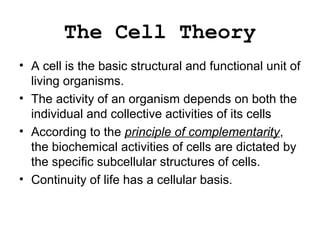
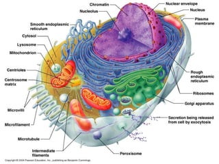
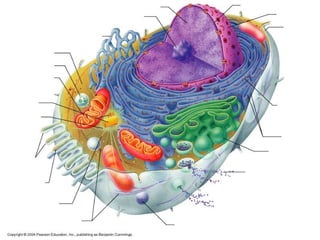
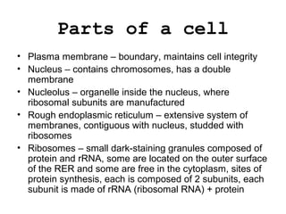
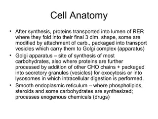
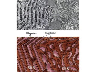
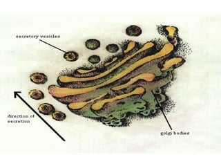
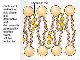
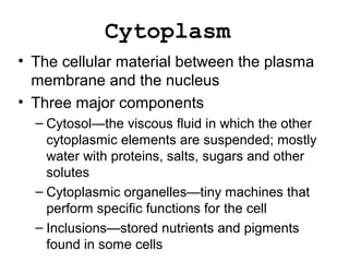

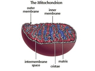
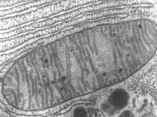
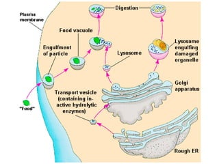
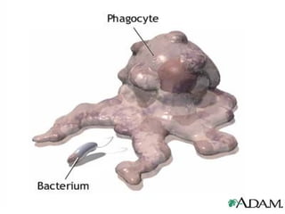
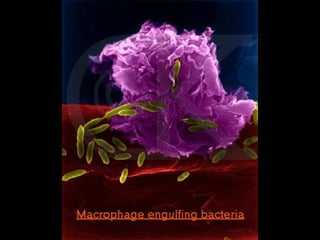
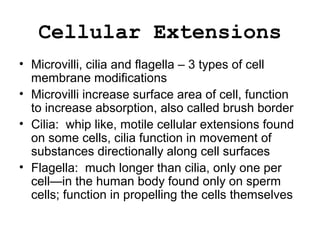
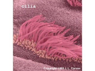
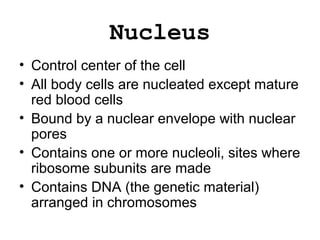
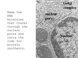
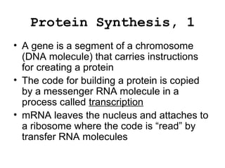

The document summarizes key aspects of cell structure and function according to the cell theory. It describes the basic components of cells, including the plasma membrane, nucleus, cytoplasm, and various organelles. It explains that cells are the basic structural and functional units of living organisms, and that their biochemical activities depend on their specific subcellular structures. The key cellular processes of protein synthesis and transport are also summarized.


