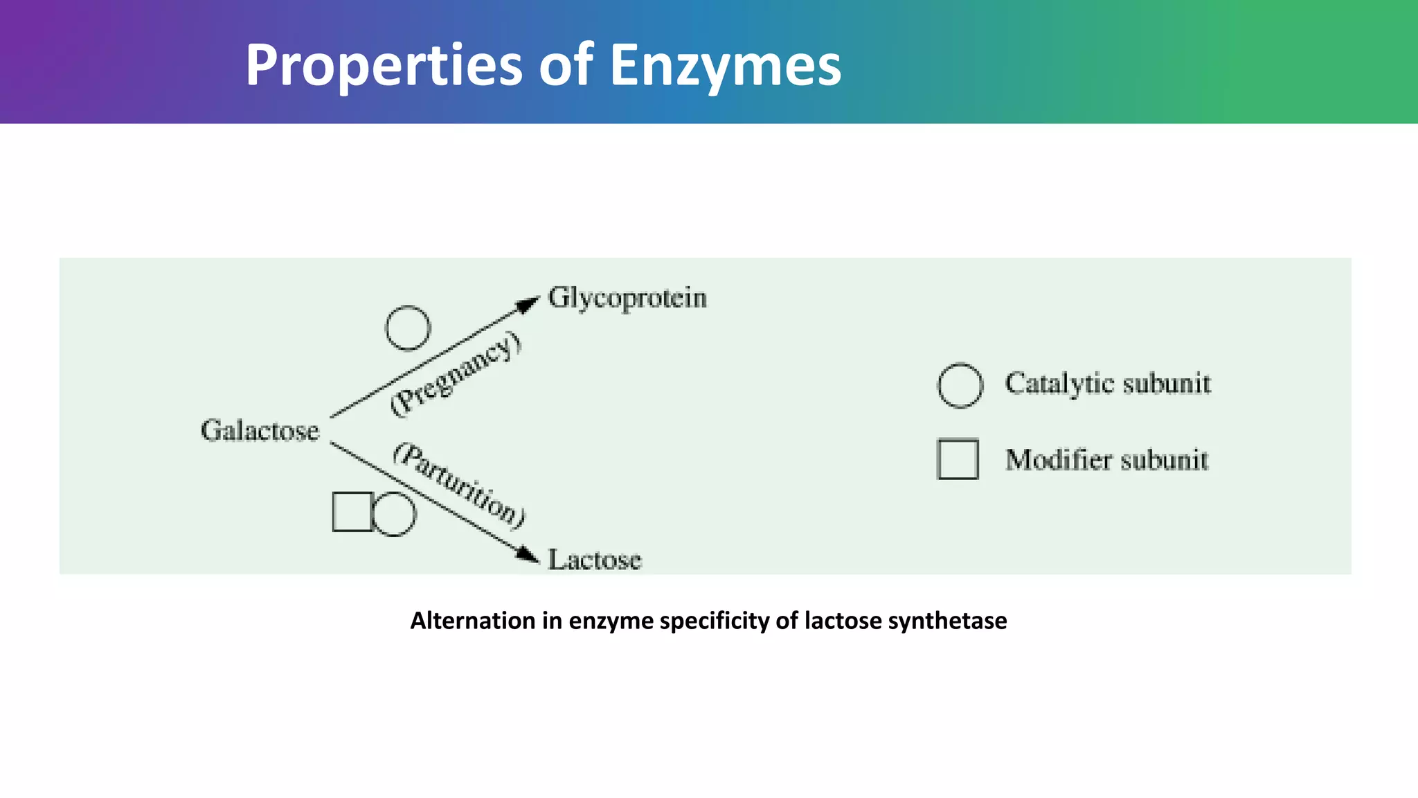
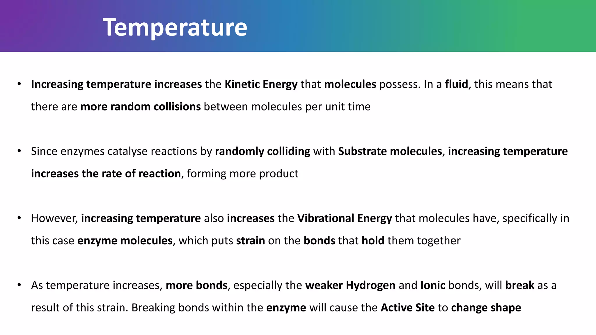
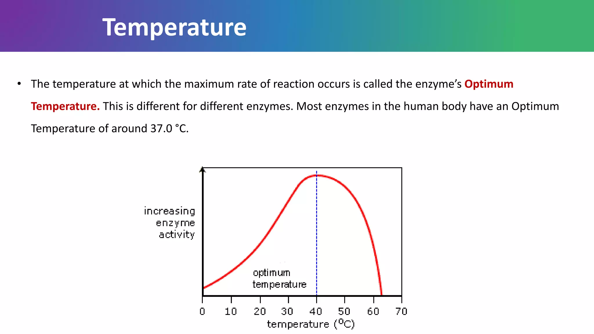
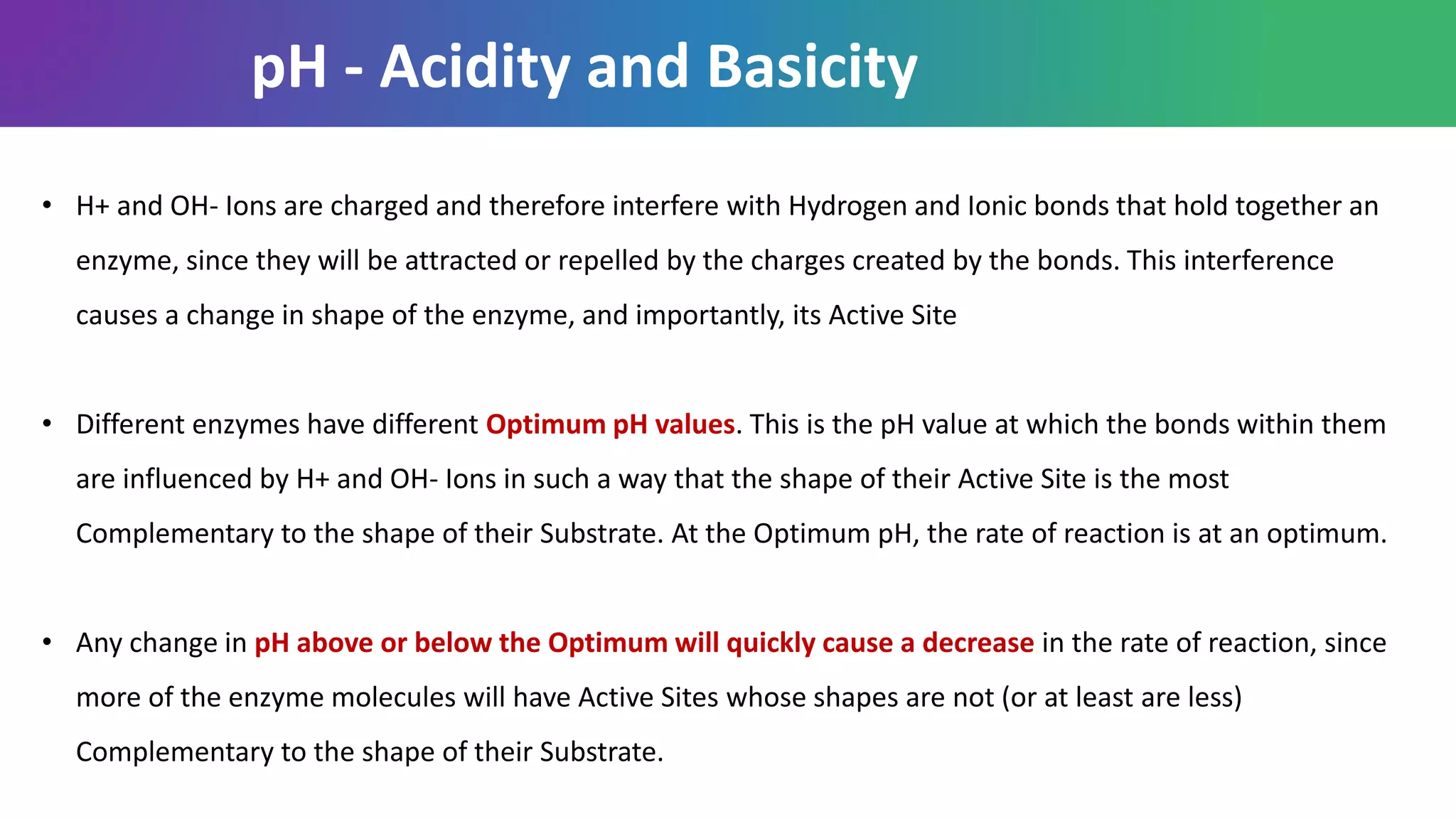
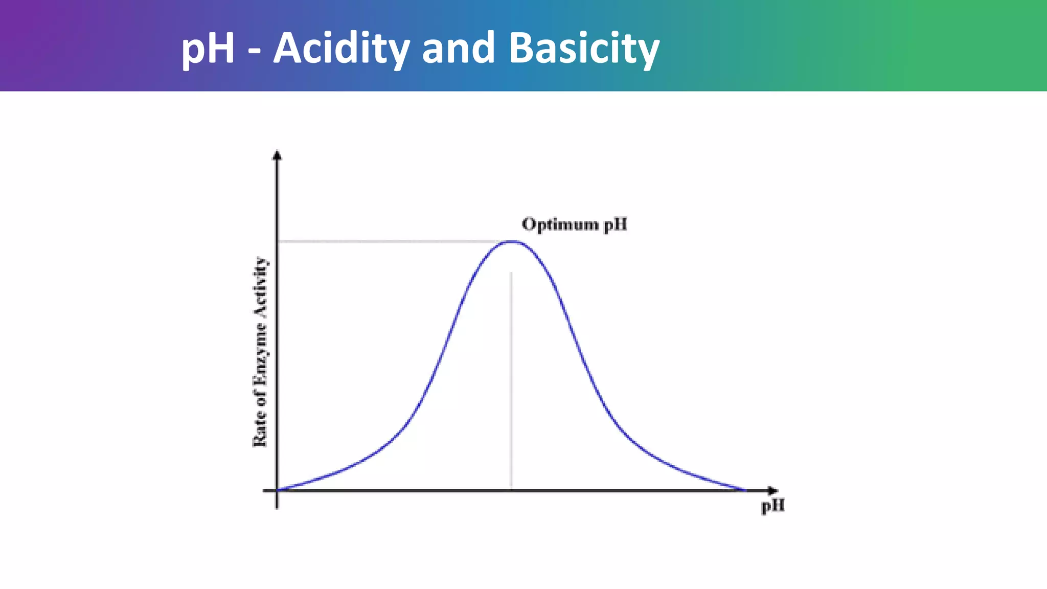
An enzyme is a substance that acts as a catalyst in living organisms, regulating the rate at which chemical reactions proceed without itself being altered in the process. The biological processes that occur within all living organisms are chemical reactions, and most are regulated by enzymes
Active sites of the enzyme is that point where substrate molecule bind for the chemical reaction. It is generally found on the surface of enzyme and in some enzyme it is a “Pit” like structure
The active site is a three-dimensional cleft formed by groups that come from different parts of the amino acid sequence
The active site takes up a relatively small part of the total volume of an enzyme
Active sites are clefts or crevices
Substrates are bound to enzymes by multiple weak attractions.
The specificity of binding depends on the precisely defined arrangement of atoms in an active site.
Enzymes properties, nomenclature and classificationJasmineJuliet
Enzymes - Definition, Introduction about biocatalysts, Properties of enzymes, Specificity, capacity for regulation, Example for enzyme at specific pH, Nomenclature of enzymes, Systematic name, common name, enzyme commission number, Classification of enzymes: Oxidoreductase, Transferase, lyases, ligases, isomerases, hydrolases.
Enzyme inhibition is explained with its kinetics, animations showing mechanism of inhibitors action, examples of inhibitors are explained in detail with Enzyme inhibited.
by Dr. N. Sivaranjani, MD
Enzymes definitions, types & classificationJasmineJuliet
Enzyme - Introduction, Biocatalysts, Definition of enzymes, Types of enzymes, classification of enzyme, Nomenclature of enzymes, EC number, Types of enzymes with examples, and reaction.
Coenzyme - Introduction, Definition, Examples for coenzyme, reaction catalysed by coenzyme, Types of coenzymes - cosubstrate and prosthetic group coenzymes, second type of classification of coenzyme- hydrogen group transfer , other than hydrogen group transfer.
In this title include the the definition of the the enzymes, their naming , classification, inhibition, and regulation and clinical applications are briefly explained.
Introduction
Definition
Historical aspects
Nomenclature of enzymes on the basis of
1. Substrate acted
2. Reaction catalyzed
3. substrate act upon and type of reaction catalyzed
Classification of enzymes
Oxidoreductase
Transferase
Hydrolase
Lyase
Isomerase
Ligase
Property of enzyme
Structure of enzyme
Mechanism of enzyme action
Lock and key model
Induced fit model
factors affecting enzyme activity
Control of enzyme action
Conclusion
Reference
The aqueous humour is a transparent, watery fluid similar to plasma, but containing low protein concentrations. It is secreted from the ciliary epithelium, a structure supporting the lens
The cornea is the transparent front part of the eye that covers the iris, pupil, and anterior chamber. The cornea, with the anterior chamber and lens, refracts light, with the cornea accounting for approximately two-thirds of the eye's total optical power.
Lens is a transparent, biconvex, crystalline structure placed between iris and the vitreous in a saucer-shaped depression, the patellar fossa. The lens is a crystalline structure that is avascular and is devoid of nerves and connective tissue
It consists of three distinct part:
Lens capsule
Anterior lens epithelium, and
Lens substance or lens fibres
Small amounts of vitamins are required in the diet to promote growth, reproduction, and health. Vitamins A, D, E, and K are called the fat-soluble vitamins, because they are soluble in organic solvents and are absorbed and transported in a manner similar to that of fats.
Water soluble vitamins include Vitamin C and the vitamin B complex: thiamin (B1), riboflavin (B2), niacin (B3), pantothenic acid (B5), Vitamin B6, biotin (B7), folic acid (B9), Vitamin B12. Vitamin A in its Beta-Carotene form is also water-soluble.
The tear film is a complex mixture of substances secreted from multiple sources on the ocular surface, including the lacrimal gland, the accessory lacrimal glands, the meibomian glands, and the goblet cells.
A picornavirus is a virus belonging to the family Picornaviridae, a family of viruses in the order Picornavirales. Vertebrates, including humans, serve as natural hosts. Picornaviruses are nonenveloped viruses that represent a large family of small, cytoplasmic, plus-strand RNA viruses with a 30-nm icosahedral capsid.
Poxviruses are brick or oval-shaped viruses with large double-stranded DNA genomes. Poxviruses exist throughout the world and cause disease in humans and many other types of animals. Poxvirus infections typically result in the formation of lesions, skin nodules, or disseminated rash.
Leptospirosis is a bacterial disease that affects humans and animals. It is caused by bacteria of the genus Leptospira. In humans, it can cause a wide range of symptoms, some of which may be mistaken for other diseases. Some infected persons, however, may have no symptoms at all.
The human immunodeficiency virus (HIV) is a lentivirus (a subgroup of retrovirus) that causes HIV infection and over time acquired immunodeficiency syndrome (AIDS).
Treponema is a genus of spiral-shaped bacteria. The major treponeme species of human pathogens is Treponema pallidum, whose subspecies are responsible for diseases such as syphilis, bejel, and yaws.
Haemophilus is the name of a group of bacteria. There are several types of Haemophilus. They can cause different types of illnesses involving breathing, bones and joints, and the nervous system. One common type, Hib (Haemophilus influenzae type b), causes serious disease. It usually strikes children under 5 years old
Moraxella is a genus of Gram-negative bacteria in the Moraxellaceae family. It is named after the Swiss ophthalmologist Victor Morax. The organisms are short rods, coccobacilli, or as in the case of Moraxella catarrhalis, diplococci in morphology, with asaccharolytic, oxidase-positive, and catalase-positive properties
Pseudomonas is a type of bacteria that can cause infections. Pseudomonas is a common genus of bacteria, which can create infections in the body under certain circumstances. There are many different types of Pseudomonas bacteria
Neisseria gonorrhoeae is the obligate human pathogen that causes the sexually transmitted disease (STD) gonorrhea. This Gram-negative diplococci/gonococci does not infect other animals or experimental animals and does not survive freely in the environment. The gonococcal infection occurs in the upper or lower tract, pharynx, ophthalmic area, rectum, and bloodstream. During the 1980’s gonorrhea was also referred to as “the clap” when public awareness was quite minimal. This was one of the venereal diseases prostitutes hoped to contract since it resulted in infertility by pelvic inflammatory disease (PID). As documentation, diagnostic testing, and public awareness improved, there has been a decline in incidence reports, however, it is still considered a very common infectious disease.
Meningococci are a type of bacteria that cause serious infections. The most common infection is meningitis, which is an inflammation of the thin tissue that surrounds the brain and spinal cord. Meningococci can also cause other problems, including a serious bloodstream infection called sepsis. In its early stages, you may have flu-like symptoms and a stiff neck. But the disease can progress quickly and can be fatal. Early diagnosis and treatment are extremely important. Lab tests on your blood and cerebrospinal fluid can tell if you have it. Treatment is with antibiotics. Since the infection spreads from person to person, family members may also need to be treated.
A vaccine can prevent meningococcal infections.
Diphtheria is an infection caused by the bacterium Corynebacterium diphtheriae. Diphtheria causes a thick covering in the back of the throat. It can lead to difficulty breathing, heart failure, paralysis, and even death. CDC recommends vaccines for infants, children, teens and adults to prevent diphtheria. The presentation consists of basic concepts regarding the bacteria and its infection. It has explanation in detail about signs and symptoms of Diptheria
Contraindications, Adverse reactions and ocular nutritional supplementsArun Geetha Viswanathan
utritional supplements comprise a great deal of the products available over the counter in most pharmacies. Although most vitamin supplements are relatively harmless—except for the fat soluble ones A, D, E, and K—they are not the only supplements available to patients. Some of these other, non-vitamin supplements can actually be harmful to patients and often they have been proven to be ineffective. This doesn’t mean that patients will stop taking them though, which in turn leaves the potential for contraindications of nutritional supplements with prescription-based drugs wide open.
Ageing is a gradual process that takes place over many decades. Most theories of ageing relate to impaired DNA replication and loss of cell viability and hence the viability of the body’s organs. Ageing is often accompanied by socioeconomic changes that can have a great impact on the nutritional needs and status of elderly individuals. The incidence of disability increases with ageing, with over a third of the elderly population limited by chronic conditions and unable to carry on normal daily living activity
Every component of the eye is vulnerable to damage from ROI, particularly retina. There are several reasons for the vulnerability of the retina, including high concentrations of polyunsaturated fatty acid (PUFA), constant exposure to visible light, high consumption of oxygen, an abundance of photosensitisers in the neurosensory retina and the RPE, the process of phagocytosis by the RPE which is known to generate hydrogen peroxide.
Professional air quality monitoring systems provide immediate, on-site data for analysis, compliance, and decision-making.
Monitor common gases, weather parameters, particulates.
(May 29th, 2024) Advancements in Intravital Microscopy- Insights for Preclini...Scintica Instrumentation
Intravital microscopy (IVM) is a powerful tool utilized to study cellular behavior over time and space in vivo. Much of our understanding of cell biology has been accomplished using various in vitro and ex vivo methods; however, these studies do not necessarily reflect the natural dynamics of biological processes. Unlike traditional cell culture or fixed tissue imaging, IVM allows for the ultra-fast high-resolution imaging of cellular processes over time and space and were studied in its natural environment. Real-time visualization of biological processes in the context of an intact organism helps maintain physiological relevance and provide insights into the progression of disease, response to treatments or developmental processes.
In this webinar we give an overview of advanced applications of the IVM system in preclinical research. IVIM technology is a provider of all-in-one intravital microscopy systems and solutions optimized for in vivo imaging of live animal models at sub-micron resolution. The system’s unique features and user-friendly software enables researchers to probe fast dynamic biological processes such as immune cell tracking, cell-cell interaction as well as vascularization and tumor metastasis with exceptional detail. This webinar will also give an overview of IVM being utilized in drug development, offering a view into the intricate interaction between drugs/nanoparticles and tissues in vivo and allows for the evaluation of therapeutic intervention in a variety of tissues and organs. This interdisciplinary collaboration continues to drive the advancements of novel therapeutic strategies.
The increased availability of biomedical data, particularly in the public domain, offers the opportunity to better understand human health and to develop effective therapeutics for a wide range of unmet medical needs. However, data scientists remain stymied by the fact that data remain hard to find and to productively reuse because data and their metadata i) are wholly inaccessible, ii) are in non-standard or incompatible representations, iii) do not conform to community standards, and iv) have unclear or highly restricted terms and conditions that preclude legitimate reuse. These limitations require a rethink on data can be made machine and AI-ready - the key motivation behind the FAIR Guiding Principles. Concurrently, while recent efforts have explored the use of deep learning to fuse disparate data into predictive models for a wide range of biomedical applications, these models often fail even when the correct answer is already known, and fail to explain individual predictions in terms that data scientists can appreciate. These limitations suggest that new methods to produce practical artificial intelligence are still needed.
In this talk, I will discuss our work in (1) building an integrative knowledge infrastructure to prepare FAIR and "AI-ready" data and services along with (2) neurosymbolic AI methods to improve the quality of predictions and to generate plausible explanations. Attention is given to standards, platforms, and methods to wrangle knowledge into simple, but effective semantic and latent representations, and to make these available into standards-compliant and discoverable interfaces that can be used in model building, validation, and explanation. Our work, and those of others in the field, creates a baseline for building trustworthy and easy to deploy AI models in biomedicine.
Bio
Dr. Michel Dumontier is the Distinguished Professor of Data Science at Maastricht University, founder and executive director of the Institute of Data Science, and co-founder of the FAIR (Findable, Accessible, Interoperable and Reusable) data principles. His research explores socio-technological approaches for responsible discovery science, which includes collaborative multi-modal knowledge graphs, privacy-preserving distributed data mining, and AI methods for drug discovery and personalized medicine. His work is supported through the Dutch National Research Agenda, the Netherlands Organisation for Scientific Research, Horizon Europe, the European Open Science Cloud, the US National Institutes of Health, and a Marie-Curie Innovative Training Network. He is the editor-in-chief for the journal Data Science and is internationally recognized for his contributions in bioinformatics, biomedical informatics, and semantic technologies including ontologies and linked data.
Introduction:
RNA interference (RNAi) or Post-Transcriptional Gene Silencing (PTGS) is an important biological process for modulating eukaryotic gene expression.
It is highly conserved process of posttranscriptional gene silencing by which double stranded RNA (dsRNA) causes sequence-specific degradation of mRNA sequences.
dsRNA-induced gene silencing (RNAi) is reported in a wide range of eukaryotes ranging from worms, insects, mammals and plants.
This process mediates resistance to both endogenous parasitic and exogenous pathogenic nucleic acids, and regulates the expression of protein-coding genes.
What are small ncRNAs?
micro RNA (miRNA)
short interfering RNA (siRNA)
Properties of small non-coding RNA:
Involved in silencing mRNA transcripts.
Called “small” because they are usually only about 21-24 nucleotides long.
Synthesized by first cutting up longer precursor sequences (like the 61nt one that Lee discovered).
Silence an mRNA by base pairing with some sequence on the mRNA.
Discovery of siRNA?
The first small RNA:
In 1993 Rosalind Lee (Victor Ambros lab) was studying a non- coding gene in C. elegans, lin-4, that was involved in silencing of another gene, lin-14, at the appropriate time in the
development of the worm C. elegans.
Two small transcripts of lin-4 (22nt and 61nt) were found to be complementary to a sequence in the 3' UTR of lin-14.
Because lin-4 encoded no protein, she deduced that it must be these transcripts that are causing the silencing by RNA-RNA interactions.
Types of RNAi ( non coding RNA)
MiRNA
Length (23-25 nt)
Trans acting
Binds with target MRNA in mismatch
Translation inhibition
Si RNA
Length 21 nt.
Cis acting
Bind with target Mrna in perfect complementary sequence
Piwi-RNA
Length ; 25 to 36 nt.
Expressed in Germ Cells
Regulates trnasposomes activity
MECHANISM OF RNAI:
First the double-stranded RNA teams up with a protein complex named Dicer, which cuts the long RNA into short pieces.
Then another protein complex called RISC (RNA-induced silencing complex) discards one of the two RNA strands.
The RISC-docked, single-stranded RNA then pairs with the homologous mRNA and destroys it.
THE RISC COMPLEX:
RISC is large(>500kD) RNA multi- protein Binding complex which triggers MRNA degradation in response to MRNA
Unwinding of double stranded Si RNA by ATP independent Helicase
Active component of RISC is Ago proteins( ENDONUCLEASE) which cleave target MRNA.
DICER: endonuclease (RNase Family III)
Argonaute: Central Component of the RNA-Induced Silencing Complex (RISC)
One strand of the dsRNA produced by Dicer is retained in the RISC complex in association with Argonaute
ARGONAUTE PROTEIN :
1.PAZ(PIWI/Argonaute/ Zwille)- Recognition of target MRNA
2.PIWI (p-element induced wimpy Testis)- breaks Phosphodiester bond of mRNA.)RNAse H activity.
MiRNA:
The Double-stranded RNAs are naturally produced in eukaryotic cells during development, and they have a key role in regulating gene expression .
Cancer cell metabolism: special Reference to Lactate PathwayAADYARAJPANDEY1
Normal Cell Metabolism:
Cellular respiration describes the series of steps that cells use to break down sugar and other chemicals to get the energy we need to function.
Energy is stored in the bonds of glucose and when glucose is broken down, much of that energy is released.
Cell utilize energy in the form of ATP.
The first step of respiration is called glycolysis. In a series of steps, glycolysis breaks glucose into two smaller molecules - a chemical called pyruvate. A small amount of ATP is formed during this process.
Most healthy cells continue the breakdown in a second process, called the Kreb's cycle. The Kreb's cycle allows cells to “burn” the pyruvates made in glycolysis to get more ATP.
The last step in the breakdown of glucose is called oxidative phosphorylation (Ox-Phos).
It takes place in specialized cell structures called mitochondria. This process produces a large amount of ATP. Importantly, cells need oxygen to complete oxidative phosphorylation.
If a cell completes only glycolysis, only 2 molecules of ATP are made per glucose. However, if the cell completes the entire respiration process (glycolysis - Kreb's - oxidative phosphorylation), about 36 molecules of ATP are created, giving it much more energy to use.
IN CANCER CELL:
Unlike healthy cells that "burn" the entire molecule of sugar to capture a large amount of energy as ATP, cancer cells are wasteful.
Cancer cells only partially break down sugar molecules. They overuse the first step of respiration, glycolysis. They frequently do not complete the second step, oxidative phosphorylation.
This results in only 2 molecules of ATP per each glucose molecule instead of the 36 or so ATPs healthy cells gain. As a result, cancer cells need to use a lot more sugar molecules to get enough energy to survive.
introduction to WARBERG PHENOMENA:
WARBURG EFFECT Usually, cancer cells are highly glycolytic (glucose addiction) and take up more glucose than do normal cells from outside.
Otto Heinrich Warburg (; 8 October 1883 – 1 August 1970) In 1931 was awarded the Nobel Prize in Physiology for his "discovery of the nature and mode of action of the respiratory enzyme.
WARNBURG EFFECT : cancer cells under aerobic (well-oxygenated) conditions to metabolize glucose to lactate (aerobic glycolysis) is known as the Warburg effect. Warburg made the observation that tumor slices consume glucose and secrete lactate at a higher rate than normal tissues.
Observation of Io’s Resurfacing via Plume Deposition Using Ground-based Adapt...Sérgio Sacani
Since volcanic activity was first discovered on Io from Voyager images in 1979, changes
on Io’s surface have been monitored from both spacecraft and ground-based telescopes.
Here, we present the highest spatial resolution images of Io ever obtained from a groundbased telescope. These images, acquired by the SHARK-VIS instrument on the Large
Binocular Telescope, show evidence of a major resurfacing event on Io’s trailing hemisphere. When compared to the most recent spacecraft images, the SHARK-VIS images
show that a plume deposit from a powerful eruption at Pillan Patera has covered part
of the long-lived Pele plume deposit. Although this type of resurfacing event may be common on Io, few have been detected due to the rarity of spacecraft visits and the previously low spatial resolution available from Earth-based telescopes. The SHARK-VIS instrument ushers in a new era of high resolution imaging of Io’s surface using adaptive
optics at visible wavelengths.
A brief information about the SCOP protein database used in bioinformatics.
The Structural Classification of Proteins (SCOP) database is a comprehensive and authoritative resource for the structural and evolutionary relationships of proteins. It provides a detailed and curated classification of protein structures, grouping them into families, superfamilies, and folds based on their structural and sequence similarities.
1. Enzyme
Properties / Mode of Action / Classification /
Examples of Coenzymes / Factors effecting
enzyme activity / Michaelis Menton equation
(No derivation)
2. Enzyme
An enzyme is a protein or RNA produced by living cells, which is highly specific and
highly catalytic to its substrates. Enzymes are a very important type of macromolecular
biological catalysts. Due to the action of enzymes, chemical reactions in organisms can
also be carried out efficiently and specifically under mild conditions.
3. History of Enzyme
• The term ‘enzyme’ was coined in 1878 by Friedrich Wilhelm
Kuhne
• ‘biological catalysts’ that had previously been called
‘ferments’
• “manifestations of nature’s impatience”.
• The name ‘enzyme’ (en (G) = in ; zyme (G) = yeast) literally
means ‘in yeast’
• Because of most recognizable reaction popularly known as
alcohol fermentation by zymase enzyme in yeast
Friedrich Wilhelm Kuhne
4. History of Enzyme
Dubrunfaut
(1830)who prepared
malt extract from
germinating barley
seeds.
1833, Payen and
Persoz prepared an
enzyme, the
diastase (now
known as amylase),
from malt extract
Horace de Saussure
prepared a
substance from
germinating wheat
which acted like
diastase
Theodor Schwann
succeeded in
extracting pepsin
and later trypsin
1837, Jönes Jacob
Berzelius recognized
with remarkable
foresight the
catalytic nature
Pasteur (1857)
demonstrated that
alcoholic
fermentation was
brought about by
the action of living
yeast cells
James B. Sumner
(1926)
at Cornell University,
isolated and purified an
enzyme, the urease,
from jack bean
(Canavalia ensiformis),
thus confirming the
proteinaceous nature of
the enzymes
5. Difference from catalysts
• Like catalysts, the enzymes do not alter the chemical equilibrium point of a reversible reaction but only the
speed of the reaction is changed
• Differ from catalysts in being the biological products, i.e., produced from the living cells.
• the enzymes are all protein and, unlike catalysts, cannot last indefinitely in a reaction system since they,
being colloidal in nature, often become damaged or inactivated by the reactions they catalyze. they must
be replaced constantly by further synthesis in the body.
• unlike catalysts, most individual enzymes are very specific in that they act either on a single or at the most
on some structurally related substrates.
6. Nomenclature
• Enzymes are generally named according to the reaction they catalyze or by suffixing “ase”
after the name of substrate
• The International Union of Biochemistry and Molecular Biology developed a
nomenclature for enzymes
• Each enzyme is described by a sequence of four numbers preceded by "EC". EC denotes
Enzyme Commission and the number of enzyme is called EC numbers.
• When classified, each enzyme is assigned the EC number, in the form of digits separated
by periods. The first number categorizes the enzyme based on its reaction.
7. Nomenclature
• The first three numbers represent the class, subclass and sub-subclass to which an
enzyme belongs, and the fourth digit is a serial number to identify the particular enzyme
within a sub-subclass.
• The class, subclass and sub-subclass provide additional information about the reaction
classified. For example, in the case of EC 1.2.3.4, the digits indicate that the enzyme is an
oxidoreductase (class 1), that it acts on the aldehyde or oxo group of donors (subclass 2),
that oxygen is an acceptor (sub-subclass 3) and that it was the fourth enzyme classified in
this sub-subclass (serial number 4).
• The last printed list of enzymes appeared in the year 1992. Since then it has been updated
and maintained online.
8. Nomenclature
EC 1.2.3.4
EC: denotes Enzyme
commission
class 1: Oxidoreductase
subclass 2: acts on
the aldehyde or oxo
group of donors
sub-subclass 3: oxygen is an
acceptor
serial number 4:
fourth enzyme
classified in this sub-
subclass
9. Classification of Enzymes
The 7 major classes of enzymes with some important examples from some subclasses are
described below :
1. Oxidoreductases
2. Transferases
3. Hydrolases
4. Lyases or Desmolases
5. Isomerases
6. Ligases or Synthetases
7. Translocases
10. 1. Oxidoreductases EC1
• This class comprises the enzymes which were earlier called dehydrogenases, oxidases,
peroxidases, hydroxylases, oxygenases etc
• The group, in fact, includes those enzymes which bring about oxidation-reduction reactions
between two substrates
11. 1. Oxidoreductases EC1
• Catalyze redox reaction and can be categorized into oxidase and reductase
• More precisely, they catalyze electron transfer reactions. In this class are included the
enzymes catalyzing oxidoreductions of CH—OH, C=O, CH—CH, CH—NH2 and CH=NH
groups
• Alcohol dehydrogenase, Acetyl-CoA dehydrogenase, Cytochrome oxidase, Catalase
12. 2. Transferases EC2
• Catalyze the transfer or exchange of certain groups among some substrates
• In these are included the enzymes catalyzing the transfer of one-carbon groups, aldehydic
or ketonic residues and acyl, glycosyl, alkyl, phosphorus or sulfur-containing groups
• Choline acetyltransferase, Phosphorylase, Hexokinase
13. 3. Hydrolases EC3
• Accelerate the hydrolysis of substrates
• These catalyze the hydrolysis of their substrates by adding constituents of
• water across the bond they split
• The substrates include ester, glycosyl, ether, peptide, acid-anhydride, C—C, halide and P—N
bonds
• Lipase, Beta-galactosidase, Arginase, Trypsin. Pepsin, plasmin,
14. 4. Lyases EC4
• Promote the removal of a group from the substrate to leave a double bond reaction or
catalyze its reverse reaction
• In these are included the enzymes acting on C—C, C—O, C—N, C—S and C—halide bonds
• Aldolase, Fumarase, Histidase
15. 5. Isomerases EC5
• Facilitate the conversion of isomers, geometric isomers or optical isomers
• Alanine racemase, Cis-trans isomerases. Retinine isomerase, Glucosephosphate isomerase
16. 6. Ligases EC6
• Catalyze the synthesis of two molecular substrates into one molecular compound with the
release energy
• These are the enzymes catalyzing the linking together of two compounds utilizing the
energy made available due to simultaneous breaking of a pyrophosphate bond in ATP or a
similar compound
• This category includes enzymes catalyzing reactions forming C—O, C—S, C—N and C—C
bonds
• Acetyl-CoA synthetase, Glutamine synthetase, Acetyl-CoA carboxylase
17. 7. Translocase EC7
• Catalyze the movement of ions or molecules across membranes or their separation within
membranes
• he reaction is designated as a transfer from “side 1” to “side 2”
• Translocases are the most common secretion system in Gram positive bacteria
• Translocase of the outer membrane (TOM) can work in conjunction with translocase of the
inner membrane (TIM) to transport proteins into the mitochondrion
18. Properties of Enzymes
1. Colloidal Nature
• Enzyme molecules are of giant size. Their molecular weights range from 12,000 to over 1
million
• They are, therefore, very large compared with the substrates or functional group they act
upon
• It has been observed that the molecular weights of many enzymes prove to be
approximately an n-fold multiple (where n is an integer) of 17,500 which is found to be an
unit in most proteins
The enzymes possess many outstanding characteristics. These are:
19. Properties of Enzymes
1. Colloidal Nature
• On account of their large size, the enzyme molecules possess extremely low rates of
diffusion and form colloidal systems in water
• Being colloidal in nature, the enzymes are nondialyzable although some contain dialyzable
or dissociable component in the form of coenzyme.
20. Properties of Enzymes
2. Catalytic Nature or Effectiveness
• An universal feature of all enzymatic reactions is the virtual absence of any side products
• They are recovered as such without undergoing any qualitative or quantitative change. This
is the reason why they, in very small amounts, are capable of catalyzing the transformation
of a large quantity of substrate
• The catalytic potency of enzymes is exceedingly great
21. Properties of Enzymes
2. Catalytic Nature or Effectiveness
• The catalytic power of an enzyme is measured by the turnover number or molecular
activity
The number of substrate molecules converted into product per unit
time, when the enzyme is fully saturated with substrate
• The value of turnover number varies with different enzymes and depends upon the
conditions in which the reaction is taking place
• However, for most enzymes, the turnover numbers fall between 1 to 104 per second
22. Properties of Enzymes
3. Specificity of Enzyme Action
• With few exceptions, the enzymes are specific in their action
• Their specificity lies in the fact that they may act
(a) on one specific type of substrate molecule
(b) on a group of structurally-related compounds
(c) on only one of the two optical isomers of a compound
(d) on only one of the two geometrical isomers
23. Properties of Enzymes
3. Specificity of Enzyme Action
• Accordingly, four patterns of enzyme specificity have been recognized :
A. Absolute specificity: Some enzyme are capable of acting on only one substrate
• For example, urease acts only on urea to produce ammonia and carbon dioxide
• Similarly, carbonic anhydrase brings about the union of carbon dioxide with water to form
carbonic acid
24. Properties of Enzymes
B. Group specificity: Some other enzymes are capable of catalyzing the reaction of a
structurally related group of compounds.
• For example, lactic dehydrogenase (LDH) catalyzes the interconversion of pyruvic and lactic
acids and also of a number of other structurally-related compounds.
25. Properties of Enzymes
C. Optical specificity: The most striking aspect of specificity of enzymes is that a particular
enzyme will react with only one of the two optical isomers
• For example, arginase acts only on Larginine and not on its D-isomer. Similarly, D-amino
acid oxidase oxidizes the D-amino acids only to the corresponding keto acids
• Although, the enzymes exhibit optical specificity, some enzymes, however, interconvert
the two optical isomers of a compound
• For example, alanine racemase catalyzes the interconversion between L- and D-alanine
26. Properties of Enzymes
D. Geometrical specificity: Some enzymes exhibit specificity towards the cis and trans forms.
As an example, fumarase catalyzes the interconversion of fumaric and malic acids
• It does not react with maleic acid which is the cis isomer of fumaric acid or with D-malic
acid.
27. Properties of Enzymes
• The degree of specificity of the enzymes for substrate is usually high and sometimes
virtually absolute
• Proteolytic enzymes catalyze the hydrolysis of a peptide bond
• Many proteolytic enzymes (pepsin, trypsin, chymotrypsin) catalyze a different but related
reaction, namely the hydrolysis of an ester bond
• These enzymes vary markedly in their degree of specificity. For example, subtilisin, a
bacterial enzyme, does not discriminate the nature of the side chains adjacent to the
peptide bond to be cleaved
28. Properties of Enzymes
• Another enzyme pepsin prefers bonds involving the carboxyl and amino groups of
dicarboxylic and aromatic amino acids respectively. Since the bonds attached are usually
located in the interior of the protein substrate, pepsin is called an endopeptidase
• Trypsin, likewise, is an endopeptidase but is quite specific in that it splits peptide bonds in
which carboxylic group is contributed by either lysine or arginine only
• Chymotrypsin preferentially splits peptide bonds in which the carboxyl group is from an
aromatic amino acid
29. Properties of Enzymes
Alteration of enzyme specificity:
• The specificity of some enzymes is altered by physiological behavior
• Lactose synthetase catalyzes the synthesis of lactose (a sugar consisting of a galactose and
a glucose residue) in the mammary glands
• It consists of a catalytic subunit and a modifier subunit
• The catalytic subunit alone cannot synthesize lactose. Instead, it has a different role of
catalyzing the attachment of galactose to proteins that contain a covalently linked
carbohydrate chain
30. Properties of Enzymes
• The modifier subunit alters the specificity of the catalytic subunit so that it links galactose
to glucose to form lactose. The level of modifier subunit is under hormonal control.
• During pregnancy, the catalytic subunit is formed in the mammary glands and very little
modifier subunit is formed
• But at the time of childbirth ( = parturition), the hormonal levels change significantly and
the modifier subunit is synthesized in great quantities, thus resulting in the production of
large amounts of lactose.
32. Active Site
As the substrate molecules are comparatively much smaller than the enzyme
molecules, there should be some specific regions or sites on the enzyme for
binding with the substrate. Such sites of attachment are variously called as
‘active sites’ or ‘catalytic sites’ or ‘substrate sites’.
33. Properties of Active Site
1. The active site occupies a relatively small portion of the enzyme molecule
2. The active site is neither a point nor a line or even a plane but is a 3- dimensional entity. It is
made up of groups that come from different parts of the linear amino acid sequence. For
example lysozyme has 6 subsites in the active site. The amino acid residues located at the
active site are 35, 52, 59, 62, 63 and 107
3. Usually the arrangement of atoms in the active site is well defined, resulting in a marked
specificity of the enzymes. Although cases are known where the active site changes its
configuration in order to bind a substance which is only slightly different in structure from its
own substrate
34. Properties of Active Site
4. The active site binds the substrate molecule by relatively weak forces
5. The active sites in the enzyme molecules are grooves or crevices from which water is largely
excluded. It contains amino acids such as aspartic acid, glutamic acid, lysine serine etc. The
side chain groups like -- COOH, --NH2, --CH2OH etc., serve as catalytic groups in the active
site. Besides, the crevice creates a micro-environment in which certain polar residues acquire
special properties which are essential for catalysis
35. Properties of Active Site
4. The active site binds the substrate molecule by relatively weak forces
5. The active sites in the enzyme molecules are grooves or crevices from which water is largely
excluded. It contains amino acids such as aspartic acid, glutamic acid, lysine serine etc. The
side chain groups like -- COOH, --NH2, --CH2OH etc., serve as catalytic groups in the active
site. Besides, the crevice creates a micro-environment in which certain polar residues acquire
special properties which are essential for catalysis
36. Fischer’s Lock and Key Model
• also known as template model - proposed by Emil Fischer in 1898
• the union between the substrate and the enzyme takes place at the active site more or less in
a manner in which a key fits a lock and results in the formation of an enzyme substrate
complex
37. Fischer’s Lock and Key Model
• In fact, the enzyme-substrate union depends on a reciprocal fit between the molecular
structure of the enzyme and the substrate
• And as the two molecules (that of the substrate and the enzyme) are involved, this
hypothesis is also known as the concept of intermolecular fit
• The enzyme-substrate complex is highly unstable and almost immediately this complex
decomposes to produce the end products of the reaction and to regenerate the free enzyme.
• The enzyme-substrate union results in the release of energy. It is this energy which, in fact,
raises the energy level of the substrate molecule, thus inducing the activated state
• In this activated state, certain bonds of the substrate molecule become more susceptible to
cleavage.
39. Koshland’s Induced Fit Model
• unfortunate feature of Fischer’s model is the rigidity of the active site
• Koshland presumed that the enzyme molecule does not retain its original shape and
structure. But the contact of the substrate induces
• some configurational or geometrical changes in the active site of the enzyme molecule.
• Consequently, the enzyme molecule is made to fit completely the configuration and active
centres of the substrate
• At the same time, other amino acid residues may become buried in the interior of the
molecule
40. Koshland’s Induced Fit Model
• As to the sequence of events during the conformational changes, 3 possibilities exist
1. The enzyme may first undergo a conformational change, then bind substrate
2. An alternative pathway is that the substrate may first be bound and then a
conformational change may occur
3. Both the processes may occur simultaneously with further isomerization to the final
conformation
• Koshland’s model has now gained much experimental support. Conformational changes
during substrate binding and catalysis have been demonstrated for various enzymes such as
phosphoglucomutase, creatine kinase, carboxypeptidase
42. Activation Energy
• All the chemical reactions in a biological system have an energy barrier which prevents
reactions from proceeding in an uncontrolled and spontaneous manner. The input of energy
required to break this energy barrier or to start a reaction is called the activation energy
• Enzymes lower the activation energy of a reaction - that is the required amount of energy
needed for a reaction to occur. They do this by binding to a substrate and holding it in a way
that allows the reaction to happen more efficiently.
44. Activation Energy
• It is important to remember that enzymes do not change the reaction’s ∆G
• In other words, they do not change whether a reaction is exergonic (spontaneous) or
endergonic
• This is because they do not change the reactants’ or products’ free energy
• They only reduce the activation energy required to reach the transition state
45. Coenzymes
• Enzymes functional groups takes part in acid-base reaction, to form transient bonds with
substrate or take part in charge – charge interactions. They are less stable for catalyzing
Redox reaction or transfer of a chemical group.
• Such reactions are carried out by small molecules called cofactors
• Cofactors may be metal ions, such as the Zn2+ required for the catalytic activity of
carboxypeptidase A, or organic molecules known as coenzymes, such as the NAD+ in YADH
46. Coenzymes
• Some cofactors, for instance NAD+, are but transiently associated with a given enzyme
molecule, so that, in effect, they function as cosubstrates
• Other cofactors, known as prosthetic groups, are essentially permanently associated with
their protein, often by covalent bonds
• For example, the heme prosthetic group of hemoglobin is tightly bound to its protein through
extensive hydrophobic and hydrogen bonding interactions together with a covalent bond
between the heme Fe2+ ion and His F8
47. Coenzymes
• Coenzymes are chemically changed by the enzymatic reactions in which they participate.
Thus, in order to complete the catalytic cycle, the coenzyme must be returned to its original
state. For prosthetic groups, this can occur only in a separate phase of the enzymatic reaction
sequence
• For transiently bound coenzymes, such as NAD+, however, the regeneration reaction may be
catalyzed by a different enzyme
48. Coenzymes
• A catalytically active enzyme–cofactor complex is called a holoenzyme (Greek: holos, whole).
• The enzymatically inactive protein resulting from the removal of a holoenzyme’s cofactor is
referred to as an apoenzyme (Greek: apo, away)
𝐀𝐩𝐨𝐞𝐧𝐳𝐲𝐦𝐞 𝐢𝐧𝐚𝐜𝐭𝐢𝐯𝐞 + 𝐜𝐨𝐟𝐚𝐜𝐭𝐨𝐫 ⇔ 𝐡𝐨𝐥𝐨𝐞𝐧𝐳𝐲𝐦𝐞 (𝐚𝐜𝐭𝐢𝐯𝐞)
51. Michaelis Menten Hypothesis
Leonor Michaelis and Maud L. Menten (1913), while studying the hydrolysis of sucrose catalyzed
by the enzyme invertase, proposed this theory. Their theory is, however, based on the following
assumptions :
1. Only a single substrate and a single product are involved
2. The process proceeds essentially to completion
3. The concentration of the substrate is much greater than that of the enzyme in the system
4. An intermediate enzyme-substrate complex is formed
5. The rate of decomposition of the substrate is proportional to the concentration of the
enzyme substrate complex
52. • The theory postulates that the enzyme (E) forms a weakly-bonded complex (ES) with the
substrate (S)
• This enyzme-substrate complex, on hydrolysis, decomposes to yield the reaction product (P)
and the free enzyme (E)
Michaelis Menten Hypothesis
𝐄 + 𝐒 ⇔ 𝐄𝐒 → 𝐄 + 𝐏
𝑽
𝑽 𝒎𝒂𝒙
=
𝑺
𝑲 𝒎 + 𝑺
𝑽 =
𝑽 𝒎𝒂𝒙 × 𝑺
𝑲 𝒎 + 𝑺
Michaelis-Menten equation
𝑲 𝒎 = 𝑺
𝑽 𝒎𝒂𝒙
𝑽
− 𝟏or
54. Michaelis Menten Hypothesis
𝑽 =
𝑽 𝒎𝒂𝒙 × 𝑺
𝑲 𝒎 + 𝑺
V = Velocity/Speed of reaction
Vmax = Max. Velocity of reaction
S = Substrate concentration
Km= Michaelis Menten Constant
55. • This can be used to calculate Km after
experimentally determining the reaction
rates at various substrate concentrations
• This equilibrium constant (Km) is usually
called Michaelis constant. It is a measure
of the affinity of an enzyme for its
substrate.
Michaelis Menten Hypothesis
𝑽 =
𝑽 𝒎 × 𝑺
𝑲 𝒎 + 𝑺
56. • When V = ½Vm, Km is numerically equal to the substrate concentration or in other words
Km is equal to the concentration of the substrate which gives half the numerical maximal
velocity, Vm
• It is noteworthy that for any enzyme-substrate system, Km has a characteristic value
which is independent of the enzyme concentration.
Michaelis Menten Hypothesis
57. • Km value is used as a measure of an enzyme’s affinity for its substrate. The lower the Km
value the higher the enzyme’s affinity for the substrate and vice versa
• Km value also provides an idea of the strength of binding of the substrate to the enzyme
molecule. The lower the Km value the more tightly bound the substrate is to the enzyme
for the reaction to be catalyzed and vice versa.
• Km value indicates the lowest concentration of the substrate [S] the enzyme can
recognize before reaction catalysis can occur.
Significance of Km and Vmax value
58. • An enzyme's Km describes the substrate concentration at which half the enzyme's active
sites are occupied by substrate
• Km value is also used as a measure of the substrate concentration [S] when the reaction
rate half maximal velocity (50%). i.e Km = [S] at ½ Vmax.
• The maximal rate (Vmax) reveals the turnover number of an enzyme i.e. the number of
substrate molecules being catalysed per second. This varies considerably from 10 in the
case of lysozyme to 600,000 in the case of carbonic anhydrase.
Significance of Km and Vmax value
59. The activity of an Enzyme is affected by its environmental conditions. Changing these alter
the rate of reaction caused by the enzyme. In nature, organisms adjust the conditions of
their enzymes to produce an Optimum rate of reaction, where necessary, or they may have
enzymes which are adapted to function well in extreme conditions where they live.
Factors affecting Enzyme Activity
60. • Increasing temperature increases the Kinetic Energy that molecules possess. In a fluid, this means that
there are more random collisions between molecules per unit time
• Since enzymes catalyse reactions by randomly colliding with Substrate molecules, increasing temperature
increases the rate of reaction, forming more product
• However, increasing temperature also increases the Vibrational Energy that molecules have, specifically in
this case enzyme molecules, which puts strain on the bonds that hold them together
• As temperature increases, more bonds, especially the weaker Hydrogen and Ionic bonds, will break as a
result of this strain. Breaking bonds within the enzyme will cause the Active Site to change shape
Temperature
61. • This change in shape means that the Active Site is less Complementary to the shape of the Substrate, so
that it is less likely to catalyse the reaction. Eventually, the enzyme will become Denatured and will no
longer function
• As temperature increases, more enzymes’ molecules’ Active Sites’ shapes will be less Complementary to
the shape of their Substrate, and more enzymes will be Denatured. This will decrease the rate of reaction
• In summary, as temperature increases, initially the rate of reaction will increase, because of increased
Kinetic Energy. However, the effect of bond breaking will become greater and greater, and the rate of
reaction will begin to decrease.
Temperature
62. • The temperature at which the maximum rate of reaction occurs is called the enzyme’s Optimum
Temperature. This is different for different enzymes. Most enzymes in the human body have an Optimum
Temperature of around 37.0 °C.
Temperature
63. • H+ and OH- Ions are charged and therefore interfere with Hydrogen and Ionic bonds that hold together an
enzyme, since they will be attracted or repelled by the charges created by the bonds. This interference
causes a change in shape of the enzyme, and importantly, its Active Site
• Different enzymes have different Optimum pH values. This is the pH value at which the bonds within them
are influenced by H+ and OH- Ions in such a way that the shape of their Active Site is the most
Complementary to the shape of their Substrate. At the Optimum pH, the rate of reaction is at an optimum.
• Any change in pH above or below the Optimum will quickly cause a decrease in the rate of reaction, since
more of the enzyme molecules will have Active Sites whose shapes are not (or at least are less)
Complementary to the shape of their Substrate.
pH - Acidity and Basicity
64. • Small changes in pH above or below the Optimum do not cause a permanent change to the enzyme, since
the bonds can be reformed. However, extreme changes in pH can cause enzymes to Denature and
permanently lose their function.
• Enzymes in different locations have different Optimum pH values since their environmental conditions may
be different. For example, the enzyme Pepsin functions best at around pH2 and is found in the stomach,
which contains Hydrochloric Acid (pH2).
pH - Acidity and Basicity
66. • Changing the Enzyme and Substrate concentrations affect the rate of reaction of an enzyme-catalysed
reaction. Controlling these factors in a cell is one way that an organism regulates its enzyme activity and so
its Metabolism.
• Changing the concentration of a substance only affects the rate of reaction if it is the limiting factor: that is,
it the factor that is stopping a reaction from preceding at a higher rate.
Concentration
67. • If it is the limiting factor, increasing concentration will increase the rate of reaction up to a point, after which
any increase will not affect the rate of reaction. This is because it will no longer be the limiting factor and
another factor will be limiting the maximum rate of reaction.
• As a reaction proceeds, the rate of reaction will decrease, since the Substrate will get used up. The highest
rate of reaction, known as the Initial Reaction Rate is the maximum reaction rate for an enzyme in an
experimental situation.
Concentration
68. • Increasing Substrate Concentration increases the rate of reaction. This is because more substrate molecules
will be colliding with enzyme molecules, so more product will be formed.
• However, after a certain concentration, any increase will have no effect on the rate of reaction, since
Substrate Concentration will no longer be the limiting factor. The enzymes will effectively become saturated,
and will be working at their maximum possible rate.
Substrate Concentration
69. • Increasing Enzyme Concentration will increase the rate of reaction, as more enzymes will be colliding with
substrate molecules.
• However, this too will only have an effect up to a certain concentration, where the Enzyme Concentration is
no longer the limiting factor.
Enzyme Concentration
70. • Enzyme inhibitors are substances which alter the catalytic action of the enzyme and consequently slow down, or in some
cases, stop catalysis
• There are three common types of enzyme inhibition - competitive, non-competitive and substrate inhibition
• Competitive inhibition occurs when the substrate and a substance resembling the substrate are both added
to the enzyme. A theory called the "lock-key theory" of enzyme catalysts can be used to explain why
inhibition occurs.
Inhibitors
72. • The lock and key theory utilizes the concept of an "active site." The concept holds that one particular portion of the
enzyme surface has a strong affinity for the substrate
• The substrate is held in such a way that its conversion to the reaction products is more favorable. If we consider the
enzyme as the lock and the substrate - the key is inserted in the lock, is turned, and the door is opened and the reaction
proceeds
• However, when an inhibitor which resembles the substrate is present, it will compete with the substrate for the position
in the enzyme lock. When the inhibitor wins, it gains the lock position but is unable to open the lock
• Hence, the observed reaction is slowed down because some of the available enzyme sites are occupied by the inhibitor.
If a dissimilar substance which does not fit the site is present, the enzyme rejects it, accepts the substrate, and the
reaction proceeds normally.
Inhibitors
73. • Non-competitive inhibitors are considered to be
substances which when added to the enzyme alter
the enzyme in a way that it cannot accept the
substrate
Inhibitors
74. • Substrate inhibition will sometimes occur when
excessive amounts of substrate are present.
• Figure 11 shows the reaction velocity
decreasing after the maximum velocity has
been reached.
Inhibitors
75. • Additional amounts of substrate added to the
reaction mixture after this point actually decrease
the reaction rate
• This is thought to be due to the fact that there are
so many substrate molecules competing for the
active sites on the enzyme surfaces that they block
the sites and prevent any other substrate
molecules from occupying them.
Inhibitors