Download as PDF, PPTX


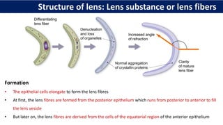


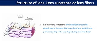


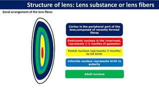
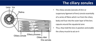


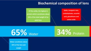


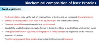


The document provides a detailed description of the structure and functions of the lens in the eye, including its biochemistry, shapes, dimensions, and components such as the lens capsule, anterior lens epithelium, and lens fibers. It discusses the lens's growth and changes with age, including weight, refractive power, and protein composition, emphasizing the importance of crystallins for maintaining transparency. The lens is characterized as a crystalline, avascular, and nerve-free structure, intricately connected to the ciliary body by zonules.


