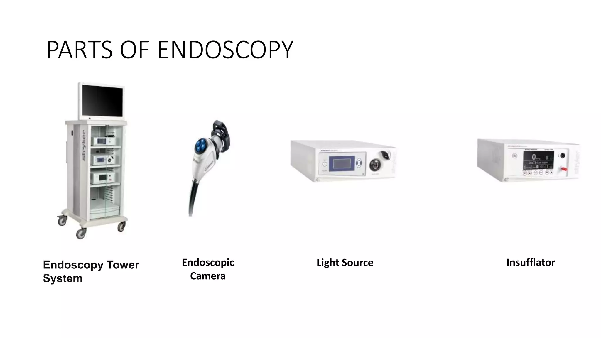
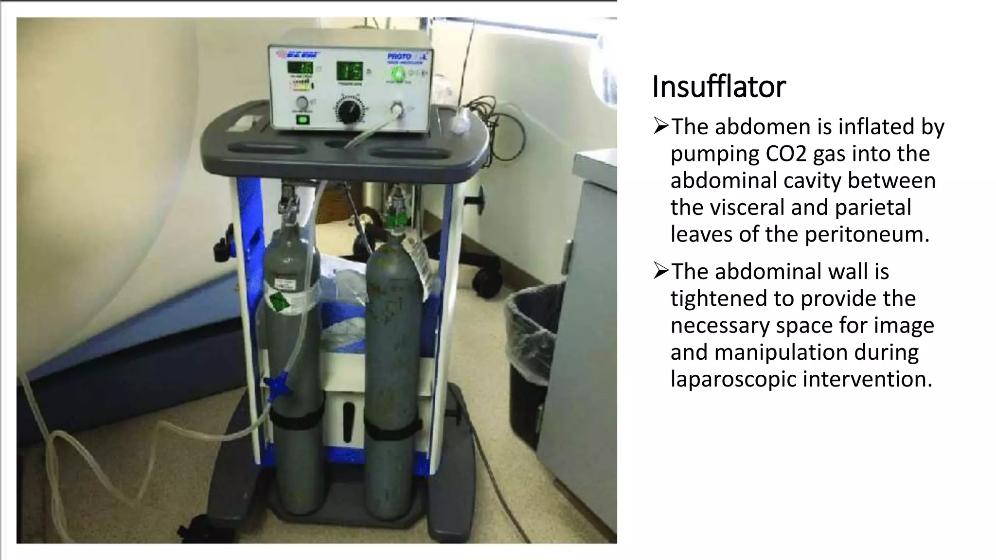
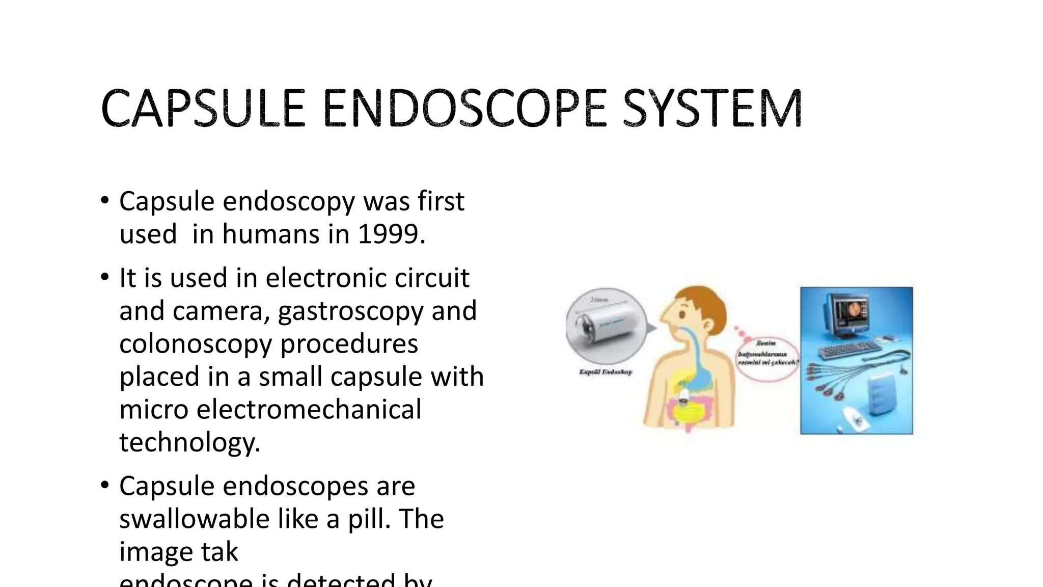
ENDOSCOPY
ALİ ÖZCAN ÜNAL
NECDET GÜVEN
MAHMUT KAMALAK
What is the Endoscopy ?
DEFINITION:
• An endoscopy is a procedure in which your doctor uses specialized
instruments to view and operate on the internal organs and vessels of
your body.
HISTORY
• The name endoscopy is a general expression. Modern endoscopy has
come to the present, with significant advances in a process that
began in 1959 with Harold H. HOPKINS (1918-1994) finding the rod-
shaped optical system and in 1960 with Karl STORZ (1911-1996)
adding the fiber optic light system. .
Why do we need an endoscopy?
• Endoscopy is typically used to:
• help your doctor determine the cause of any abnormal symptoms you’re
having
• remove a small sample of tissue, which can then be sent to a lab for further
testing; this is called an endoscopic biopsy
• help your doctor see inside the body during a surgical procedure, such as
repairing a stomach ulcer, or removing gallstones or tumors
Your doctor may order an endoscopy if you’re having symptoms of any of the
following conditions:
• inflammatory bowel diseases (IBD), such as ulcerative colitis (UC) and Crohn’s disease
• stomach ulcer
• chronic constipation
• pancreatitis
• gallstones
• unexplained bleeding in the digestive tract
• tumors
• infections
• blockage of the esophagus
• gastroesophageal reflux disease (GERD)
• hiatal hernia
• unusual vaginal bleeding
• blood in your urine
• other digestive tract issues
Where is endoscopy used in the body?
TYPES OF
ENDOSCOPY
PARTS OF ENDOSCOPY
Endoscopic
Camera
Light Source InsufflatorEndoscopy Tower
System
PROCESS OF ENDOSCOPY TOWER SYSTEM
• Endoscopy Tower System offers facilities a simple, high-quality
solution for all of their endoscopic imaging needs. Each component —
camera, light source, monitor and insufflator — fits conveniently into
the mobile cart for maximum portability.
Endoscopıc Camera
Ø The basic camera system consists of three
parts:
• A.Camera Head
• B.Processor (CCU)
• C.Monitor
A. Camera Head
ØThe camera head is the part
where the telescope is
attached, the image is
processed and sent to the
camera control unit (CCU).
ØCamera head has different
functions control buttons :
• I.Optical zoom
• II.Focus settings
CCD Sensors (Charged
Coupled Devıce)
• The camera head has a CCD sensor.
• CCDs are sensors used in digital cameras and
video cameras to record still and moving
images.
• The CCD sensor sends the image to the
CCU as analog or digital.
• A single sensor or three sensor structure on
the camera head affects image quality.
CCD sensor Study
• On the CCD sensor front
panel, there are photo
diodes that detect light
signals and transmit
electrical signals.
• The value of the voltage
obtained in the photo diode
is directly proportional to
the value of the light
intensity falling on the photo
diode.
• When the light intensity
falling on the photo diode
increases, the
voltage transmitted by each
photo diode increases
The most basIc terms to learn about a camera and choose
the rıght camera ın use:
• Sensitivity
• Signal to noise ratio
• Automatic Gain Control
• Resolution
CMOS sensor
• •A CMOS sensor is an
electronic sensor that
converts photons to electrons
for digital processing.
Dıfferences between CCD and CMOS sensors
Telescope-optıc endoscope
• Telescopes are used to obtain the desired image
in the body and to perform the necessary
interventions and operations with the help of
this image.
• •Telescopes are named according to the area
they are used in and are designed in a different
structure according to the place of use.
• •Basically, telescopes are collected under two
headings. These are rigid telescopes and
flexible telescopes.
Rıgıd Telescopes
• Rigid telescopes are endoscopes that are not
flexible in structure.
• It consist of these parts:
• I.Eye part (ocular)
• II.Light source (fiber optic cable)port
• III.Lens
FlexIble Telescope
• Flexible telescopes conduct image and light transmission with fiber
optics.
• Since it has a folding structure, it is used for imaging and surgical
intervention in many parts of the body.
• Flexible telescopes can be used conventionally.
• The flexible telescope has a cold light channel, biopsy channel, image
channel and anti-fog air channel for image surface cleaning.
LİGHT SOURCE
1. One of the two main endoscope cables carries light from a
bright lamp in the operating room into the body, illuminating
the cavity where the endoscope has been inserted.
2. The light bounces along the walls of the cable into the patient's
body cavity.
3. The diseased or injured part of the patient's body is illuminated
by the light shining in.
4. Light reflected off the body part travels back up a separate
fiber-optic cable, bouncing off the glass walls as it goes.
5. The light shines into the physician's eyepiece so he or she can
see what's happening inside the patient's body. Sometimes the
fiber-optic cable is directed into a video camera (which displays
what's happening on a television monitor) or a CCD (which can
capture images like a digital camera or feed them into a
computer for various kinds of image enhancement).
Light Source
Ø Since lighting with bulbs at the end of
the endoscope or laparoscope causes
heat trauma, the concept of cold light
was born with the use of fiber optic
glass fibers.
 Cold light source cable is two types:
1. Glass fiber cable
2. Crystal liquid cable
Insufflator
The abdomen is inflated by
pumping CO2 gas into the
abdominal cavity between
the visceral and parietal
leaves of the peritoneum.
The abdominal wall is
tightened to provide the
necessary space for image
and manipulation during
laparoscopic intervention.
RISKS
• The main risks are infection,
over-sedation, perforation, or
a tear of the stomach or
esophagus linig and bleeding.
Although perforation generally
requires surgery, certain cases
may be treated with antibiotics
and intravenous fluids.
• Occasionally, the site of the
sedative injection may become
Trends and
future
• Capsule endoscopy was first
used in humans in 1999.
• It is used in electronic circuit
and camera, gastroscopy and
colonoscopy procedures
placed in a small capsule with
micro electromechanical
technology.
• Capsule endoscopes are
swallowable like a pill. The
image taken with the
Da Vinci SurgIcal
System
• In emerging systems, endoscopic
methods made by remote controlled
robotic arms are used instead of the
doctor's control of endoscopy. While
the doctor is in the control room, the
patient is at the operating table.
• Not really a robot!
Master-slave system – the surgeon
directly initiates all the movements off
the robotic instruments in real time.
Da Vinci Surgical System
• IMAGING
• Double lenses laparoscope
• 3D , high definition, binocular view
• 10-15X magnification
Da Vinci Surgical System
DaVinci Offers
• Improved dexterity
• Better control
• Better precision
Advantages
• Reduced hospital stay
• Less staff required
• Less risky
!!!! Of course, there are some
limitations!!!!
QUESTIONS 3)Which type of system does Da
Vinci have ?
A)Master-Master system
B)Master-Slave system
C)Slave-Slave system
ANSWER IS B
references
• https://www.1stvision.com/machine-
vision-solutions/2019/07/benefits-of-
cmos-based-machine-vision-cameras-vs-
ccd.html
• http://www.megep.meb.gov.tr/mte_progr
am_modul/moduller_pdf/Endoskopik
• https://www.explainthatstuff.com/endosc
opes.html
• https://www.slideshare.net/sathishsak/en
doscopy-82816596
• www.artroskopi.org
• www.biyomedikal.org
• www.gata.edu.tr
• www.tse.org.tr
Endoscopy

Endoscopy

  • 1.
  • 2.
    What is theEndoscopy ? DEFINITION: • An endoscopy is a procedure in which your doctor uses specialized instruments to view and operate on the internal organs and vessels of your body.
  • 3.
    HISTORY • The nameendoscopy is a general expression. Modern endoscopy has come to the present, with significant advances in a process that began in 1959 with Harold H. HOPKINS (1918-1994) finding the rod- shaped optical system and in 1960 with Karl STORZ (1911-1996) adding the fiber optic light system. .
  • 4.
    Why do weneed an endoscopy? • Endoscopy is typically used to: • help your doctor determine the cause of any abnormal symptoms you’re having • remove a small sample of tissue, which can then be sent to a lab for further testing; this is called an endoscopic biopsy • help your doctor see inside the body during a surgical procedure, such as repairing a stomach ulcer, or removing gallstones or tumors
  • 5.
    Your doctor mayorder an endoscopy if you’re having symptoms of any of the following conditions: • inflammatory bowel diseases (IBD), such as ulcerative colitis (UC) and Crohn’s disease • stomach ulcer • chronic constipation • pancreatitis • gallstones • unexplained bleeding in the digestive tract • tumors • infections • blockage of the esophagus • gastroesophageal reflux disease (GERD) • hiatal hernia • unusual vaginal bleeding • blood in your urine • other digestive tract issues
  • 6.
    Where is endoscopyused in the body?
  • 7.
  • 8.
    PARTS OF ENDOSCOPY Endoscopic Camera LightSource InsufflatorEndoscopy Tower System
  • 9.
    PROCESS OF ENDOSCOPYTOWER SYSTEM • Endoscopy Tower System offers facilities a simple, high-quality solution for all of their endoscopic imaging needs. Each component — camera, light source, monitor and insufflator — fits conveniently into the mobile cart for maximum portability.
  • 10.
    Endoscopıc Camera Ø Thebasic camera system consists of three parts: • A.Camera Head • B.Processor (CCU) • C.Monitor
  • 11.
    A. Camera Head ØThecamera head is the part where the telescope is attached, the image is processed and sent to the camera control unit (CCU). ØCamera head has different functions control buttons : • I.Optical zoom • II.Focus settings
  • 12.
    CCD Sensors (Charged CoupledDevıce) • The camera head has a CCD sensor. • CCDs are sensors used in digital cameras and video cameras to record still and moving images. • The CCD sensor sends the image to the CCU as analog or digital.
  • 13.
    • A singlesensor or three sensor structure on the camera head affects image quality.
  • 14.
    CCD sensor Study •On the CCD sensor front panel, there are photo diodes that detect light signals and transmit electrical signals. • The value of the voltage obtained in the photo diode is directly proportional to the value of the light intensity falling on the photo diode. • When the light intensity falling on the photo diode increases, the voltage transmitted by each photo diode increases
  • 15.
    The most basIcterms to learn about a camera and choose the rıght camera ın use: • Sensitivity • Signal to noise ratio • Automatic Gain Control • Resolution
  • 16.
    CMOS sensor • •ACMOS sensor is an electronic sensor that converts photons to electrons for digital processing.
  • 17.
    Dıfferences between CCDand CMOS sensors
  • 18.
    Telescope-optıc endoscope • Telescopesare used to obtain the desired image in the body and to perform the necessary interventions and operations with the help of this image. • •Telescopes are named according to the area they are used in and are designed in a different structure according to the place of use. • •Basically, telescopes are collected under two headings. These are rigid telescopes and flexible telescopes.
  • 19.
    Rıgıd Telescopes • Rigidtelescopes are endoscopes that are not flexible in structure. • It consist of these parts: • I.Eye part (ocular) • II.Light source (fiber optic cable)port • III.Lens
  • 20.
    FlexIble Telescope • Flexibletelescopes conduct image and light transmission with fiber optics. • Since it has a folding structure, it is used for imaging and surgical intervention in many parts of the body. • Flexible telescopes can be used conventionally. • The flexible telescope has a cold light channel, biopsy channel, image channel and anti-fog air channel for image surface cleaning.
  • 21.
    LİGHT SOURCE 1. Oneof the two main endoscope cables carries light from a bright lamp in the operating room into the body, illuminating the cavity where the endoscope has been inserted. 2. The light bounces along the walls of the cable into the patient's body cavity. 3. The diseased or injured part of the patient's body is illuminated by the light shining in. 4. Light reflected off the body part travels back up a separate fiber-optic cable, bouncing off the glass walls as it goes. 5. The light shines into the physician's eyepiece so he or she can see what's happening inside the patient's body. Sometimes the fiber-optic cable is directed into a video camera (which displays what's happening on a television monitor) or a CCD (which can capture images like a digital camera or feed them into a computer for various kinds of image enhancement).
  • 22.
    Light Source Ø Sincelighting with bulbs at the end of the endoscope or laparoscope causes heat trauma, the concept of cold light was born with the use of fiber optic glass fibers.  Cold light source cable is two types: 1. Glass fiber cable 2. Crystal liquid cable
  • 23.
    Insufflator The abdomen isinflated by pumping CO2 gas into the abdominal cavity between the visceral and parietal leaves of the peritoneum. The abdominal wall is tightened to provide the necessary space for image and manipulation during laparoscopic intervention.
  • 24.
    RISKS • The mainrisks are infection, over-sedation, perforation, or a tear of the stomach or esophagus linig and bleeding. Although perforation generally requires surgery, certain cases may be treated with antibiotics and intravenous fluids. • Occasionally, the site of the sedative injection may become
  • 25.
  • 26.
    • Capsule endoscopywas first used in humans in 1999. • It is used in electronic circuit and camera, gastroscopy and colonoscopy procedures placed in a small capsule with micro electromechanical technology. • Capsule endoscopes are swallowable like a pill. The image taken with the
  • 27.
    Da Vinci SurgIcal System •In emerging systems, endoscopic methods made by remote controlled robotic arms are used instead of the doctor's control of endoscopy. While the doctor is in the control room, the patient is at the operating table. • Not really a robot! Master-slave system – the surgeon directly initiates all the movements off the robotic instruments in real time.
  • 28.
    Da Vinci SurgicalSystem • IMAGING • Double lenses laparoscope • 3D , high definition, binocular view • 10-15X magnification
  • 29.
    Da Vinci SurgicalSystem DaVinci Offers • Improved dexterity • Better control • Better precision Advantages • Reduced hospital stay • Less staff required • Less risky !!!! Of course, there are some limitations!!!!
  • 30.
    QUESTIONS 3)Which typeof system does Da Vinci have ? A)Master-Master system B)Master-Slave system C)Slave-Slave system
  • 31.
  • 32.
    references • https://www.1stvision.com/machine- vision-solutions/2019/07/benefits-of- cmos-based-machine-vision-cameras-vs- ccd.html • http://www.megep.meb.gov.tr/mte_progr am_modul/moduller_pdf/Endoskopik •https://www.explainthatstuff.com/endosc opes.html • https://www.slideshare.net/sathishsak/en doscopy-82816596 • www.artroskopi.org • www.biyomedikal.org • www.gata.edu.tr • www.tse.org.tr