Download to read offline


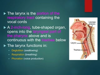
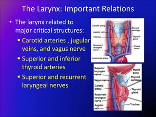
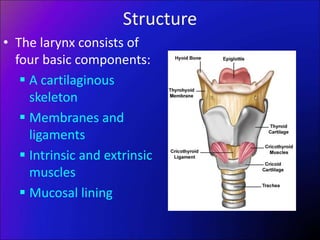
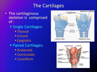
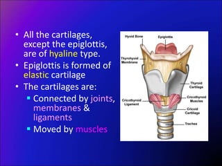
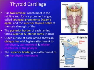
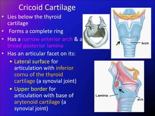
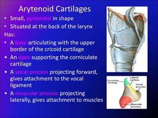
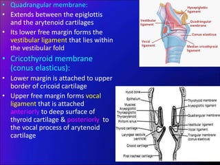
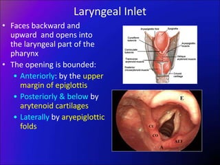
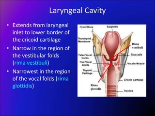
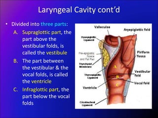
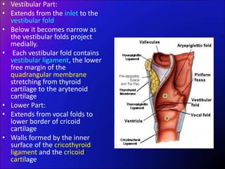
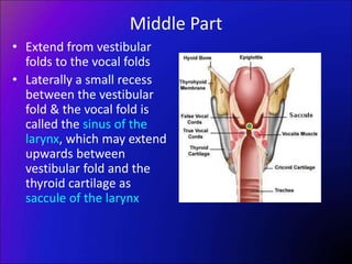

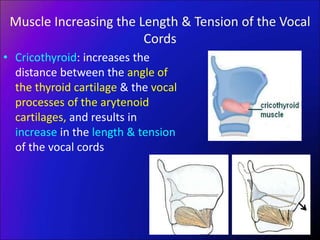
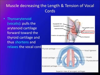
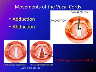
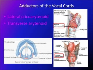
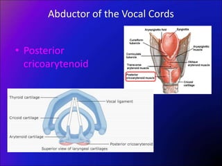
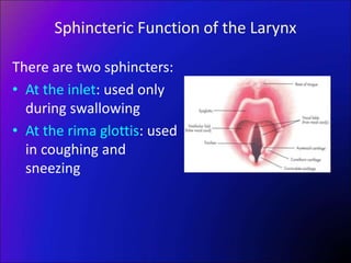
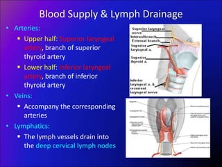
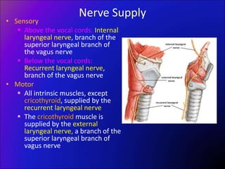

The larynx is a hollow structure located in the neck that contains the vocal cords. It functions in respiration, swallowing, and voice production. The larynx has a cartilaginous skeleton including the thyroid, cricoid, arytenoid, and epiglottic cartilages connected by membranes and ligaments. It is supplied by nerves including the recurrent laryngeal nerve which controls the vocal cords. During phonation, expired air causes the vocal cords to vibrate and resonate within the larynx and pharynx to produce voice.


![Applied Anatomy &Physiology of Cranial nerves [Autosaved].pptx](https://cdn.slidesharecdn.com/ss_thumbnails/appliedanatomyphysiologyofcranialnervesautosaved-250912155808-c3374f2c-thumbnail.jpg?width=640&height=640&fit=bounds)


![PERI-PROSTHETIC FRACTURE NAIL-PLATE CONSTRUCT [NPC].pptx](https://cdn.slidesharecdn.com/ss_thumbnails/drarunkumardrmohamedashrafperiprostheticfrasturenail-plateconstructnpc-260209164459-7e9d15a1-thumbnail.jpg?width=640&height=640&fit=bounds)


