


![EUS features of chronic pancreatitis: Rosemont's Criteria
Parenchymal criteria Ductal criteria
Major Hyperechoic foci (>2 mm in length/width) MPD calculi (echogenic structure[s] within the
MPD)
Lobularity (≥ 13 contiguous lobules
'honeycombing')
Minor Cyst (anechoic, round/elliptical with or without
septations)
Dilated duct (≥ 3.5 mm in body or >1.5 mm in tail)
Hyperechoic strands (≥ 3 mm in at least 2 different
directions with respect to the imaged plane)
Irregular MPD contour (uneven or irregular
outline and ectatic course)
Dilated side branch (>3 tubular anechoic
structures each measuring ≥1 mm in width,
budding from the MPD)
Hyperechoic MPD wall (echogenic, distinct
structure >50% of entire MPD in the body
and tail)](https://image.slidesharecdn.com/endoscopicmanagementinpancreaticdiseasesmini-170204200502/75/Endoscopic-management-in-pancreatic-diseases-14-2048.jpg)


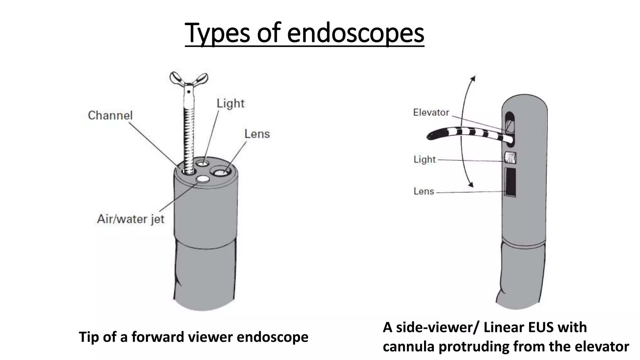
The document provides a comprehensive overview of the endoscopic management of pancreatic diseases, detailing the anatomy and embryology of the pancreas, various endoscopic procedures, and the evolution of techniques since the 1970s. Key topics include endoscopic ultrasonography, coeliac plexus neurolysis, and pancreatic function tests, focusing on their roles in diagnosing and managing pancreatic conditions. Additionally, it discusses the challenges associated with endoscopic procedures, including cannulation techniques and complications, while emphasizing advancements that enhance therapeutic capabilities.


