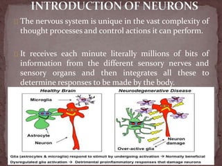
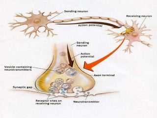
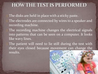
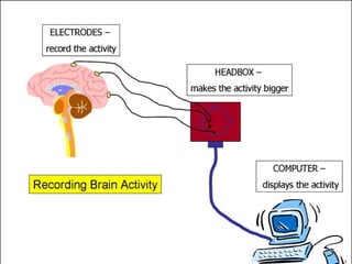
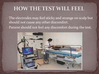
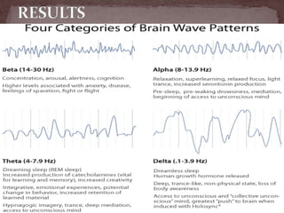
The nervous system receives millions of bits of sensory information per minute and integrates them to determine responses. The basic functional unit of the brain is neurons, which communicate via electrical signals. An electroencephalogram (EEG) records these electrical signals from the scalp using electrodes. During an EEG test, electrodes are placed on the scalp to detect brain waves which are displayed as wavy lines. EEGs can help diagnose conditions affecting brain function and electrical activity such as epilepsy, brain tumors, and sleep disorders.
3. The nervous system is unique in the vast complexity of
thought processes and control actions it can perform.
It receives each minute literally millions of bits of
information from the different sensory nerves and
sensory organs and then integrates all these to
determine responses to be made by the body.
4.
5. The Basic Functional Unit of the brain is
NEURONS..
The central nervous system contains more than 100
billion neurons.
Incoming signals enter this neuron through synapses
located mostly on the neuronal dendrites, but also on
the cell body.
A special feature of most synapses is that the signal
normally passes only in the forward direction
6.
7. The ELECTROENCEPHALOGRAM (EEG) is recording
of the electrical activity of the brain from the scalp.
Another name for EEG is brain wave test
The first recordings were made by Hans Berger in 1929.
But similar studies have been done at earlier years
around 1870’s in animals.
The wave forms recorded are thought to reflect the
activity of the surface of the brain and cortex.
8. The brain's cells produce tiny electrical signals when
they send messages to each other.
During an EEG test, small electrodes are placed on
to scalp.
They pick up brain's electrical signals and send them
to a machine called an electroencephalograph.
They records the signals as wavy lines on to a
computer screen or paper.
9. Brain cells communicate with each other by producing
tiny electrical signals, called impulses
An EEG measures this activity. The test is done by a
EEG specialist in doctor's office or at a hospital or
laboratory.
They will be asked to lie on back on a bed or in a
reclining chair.
Flat metal disks called electrodes are placed all over
scalp.
10.
11. The disks are held in place with a sticky paste.
The electrodes are connected by wires to a speaker and
recording machine.
The recording machine changes the electrical signals
into patterns that can be seen on a computer. It looks
like wavy lines.
The patient will need to lie still during the test with
their eyes closed because movement can change the
results.
12.
13. Wash hair at night before the test going to
perform.
Do not use any oils, sprays, or conditioner on hair
before this test.
Avoid all food and drinks containing caffeine for 8
hours before the test.
Patient may need to sleep during the test.
If so, patient may be asked to reduce their sleep
time the night before.
But to reduce the sleep do not drink any caffeine
containing material, energy drinks etc..
14. The electrodes may feel sticky and strange on scalp but
should not cause any other discomfort.
Patient should not feel any discomfort during the test.
15. This procedure is completely painless and can be
performed without shaving any of patient’s hair.
EEG can help diagnose a number of conditions,
including epilepsy, sleep disorders and brain
tumors.
Normal brain waves occur at a rate of up to 30 per
second, but in someone with epilepsy - the EEG
may show bursts of abnormal discharges in the
form of spikes and sharp wave patterns.
16. Sleep disorders (such
as narcolepsy)
Head injuries
Brain infection
Brain hemorrhage
Alzheimer's disease
Degeneration of brain
tissue
Metabolic conditions
that affect brain tissue
Hormonal conditions
that affect brain tissue
Certain disorders of the
central nervous system
Stroke
Brain tumor
Brain death.
17. Dementia– a group of symptoms that are
responsible for the decline of brain function.
Vertigo– the sensation that you are moving
even when you are standing still.
Brain abscess – a pus-filled swelling in the
brain that is caused by an infection.
Encephalitis – inflammation of the brain
tissue that is sometimes caused by an infection.
Cerebral infarct – brain tissue that has
died due to a blockage in blood flow.
18. An abnormal EEG doesn't automatically mean that
patient have epilepsy.
The EEGs of babies and young children can often
record irregular patterns that don't mean
anything, or the irregularities may flag previously
diagnosed neurological conditions such as cerebral
palsy.
19.
20.
21. Abnormal results on an EEG test may be due to:
Abnormal bleeding (hemorrhage)
An abnormal structure in the brain (such as a brain tumor)
Attention problems
Tissue death due to a blockage in blood flow (cerebral
infarction)
Drug or alcohol abuse
Head injury
Migraines (in some cases)
Seizure disorder (such as epilepsy or convulsions)
Sleep disorder (such as narcolepsy)
Swelling of the brain (encephalitis)
22. The procedure is very safe.
The flashing lights or fast breathing
(hyperventilation) required during the test may
trigger seizures in those with seizure disorders.
The health care provider performing the EEG is
trained to take care of patient if this happens.