Downloaded 1,217 times

![22
Professor Yasser Metwally
www.yassermetwally.com
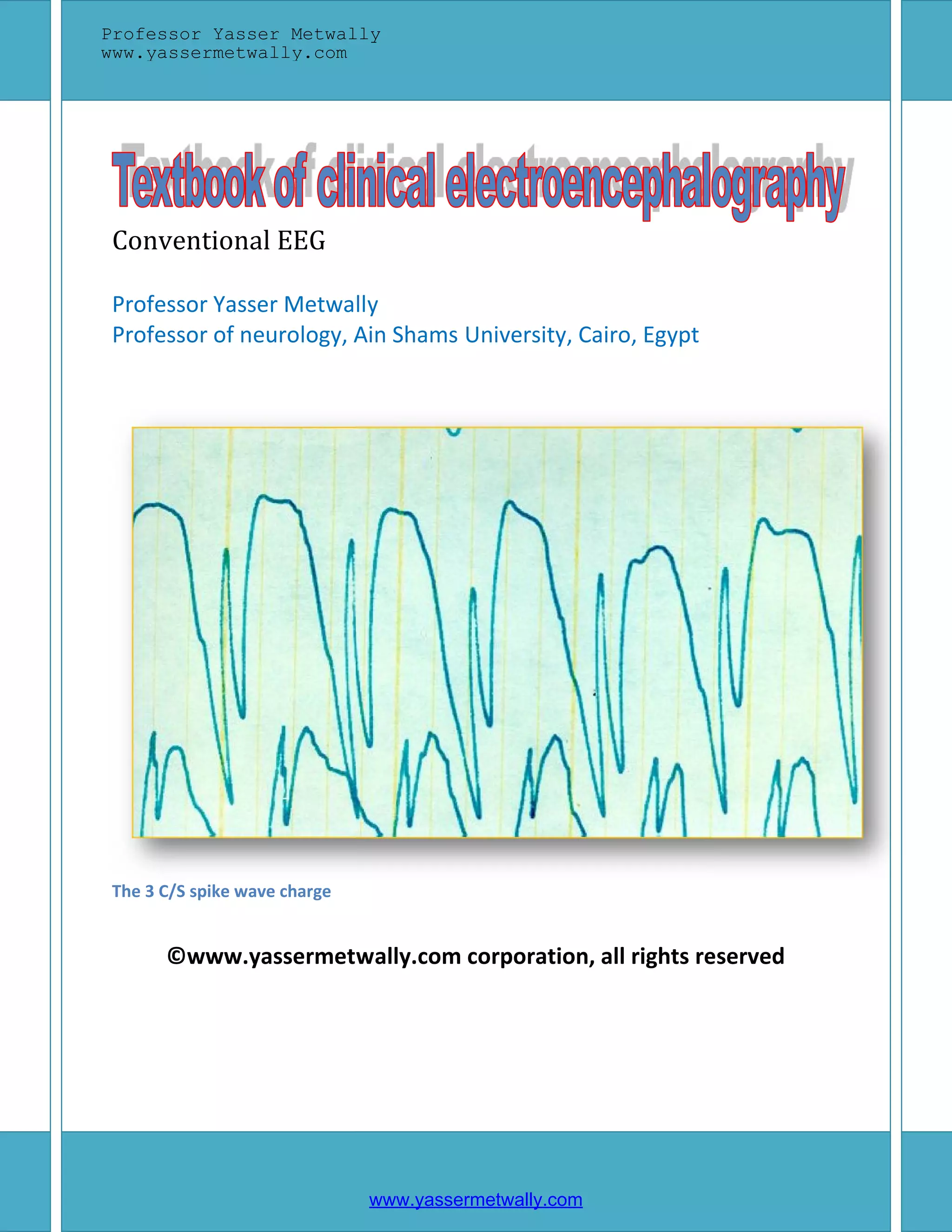
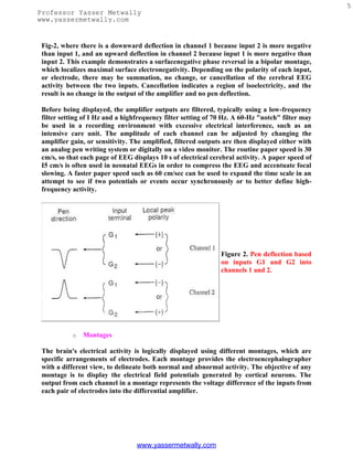
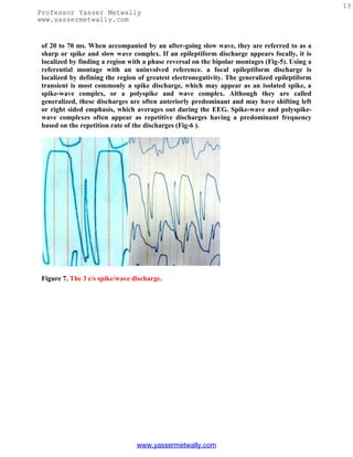
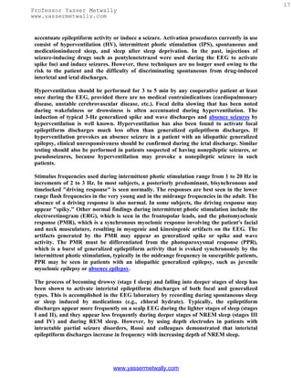
Figure 12. Examples of sharp
waves [left] and spike [right]
Table 2. Electroclinical criteria of spike/ sharp wave discharge
A spike is a transient, clearly distinguished from the background activity, with
pointed peak at conventional paper speeds and a duration from 20 to under 70
msec; the main component is generally negative. Amplitude is variable. Spikes
represent the basic element of paroxysmal activity in the EEG
A sharp wave is a transient, clearly distinguished from background activity,
with pointed peak at conventional paper speeds and duration of 70 to 200 msec.
The main component is generally negative relative to other areas.
Both spikes and sharp waves have multiphasic characters, being composed of a
sequence of a minor positive, a major negative, and a second minor positive
component is typical in most instances. The long duration of a sharp wave
permits better insight into the multiphasic character of this potential.
The spike/sharp wave potentials are reliable indicators of a potential seizure
focus because they result from the characteristic neurophysiological event "the
paroxysmal depolarization shift" (PDS). This phenomenon consists of thousands
of neurons simultaneously undergoing large depolarization with superimposed
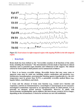
action potentials. Both synaptic events and intrinsic cellular currents have been
implicated in this process. EEG spikes/sharp waves are due to the slow
depolarization currents in the PDS. Neurons surrounding the focus are inhibited
during the paroxysmal depolarization shift, and within the focus the the
paroxysmal depolarization shift is followed by a hyperpolarization potential.
Both an increase in depolarizing events and a loss of inhibitory mechanisms can
lead to persistence and propagation of the discharge as a seizure.
Spikes and sharp waves are neurophysiologically closely related phenomena;
both of them are typical paroxysmal discharges and highly suggestive of an
epileptic seizure disorder, although both phenomena may occur in patients
without a history of seizure disorder.
The largest and most pronounced spikes are not necessarily associated with more
serious epileptic seizure disorders. On the contrary, Rolandic spikes in a child age 4 to
10 yr are very prominent; however, the seizure disorder is usually quite benign or there
may be no clinical seizures at all. low voltage spiking in the frontal or anterior temporal
regions is highly epileptogenic even though its amplitude can be so low to the point that
these spikes might be completely drowned within the background waves and
subsequently can not be easily detected.
www.yassermetwally.com](https://image.slidesharecdn.com/eeg-120912084228-phpapp02/85/Textbook-of-electroencephalography-25-320.jpg)
![25
Professor Yasser Metwally
www.yassermetwally.com
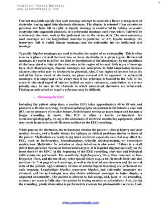
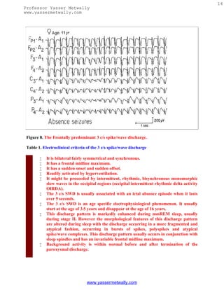
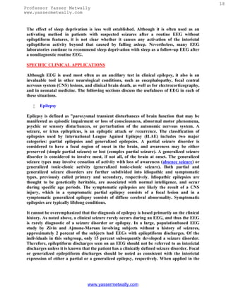
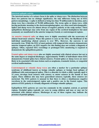
Figure 16. The intermittent rhythmic delta activity [left image] and the the polymorphic
slow wave activity [right image]
Table 3. Electrical criteria of The intermittent rhythmic delta activity.
Consists of sinusoidal waveforms of approximately 2.5 Hz that occur
intermittently in the EEG recording. It is most often symmetric but can be
lateralized.
In adults, the delta activity has a frontal predominance (frontal
intermittent rhythmic delta activity [FIRDA]). In children, it is maximal
posteriorly (occipital intermittent rhythmic delta activity [OIRDA])
The intermittent rhythmic delta activity shows visual reactivity and is
commonly suppressed in the eye open state unless the patient is comatose.
Intermittent rhythmic delta activity is associated with structural lesions,
most commonly diencephalic, infratentorial or intraventricular tumors, or
with diffuse encephalopathies.
FIRDA occurring in patients with a normal EEG background suggests
that the pattern is due to a structural lesion; when associated with EEG
background abnormalities, it is likely to be due to encephalopathy.
OIRDA is associated with absence epilepsy in children aged 6-10 years.
www.yassermetwally.com](https://image.slidesharecdn.com/eeg-120912084228-phpapp02/85/Textbook-of-electroencephalography-28-320.jpg)
![28
Professor Yasser Metwally
www.yassermetwally.com
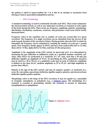
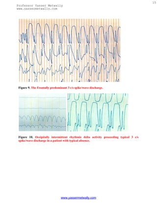
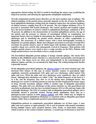
Table 4. Electrical criteria of the Polymorphic slow wave activity.
Quite variable in wave shape morphology, frequency and amplitude.
Commonly lateralized over a wide area of the scalp, persistent in eye closed, eye
open state, during all sleep stages, with no visual reactivity. Polymorphic Delta
activity that fails to persist into sleep or attenuates significantly with arousal or
eye opening is less indicative of structural pathology.
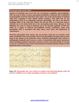
Persistent polymorphic delta activity may not precisely match the true location
of the lesion, particularly since it presumably arises from physiological deranged
neurons often lying on the margin of the destructive lesion. Persistent
polymorphic delta activity is aetiologically nonspecific and is seen in a variety of
subcortical (while matter) destructive lesions including neoplasms, infarctions,
abscesses, trauma, and haemorrhage. It can also be seen in reversible processes
such as focal ischemia in transient ischemic attacks or focal depression from a
recent seizure.
Commonly due to a subcortical white matter lesion inducing deafferentation of
the cerebral cortex.
A purely cortical lesion does not induce polymorphic slow wave activity.
Rhythmic delta activity
consists of sinusoidal waveforms of approximately 2.5 Hz that occur intermittently in the
EEG recording. It is most often symmetric but can be lateralized. In adults, the delta
activity has a frontal predominance (frontal intermittent rhythmic delta activity [FIRDA]).
In children, it is maximal posteriorly (occipital intermittent rhythmic delta activity
[OIRDA]). Intermittent rhythmic delta activity is associated with structural lesions, most
commonly diencephalic, infratentorial or intraventricular tumors, or with diffuse
encephalopathies. FIRDA occurring in patients with a normal EEG background suggests
that the pattern is due to a structural lesion; when associated with EEG background
abnormalities, it is likely to be due to encephalopathy. In cases of encephalopathy with
FIRDA, the pathophysiologic processes are believed to involve cortical and subcortical gray
matter. OIRDA is associated with absence epilepsy in children aged 6-10 years
Figure 19. The intermittent rhythmic delta activity [left image] and the the polymorphic
slow wave activity [right image]
www.yassermetwally.com](https://image.slidesharecdn.com/eeg-120912084228-phpapp02/85/Textbook-of-electroencephalography-31-320.jpg)
![29
Professor Yasser Metwally
www.yassermetwally.com
Table 5. Electrical criteria of The intermittent rhythmic delta activity.
Consists of sinusoidal waveforms of approximately 2.5 Hz that occur
intermittently in the EEG recording. It is most often symmetric but can be
lateralized.
In adults, the delta activity has a frontal predominance (frontal intermittent
rhythmic delta activity [FIRDA]). In children, it is maximal posteriorly
(occipital intermittent rhythmic delta activity [OIRDA])
The intermittent rhythmic delta activity shows visual reactivity and is commonly
suppressed in the eye open state unless the patient is comatose.
Intermittent rhythmic delta activity is associated with structural lesions, most
commonly diencephalic, infratentorial or intraventricular tumors, or with
diffuse encephalopathies.
FIRDA occurring in patients with a normal EEG background suggests that the
pattern is due to a structural lesion; when associated with EEG background
abnormalities, it is likely to be due to encephalopathy.
OIRDA is associated with absence epilepsy in children aged 6-10 years.
Focal theta activity
Is less likely to reflect a macroscopic structural lesion than is focal delta. Theta is
commonly, however, associated with a functional disturbance, such as epileptogenic
cortex, especially postictally, after amplitude suppression and focal delta have resolved. In
addition, localized theta is usually superimposed on focal delta to some degree; the relative
proportion of delta and theta reflects the size and/or severity of the underlying structural
or functional cerebral abnormality.
Epileptiform activity may be seen on the EEG in some degenerative encephalopathies that
have associated seizures. Multifocal, independent epileptiform spike discharges may be
seen in TaySachs disease, in several of the progressive myoclonic epilepsies (neuronal
ceroid lipofuscinosis, Lafora body disease, and some mitochondrial encephalomyopathies),
and in Rett syndrome. Atypical generalized spike and wave activity is present in
UnverrichtLundborg disease, which is another type of progressive myoclonic epilepsy.
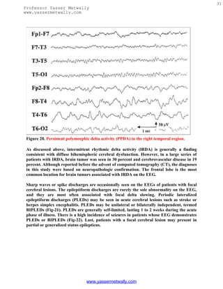
Of the inflammatory encephalopathies, distinctive EEG findings are seen in subacute
sclerosing panencephalitis (SSPE) and herpes simplex encephalitis. The clinical
presentation of SSPE includes myoclonus with progressive encephalopathy. The EEG
shows periodic, polyphasic sharp and slow wave complexes that have an interburst interval
of 4 to 10 s. As SSPE progresses, there is gradual loss of the intermixed background
frequencies, resulting in a pattern similar to burst suppression. Herpes simplex encephalitis
is the most common sporadic viral encephalitis, typically presenting with fever,
encephalopathy, and secondarily generalized seizures. The EEG commonly shows periodic
lateralized epileptiform discharges (PLEDS). which are lateralized to the side of the herpes
infection. Should both temporal lobes be involved, bilateral independent periodic
epileptiform discharges (BIPLEDS) may be seen on the EEG. Other forms of inflammatory
www.yassermetwally.com](https://image.slidesharecdn.com/eeg-120912084228-phpapp02/85/Textbook-of-electroencephalography-32-320.jpg)

![39
Professor Yasser Metwally
www.yassermetwally.com
indispensable for the diagnosis of nonconvulsive status epilepticus and for separating
epileptic from other paroxysmal (nonepileptic) episodes. There are EEG patterns
predictive of the cause of the encephalopathy (i.e., triphasic waves in metabolic
encephalopathy) or the location of the lesion (i.e., focal polymorphic delta activity in lesions
of the subcortical white matter). The various EEG characteristics of infantile, childhood,
and adult epilepsies are described as well as the EEG patterns that are morphologically
similar to interictal/ictal epileptiform discharges but unrelated to epilepsy. An EEG is most
helpful in determining the severity and, hence, the prognosis of cerebral dysfunction.
Lastly, EEG is extremely helpful in assessing normal or abnormal brain functioning in a
newborn because of the serious limitation in performing an adequate neurologic
examination on the neonate who is intubated or paralyzed for ventilatory control. Under
such circumstances, the EEG may be the only available tool to detect an encephalopathic
process or the occurrence of epileptic seizures.
Electroencephalography (EEG) is the technique of recording from the scalp the
spontaneous electrical activity of the brain and correlating it to the underlying brain
function. Since the first recording of a human EEG in 1929 by Hans Berger, improvement
in electronics and technology has made EEG one of the most widely used laboratory tests
for clinical evaluation of neurologic disorders. However, in the past three decades with
continuing advances in neuroimaging, particularly magnetic resonance imaging (MRI), the
role of clinical EEG has become restricted and progressively more focused. Its major utility
at present is in the evaluation of focal and diffuse encephalopathies, comatose conditions,
epileptic disorders, and cerebral disorders affecting neonates and infants. The present
article is not an attempt to describe EEG comprehensively in normal subjects and in
different disease processes but to highlight its usefulness/limitation and emphasize
precautions/care needed in its optimal utility. The subject will be discussed under seven
sections: EEG in normal subjects, EEG in patients with altered mental status or diffuse
encephalopathies, EEG in focal or lateralized cerebral hemispheric lesions, EEG in
paroxysmal disorders, EEG in generalized epilepsies, EEG in neonates, and EEG in status
epilepticus.
EEG In Normal Subjects
The EEG in the normal awake child and adult is well known and needs no detailed
description. The following are points of emphasis:
1. Alpha rhythm in the two hemispheres is very similar in frequency. A consistent
difference of even 0.5 to 1.0 cps on the two sides is significant; the side showing a
slower frequency may have a hemispheric dysfunction. Amplitude asymmetry is of
relatively less significance, unless the asymmetry is prominent. In general, the alpha
rhythm is higher in amplitude over the right hemisphere. If the amplitude of the
alpha rhythm on the right side is more than 1 1/2 times that on the left side, the
asymmetry is usually regarded as significant. When the alpha rhythm is over 25%
higher in amplitude on the left side than the right side, this constitutes a significant
asymmetry.[1]
www.yassermetwally.com](https://image.slidesharecdn.com/eeg-120912084228-phpapp02/85/Textbook-of-electroencephalography-42-320.jpg)
![40
Professor Yasser Metwally
www.yassermetwally.com
2. Significant theta activity (4 to 7 Hz) is present in the EEG of children and
adolescents. Delta activity in the awake tracing is rarely seen after the age of 5
years. A common EEG pattern in adolescents is the presence of intermittent delta
waves intermixed with alpha rhythm over the posterior head regions, the so-called
"slow waves of youth."
3. The EEG during non-rapid eye movement (NREM) sleep in children shows very
prominent spikelike vertex sharp transients, which are often mistaken for
epileptiform activity by EEG interpreters inexperienced with children's EEGs (Fig.
1). Similarly, positive occipital sharp transients (POSTs), when high in amplitude
and sharp in configuration, can be easily misinterpreted as abnormal spikes,
especially in linkages where occipital electrodes are connected to input terminal 2
(grid 2) of the amplifier (e.g., "double banana run").
4. In a small proportion of normal adult subjects, clearly identifiable and countable
alpha rhythm may be entirely absent. The background may consist of irregular
mixtures of low amplitude (<20 µV) activities, mostly from 5.0 to 30.0 cps without a
dominant frequency. Such low-voltage EEGs have been studied in detail.[2] The
EEG is reactive to various physiologic stimuli such as sleep, drugs, and pathologic
processes. In over half of the patients with low-voltage EEGs, hyperventilation may
bring out an alpha rhythm. During sleep, normal activities such as vertex sharp
transients and sleep spindles may be generated. It is essential that low-voltage
tracings be clearly distinguished from EEGs showing electrocerebral inactivity,
which have a grave prognosis. These EEGs lack reactivity and lability, and with
increased instrumental sensitivities show no electrical activity of cerebral origin.
Low-voltage EEGs are generally considered to be a normal variant occurring in 7 to
10% of normal subjects over the age of 20 years. The low-voltage EEG does not
correlate with neurologic or psychiatric disease.
5. Changes in the EEG during normal senescence has been described in detail.[3-5]
The most frequent change is the slowing of the alpha frequency. By the age of 70
years, the mean alpha frequency decreases to 9.0 to 9.5 cps and decreases further to
8.5 to 9.0 cps beyond the age of 80 years. In healthy elderly subjects, even at or over
the age of 100 years, the frequency of the alpha rhythm remains well above 8.0
cps.[6,7] Therefore, an average alpha frequency of less than 8.0 cps measured with
the patient fully alert must be considered abnormal in elderly patients at all ages.
6. Another EEG finding is the presence of isolated transients of irregular focal slowing
in the theta-delta frequency range over the anterior temporal region, reported in
40% of healthy elderly subjects.[4,5,8] They are most frequent over the left
temporal area particularly during drowsiness (Fig. 2). Sometimes poorly defined
sharp waves are interspersed with focal slow components. The left-sided
accentuation of this activity remains unexplained. Such intermittent slow activity,
with or without sharp components over the temporal region, has no correlation with
intellectual or cognitive functioning or presence of a seizure disorder. More recent
investigations suggest that the temporal slowing in the awake tracing may, in fact,
not be the inevitable consequence of advancing age. In neurologically and
psychologically normal septuagenarians, Katz and Horowitz[9] found that the focal
slow activity was seen in only 17% of records and when present occupied less than
1% of the tracing. Hence, intermittent temporal theta-delta activity occupying only
www.yassermetwally.com](https://image.slidesharecdn.com/eeg-120912084228-phpapp02/85/Textbook-of-electroencephalography-43-320.jpg)
![41
Professor Yasser Metwally
www.yassermetwally.com
a small proportion of the wake tracing should be considered as a normal aging
phenomena. When the temporal slow activity comprises more delta than theta slow
waves, which either recur frequently or occur in long runs and are widespread in
distribution, a dementing process or focal lesion has to be seriously considered.
Diffuse theta-delta activity in elderly subjects are likely to occur in those with
intellectual impairment.[5]
7.
Figure 1. EEG of a 2-year-old child with very prominent spikelike vertex sharp transients.
www.yassermetwally.com](https://image.slidesharecdn.com/eeg-120912084228-phpapp02/85/Textbook-of-electroencephalography-44-320.jpg)
![42
Professor Yasser Metwally
www.yassermetwally.com
Figure 2. EEG showing intermittent slow wave transients (*) in a 61-year-old subject.
EEG in Patients with Altered Mental Status or Diffuse Encephalopathies
The term encephalopathy is usually applied to patients displaying altered mental status as
a result of a diffuse disturbance of brain function. Common encephalopathies are divided
into metabolic, toxic, inflammatory (encephalitis), anoxic, and degenerative types. The
EEG in most encephalopathies shows an alteration of background activities and emergence
of varying degrees of theta-delta slowing. Remember that the EEG findings are generally
nonspecific from a differential standpoint. The EEG is unable to distinguish between
different etiologies. The main contribution of the EEG is in providing an objective measure
of severity of encephalopathy, prognosis, and effectiveness of therapy.[10]
There is a good correlation between the severity of the EEG changes, the severity of the
encephalopathy, and the clinical state of the patient. In mild encephalopathy associated
with mild clouding of consciousness and confusion, there is at first slowing of the posterior
dominant rhythm, which decreases from a higher to a lower alpha frequency and then into
the theta frequency range. More severe encephalopathy is associated with deeper levels of
coma, and the background consists mainly of high-amplitude irregular delta activity. With
further deterioration in the encephalopathy, the amplitude of all activities drop below 20
µV and the EEG may consist of relatively low-amplitude, invariant delta activity. Some
tracings reveal suppression-burst pattern where there is regular alternation of very-low-
amplitude EEG with relatively higher-amplitude EEG segments. The most extreme type of
abnormality is, of course, lack of any cerebral activity (i.e., electrocerebral inactivity).
Presence of the later three types of EEG patterns (invariant low-amplitude delta,
suppression-burst, and electrocerebral inactivity) carry a grave prognosis, if drug
intoxication can be excluded as the cause of encephalopathy. If due to drug intoxication,
www.yassermetwally.com](https://image.slidesharecdn.com/eeg-120912084228-phpapp02/85/Textbook-of-electroencephalography-45-320.jpg)
![43
Professor Yasser Metwally
www.yassermetwally.com
these severely abnormal patterns are quite reversible with treatment, with a high potential
for complete recovery of neurologic functioning.
Besides the degree of background slowing, there are two other features in the EEG that
must be evaluated to determine the severity of encephalopathy. These are spontaneous
variability of the EEG over several seconds to minutes, and reactivity to painful
stimulation. In milder encephalopathies, the EEG shows spontaneous variability during the
recording period and evidence of EEG reactivity to painful stimulation. When the EEG
shows reactivity, painful stimulation commonly results in reduction of the amplitude,
increase in frequency of the background activity, and reduction in the slow activity. There
is often a "paradoxical activation," which is a period of more severe delta slowing following
painful stimulation (Fig. 3). The presence of any type of reactivity (reduction in slow
activity or increase in the degree of slowing) on painful stimulation suggests a lower grade
of encephalopathy, whereas the EEG lacking spontaneous variability (invariant EEG) and
total lack of any reactivity to intense and prolonged stimulation suggests a severe degree of
encephalopathy.
Figure 3. EEG of an 8-year-old child with hemolytic anemia and uremia, showing
paradoxical activation characterized by increased delta slowing induced by painful
stimulation.
A grading system of EEG abnormalities in adults is shown in Table 1, similar to other
rating systems.[11,12] It is helpful in prognosis, evaluation of effectiveness of therapy, and
comparing serial EEG studies. The slow activities associated with an encephalopathy are
usually widespread and symmetrical over the two hemispheres. In children, the slowing
www.yassermetwally.com](https://image.slidesharecdn.com/eeg-120912084228-phpapp02/85/Textbook-of-electroencephalography-46-320.jpg)
![44
Professor Yasser Metwally
www.yassermetwally.com
may predominate over the posterior hemisphere, and in adults, usually over the frontal
areas. These are simply maturation-related spatial EEG features, which do not signify that
the encephalopathy is more severe posteriorly in children and anteriorly in adults.
Table 1. Grading of EEG Abnormalities in Diffuse Encephalopathy
It is unusual to see prominent focal or lateralized EEG findings with a diffuse
encephalopathy unless there is an associated focal process, such as an old infarct or tumor.
An exception is nonketotic hyperosmolar coma, a form of metabolic encephalopathy, which
is very often associated with focal clinical (e.g., focal seizures) and focal EEG findings.
Herpes simplex encephalitis and Creutzfeldt-Jakob disease (in the early stages) may also
produce lateralized EEG slowing related to unilateral emphasis of the associated pathologic
process (see below).
Another EEG pattern associated with a mild form of encephalopathy is the presence of
bursts of intermittent rhythmic delta activity (IRDA) superimposed on a more or less
normal background activity. Depending on the area of predominance, the IRDA is further
divided into frontal or occipital types. IRDA has been traditionally considered a "projected
rhythm" and a hallmark of EEG findings in patients with deep midline lesions of
diencephalic, upper brain stem, or posterior fossa locations.[13] Critical evaluations
subsequently have cast serious doubts on this classic concept because this EEG pattern has
been found in a large variety of pathological conditions and is often absent in deep midline
lesions. As a matter of fact, the most common etiology of IRDA is a mild to moderate
encephalopathy associated with some disturbance in consciousness (Fig. 4).[14]
www.yassermetwally.com](https://image.slidesharecdn.com/eeg-120912084228-phpapp02/85/Textbook-of-electroencephalography-47-320.jpg)
![45
Professor Yasser Metwally
www.yassermetwally.com
Figure 4. EEG of an
82-year-old patient
with recent history of
lethargy and
confusion, showing
frontal intermittent
rhythmic delta
activity.
Are there any unique or specific EEG features that help narrow the differential diagnosis
of diffuse encephalopathy and point toward a more specific etiology? There are a few EEG
patterns (e.g., triphasic waves, positive spikes, and periodic complexes) that, although not
commonly encountered in encephalopathic patients, when present suggest a specific
etiology for the encephalopathy. Periodic patterns are specifically encountered in anoxic
encephalopathy and certain encephalitides, whereas triphasic waves and positive spikes
characteristically occur in metabolic encephalopathies.
o Metabolic Encephalopathy
An EEG showing diffuse slowing of the background and presence of triphasic waves is
highly suggestive of a metabolic encephalopathy. triphasic waves are high amplitude (200
to 300 µV), usually bilaterally synchronous, symmetrical, and maximum in amplitude over
the frontocentral regions (Fig. 5). The most prominent component is a positive sharp wave
that is preceded by a short-duration negative sharp wave and followed by a long-duration
negative slow wave.[15] However, variations are quite common and the waveform may be
monophasic or biphasic.
www.yassermetwally.com](https://image.slidesharecdn.com/eeg-120912084228-phpapp02/85/Textbook-of-electroencephalography-48-320.jpg)
![46
Professor Yasser Metwally
www.yassermetwally.com
Figure 5. EEG of a 69-year-old patient with hepatic encephalopathy, showing triphasic
waves.
Although earlier authors[15] emphasized that the triphasic waves were highly specific for
hepatic encephalopathy, this EEG pattern has been found to correlate best with any
metabolic type of encephalopathy; hepatic, renal, and anoxic etiologies account for over
75% of EEGs with triphasic waves.[16-18] A feature of triphasic waves often stressed is the
progressive time lag (25 to 140 milliseconds) of the positive component of the triphasic
wave from the anterior to the posterior region. This feature was considered to be most
specific for hepatic etiology.[17,19] Recent studies[18] demonstrated that the time lag is
neither a consistent feature of triphasic waves, nor has any specificity with regard to the
type of metabolic encephalopathy. The "peril" is that no single feature or group of features
regarding triphasic waves distinguish hepatic from nonhepatic cases.
www.yassermetwally.com](https://image.slidesharecdn.com/eeg-120912084228-phpapp02/85/Textbook-of-electroencephalography-49-320.jpg)
![47
Professor Yasser Metwally
www.yassermetwally.com
There are a few other "pearls" regarding triphasic waves. Patients with metabolic
encephalopathies showing prominent triphasic wave activity in their EEG have an overall
poor prognosis; in one series, over two thirds died in a matter of a few months.[20]
Furthermore, triphasic waves occur essentially in adults; this pattern has been rarely
reported below the age of 20 years.[21] This is particularly true with Reyes disease, an
acute childhood encephalopathy with hepatic fatty infiltration, where triphasic waves are
absent.[22] The EEG pattern of 14 to 6 per second, positive spikes are a well-known
maturational EEG pattern normally seen in children in adolescence during NREM sleep.
The presence of positive spike bursts in comatose patients with continuous delta activity is
a unique, albeit rare, EEG pattern associated with hepatic or anoxic encephalopathy in
children (Fig. 6).[23,24]
Figure 6. EEG of a 16-year-old comatose patient with Reye's syndrome, showing 14 cps
positive spikes.
o Toxic Encephalopathy
Overdose of hypnotic-sedative drugs is a common cause of coma encountered in the
emergency room; excessive beta activity is a prominent feature in the EEG over the
anterior head regions. What is less well recognized is that with more severe intoxication,
the fast activity assumes a slower frequency (usually 10 to 13 Hz), which is widespread but
with anterior predominance. The presence of generalized theta-delta activity with
superimposed alpha frequency activity is a unique encephalographic pattern highly
characteristic of sedative drug intoxication (Fig. 7). In the absence of prominent slow
www.yassermetwally.com](https://image.slidesharecdn.com/eeg-120912084228-phpapp02/85/Textbook-of-electroencephalography-50-320.jpg)
![48
Professor Yasser Metwally
www.yassermetwally.com
activity, the anterior dominant generalized fast activity produces alpha or spindle coma
pattern in the EEG indistinguishable from that seen with severe anoxic
encephalopathy.[25,26]
Figure 7. EEG of an 18-year-
old patient with phenobarbital
intoxication, showing
generalized theta-delta activity
with superimposed beta
frequencies (A) followed in 3
days by normalization of the
EEG (B).
Very severe drug intoxication results in suppression-burst pattern or electrocerebral
inactivity. Even though these patterns signify advanced intoxication, they do not carry as
ominous a prognosis as when they occur in the setting of cardiopulmonary arrest. It has
been repeatedly demonstrated that patients with drug-induced coma may have
electrocerebral inactivity lasting over a day and may still make a full neurologic recovery.
Phencyclidine hydrochloride ("angel dust," "PCP pills") is associated with a distinctive
EEG pattern similar to that of subacute sclerosing panencephalitis (SSPE). The EEG shows
generalized sinusoidal 6.0 cps theta activity that is interrupted approximately every 4
seconds by generalized slow wave discharges.[27] A similar periodic EEG pattern is
described transiently during ketamine (a phencyclidine derivative) anesthesia.
www.yassermetwally.com](https://image.slidesharecdn.com/eeg-120912084228-phpapp02/85/Textbook-of-electroencephalography-51-320.jpg)
![49
Professor Yasser Metwally
www.yassermetwally.com
o Anoxic Encephalopathy
EEG is commonly performed in patients with anoxic encephalopathy due to
cardiopulmonary arrest for assessing the severity of cerebral insult and for prognosis.
Patients with normal or almost normal EEG tracings (grade I encephalopathy) following
an episode of cerebral anoxia have an excellent prognosis for full neurologic recovery. On
the other hand, patients with grade IV or V EEG abnormalities have a uniformly fatal
prognosis; most of these patients die without regaining consciousness. An EEG should be
obtained at least 5 or 6 hours after successful resuscitation since it takes an hour or more
for the EEG to stabilize after an anoxic episode.[12]
Besides electrocerebral inactivity, there are three other unique EEG patterns, encountered
in association with anoxic encephalopathy, that carry a poor prognosis for neurologic
recovery.
Periodic discharges in anoxic encephalopathy may be either bilaterally synchronous
periodic epileptiform discharges (BiPLEDs)[28] or independently occurring periodic
lateralized epileptiform discharges (bilateral PLEDs).[29] Both periodic EEG patterns are
often associated with myoclonic seizures (or even myoclonic status) and carry an extremely
poor prognosis and uniform mortality (Fig. 8). Vigorous antiepileptic medication treatment
of myoclonic seizures related to the two EEG patterns do not affect the ultimate prognosis.
Suppression-burst EEG pattern due to anoxic encephalopathy is at times associated with
interesting clinical phenomena; during periods of activity both eyes may open or there are
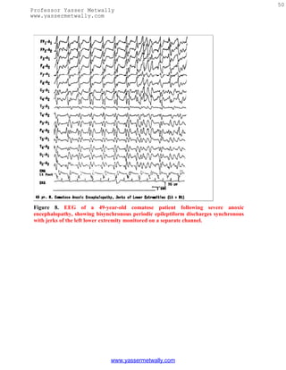
other brief body movements (Fig. 9).[30,31] Whether this is an epileptic event (a brief
myoclonic seizure) or a brain stem release phenomena remains unknown. At times these
movements may cause confusion in the minds of relatives and even treating physicians
about the patient's state of consciousness, as they may mimic volitional motor activity.
A rare EEG pattern seen in severe anoxic encephalopathy is the alpha coma pattern,
denoting the conjunction of clinical coma associated with alpha frequency activity.[32-34]
Because in such tracings the dominant frequency is alpha frequency activity without
significant slower frequencies, the EEG superficially resembles that of an "awake" person,
but there are major differences. The alpha frequency activity in alpha pattern coma is
widespread in distribution and is often prominent over the anterior head regions (Fig. 10).
Reactivity to any type of sensory stimulation is usually absent. The prognosis of alpha
pattern coma is extremely poor; all patients have either died or survived in chronic
vegetative state.
www.yassermetwally.com](https://image.slidesharecdn.com/eeg-120912084228-phpapp02/85/Textbook-of-electroencephalography-52-320.jpg)
![52
Professor Yasser Metwally
www.yassermetwally.com
Figure 10. EEG of a 77-year-old comatose patient following cardiopulmonary arrest 4 days
previously, showing "alpha coma pattern." Patient died after 2 days.
Remember that EEG findings of alpha pattern coma are also seen in the setting of
sedative/hypnotic drug intoxication[25,35] and in association with intrinsic brain stem
lesions[36] with a much more favorable prognosis.
o Cerebral Death
The EEG is being employed with increasing frequency for the determination of cerebral
death in patients with irreversible coma, particularly when organs have to be salvaged for
transplantation. It cannot be overemphasized that the absence of cerebral activity on the
EEG is only one of the criteria, and should always be considered along with the clinical
findings and blood flow studies for brain death. To properly identify very-low-voltage
cerebral activity, to distinguish physiological or instrumental artifacts, and to eliminate the
possibility of errors through malfunctioning equipment or inadequate techniques, the
American EEG Society[37] has a number of recommendations that must be followed
during EEG recordings in all cases of suspected brain death. In such "flat" tracings, EEG
activity may be obscured by very-low-amplitude fast activity due to sustained contraction
of scalp muscles, which can be eliminated by giving a short-acting neuromuscular blocking
agent (succinylcholine, 20 to 40 mg IV). This step, which is very easy to undertake, is often
overlooked to obtain a satisfactory recording in such patients.
A single EEG and a 6- to 12-hour clinical observation after an unequivocal acute cerebral
insult are minimum requirements for brain death evaluation in an adult. In young
children, the guidelines are slightly different because of the more difficult task of
www.yassermetwally.com](https://image.slidesharecdn.com/eeg-120912084228-phpapp02/85/Textbook-of-electroencephalography-55-320.jpg)
![53
Professor Yasser Metwally
www.yassermetwally.com
confirming brain death in this age group. A special task force[38] recommended the
following:
1. Brain death should not be determined until at least 7 days of age.
2. Seven days to 2 months: two examinations and two EEGs separated by at least 48
hours are required.
3. Two months to 1 year: two examinations and two EEGs separated by at least 24
hours are required.
4. Older than 1 year: similar criteria as an adult (i.e., one EEG and at least 12 hours of
observation).
Encephalitides
In viral encephalitis the severity of the EEG abnormalities generally parallel the clinical
picture, but at times the EEG may be more disorganized and slow than the mental state of
the patient may suggest. With a few exceptions, the EEG changes in different viral
encephalitides are generally nonspecific and not helpful to distinguish one etiologic agent
from another.[39]
The EEG pattern and its evolution in herpes simplex encephalitis are rather characteristic
so that the diagnosis can often be suspected by EEG findings when considered in the
proper clinical setting. The EEG in herpes simplex encephalitis may show a prominent
focal abnormality, usually a focus of polymorphic delta activity over a temporal region,
corresponding to the initial localization of pathology to the temporal lobe of the brain.[40-
42] The most characteristic EEG feature of herpes simplex encephalitis is the occurrence of
pseudo-periodic, focal or unilateral, large amplitude, sharp wave complexes that repeat at
regular intervals of 1 to 3 seconds.[40-43] These periodic lateralized epileptiform
discharges are usually expressed maximally over the involved temporal lobe (Fig. 11). This
characteristic periodic pattern is usually seen between 2 and 15 days after the onset of
illness. As the disease progresses and the other hemisphere becomes involved, the periodic
complexes may disappear on the side of initial involvement before appearing on the side
more recently involved. With bilateral involvement of the brain, periodic complexes may
occur over both hemispheres; they may then occur either synchronously or independently
over the two sides.
www.yassermetwally.com](https://image.slidesharecdn.com/eeg-120912084228-phpapp02/85/Textbook-of-electroencephalography-56-320.jpg)
![54
Professor Yasser Metwally
www.yassermetwally.com
Figure 11. EEG of a 65-year-old patient with Herpes simplex encephalitis, showing periodic
epileptiform discharges occurring over the right temporal region every 1 to 2 seconds.
The presence of unilateral or focal periodic complexes (PLEDs) is not unique for herpes
simplex encephalitis. PLEDs may occur with acute focal cerebral hemispheric processes
(e.g. infarction, brain abscess, or neoplasm).[44] Nevertheless, the presence of unilateral
periodic complexes in association with an acute febrile illness, focal seizures, and spinal
fluid pleocytosis is strongly suggestive of herpes simplex encephalitis.
SSPE, a childhood disorder that is a slow virus infection of the central nervous system due
to measles, has virtually disappeared from the United States since the introduction of
measles vaccination. The EEG in SSPE is highly specific and characterized by the presence
of high-amplitude periodic complexes that are bilateral, usually synchronous, and
symmetrical.[45-47] They are remarkably stereotyped and consist of two or more delta
waves with or without sharp wave components intermixed with them. The periodic
complexes repeat with a fair regularity every 4 to 10 seconds and there is a 1:1 relationship
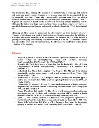
of the EEG periodic complexes to the clinical myoclonic jerks, when present (Fig. 12). In
the early stages of the disease, the periodic complexes may occur at irregular and long
intervals, and sleep may activate them. A sleep recording, therefore, is recommended in a
suspected case of SSPE in which the awake tracing has failed to reveal periodic
complexes.[48] Also, in the early stages there is asymmetry of the periodic complexes,
which may be associated with asymmetry of the myoclonic jerks that occur contralateral to
the periodic complexes.[47] Later in the disease, the periodic complexes are bilaterally
symmetrical and synchronous.
www.yassermetwally.com](https://image.slidesharecdn.com/eeg-120912084228-phpapp02/85/Textbook-of-electroencephalography-57-320.jpg)
![55
Professor Yasser Metwally
www.yassermetwally.com
Figure 12. EEG of a 16-year-old patient with subacute sclerosing panencephalitis, showing
high-amplitude generalized periodic complexes repeating at intervals of 8 to 10 seconds and
accompanied by eye jerks and myoclonic jerks of the upper extremities monitored on the
last two channels.
Creutzfeldt-Jakob disease, a prion disorder of the central nervous system (CNS), is also
characterized by a very specific EEG pattern, which consists of periodic, bilaterally
synchronous wave forms.[49] The periodic discharges take the form of diphasic or
triphasic sharp waves, which repeat regularly at a frequency close to one per second. There
is a fairly close relationship between the periodic complexes and myoclonic jerks; the latter
may occur a few milliseconds before or after the electrical event.
What is less well known is the fact that in the early stages of Creutzfeldt-Jakob disease,
focal or lateralized periodic sharp waves (PLEDs) may occur,[50,51] which later evolve into
bilaterally symmetrical and synchronous periodic discharges superimposed on a "flat"
background (Fig. 13). Although the periodic EEG pattern is not pathognomonic, the
presence of periodic sharp waves occurring regularly around one per second, in association
with clinical findings of progressive dementia and myoclonus in elderly individuals,
provides strong support to the diagnosis of Creutzfeldt-Jakob disease. This characteristic
periodic pattern is reported in more than 75% of patients with histologically verified
Creutzfeldt-Jakob disease, and the pattern becomes fully established within the first 3
months of the onset of symptoms.[52,53]
www.yassermetwally.com](https://image.slidesharecdn.com/eeg-120912084228-phpapp02/85/Textbook-of-electroencephalography-58-320.jpg)
![56
Professor Yasser Metwally
www.yassermetwally.com
Figure 13. Serial EEGs of a 62-year-old patient with Creutzfeldt-Jakob disease. The first
EEG (A), obtained 2 months after the onset of dementia and progressive right hemiparesis,
shows left-sided delta activity. EEG 2 weeks later (B) shows periodic lateralized
epileptiform discharges over the left hemisphere, and an EEG taken 5 months after the
onset of illness (C) shows typical bisynchronous high-amplitude periodic complexes
superimposed on "flat" background. Myoclonic jerks monitored on the last channel are
synchronous to the periodic complexes.
Degenerative Encephalopathies
In degenerative encephalopathies, a common denominator is a disturbance in the
regulation and frequency of the background activity, but the EEG features do differ with
regard to whether the pathologic process involves predominantly cortical and/or
subcortical gray matter or cerebral white matter.[54] In disorders that primarily involve
the cerebral white matter, the EEG is characterized predominantly by the presence of
high-amplitude continuous generalized polymorphic delta activity associated with a
markedly disordered background and virtual absence of epileptiform activity or
paroxysmal discharges. Such changes are characteristically seen in all types of
leukodystrophies, Schilder's disease, and multifocal leukoencephalopathy. In diffuse
cortical gray matter encephalopathies, the EEG is characterized by abnormal background
activity that is slow, irregular, and low in amplitude. There is minimal continuous
generalized polymorphic delta activity, and paroxysmal findings are usually absent or
www.yassermetwally.com](https://image.slidesharecdn.com/eeg-120912084228-phpapp02/85/Textbook-of-electroencephalography-59-320.jpg)
![57
Professor Yasser Metwally
www.yassermetwally.com
minimal. Examples include Alzheimer's or Pick's disease. In diffuse cortical and
subcortical gray matter encephalopathies, the EEG shows generalized bilaterally
synchronous paroxysmal discharges in the form of bursts of monorhythmic delta waves or
paroxysms of slow spike wave activity superimposed on an abnormal background.
Pathological conditions include cerebromacular degeneration (e.g., Batten's disease).
Degenerative disorders with lesions predominantly below the cerebrum produce only
minimal alterations in the EEG. This is usually the case in spinocerebellar degeneration,
Parkinson's disease, progressive supranuclear palsy, and so on, where the EEG either
remains normal or shows mild nonspecific slowing of the background activity. There are
virtually no other EEG features associated with degenerative encephalopathies that have a
high correlation to a specific etiologic process. The exception is the occurrence of large-
amplitude spikes in response to single flashes or at flickering rates below three per second,
which is a highly characteristic feature of Batten's disease.[55] These large potentials may
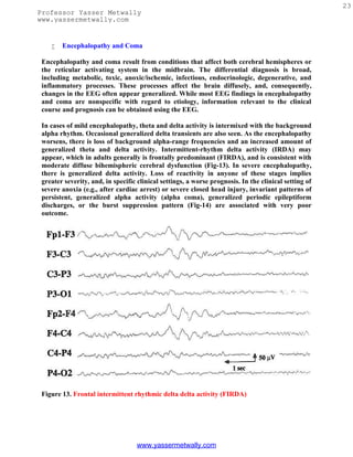
reach 50 to 500 µV, maximum over the occipital region, and sometimes associated with
myoclonic jerks of the limbs and face (Fig. 14). With advancing retinal disease and
blindness, the characteristic photic response is lost.
Figure 14. EEG
of a 6-year-old
patient with
Batten's disease,
showing high-
amplitude
spikes induced
by photic
stimulation at
one per second.
In patients with senile or presenile dementia, the EEG background shows varying degrees
of slowing and disorganization[56-58] but may remain within normal limits in individuals
with obvious intellectual impairment. A slowing of the alpha rhythm from 11 to 12 Hz to 8
to 9 Hz may represent a significant deterioration of the EEG, but in the absence of serial
studies this would remain unrecognized. Furthermore, the rate of progression of dementia
is important because patients with very slowly progressing dementia are likely to show
minimal EEG changes. Epileptiform discharges are rare in the Alzheimer's type of
presenile or senile dementia except in very advanced disease. Sharp or triphasic waves over
the posterior head regions in severely demented patients have been reported.[59] At times
these EEG waveforms may raise a suspicion of Creutzfeldt-Jakob disease; however, unlike
Creutzfeldt-Jakob disease, these discharges occur irregularly with little or no tendency
toward periodic occurrence.
Huntington's disease has a very characteristic clinical picture, and the diagnosis is
confirmed by genetic testing. There is a high incidence of abnormal EEG tracing in
Huntington's disease; the characteristic feature is the presence of a "flat tracing" with
virtual absence of rhythmic activity. Such features are reported in as high as two thirds of
www.yassermetwally.com](https://image.slidesharecdn.com/eeg-120912084228-phpapp02/85/Textbook-of-electroencephalography-60-320.jpg)
![58
Professor Yasser Metwally
www.yassermetwally.com
the patients with Huntington's disease.[60] There is absence of any EEG activity in excess
of 10 µV. In addition, the EEG is practically devoid of any rhythmic activity, not merely a
paucity of recognizable rhythms. Such "flat" EEGs in Huntington's disease need to be
differentiated from a normal variant, low-amplitude tracings in adults, which have activity
less than 20 µV and a paucity of recognizable rhythms. Hyperventilation would increase
the amount and amplitude of rhythmic activity with normal variant, whereas in patients
with Huntington's disease hyperventilation remains ineffective.[2]
EEG in Focal or Lateralized Cerebral Hemispheric Lesions
Since the advent of computerized tomography and MRI, the EEG has been utilized less for
localizing focal cerebral lesions, including brain tumors. Nevertheless, the EEG is still
extensively used to evaluate the epileptogenic potential of a focal cerebral process
demonstrated on imaging studies. The EEG shows focal or lateralizing findings in localized
lesions that involve a superficial assessable portion of a cerebral hemisphere.[61] There is
slowing and decreased amplitude of the alpha rhythm on the side of the focal cerebral
lesion. With extensive processes, the alpha rhythm disappears and is replaced by slower-
frequency activity (theta/delta). Comparable changes can occur in the anterior beta activity
and can be spontaneous or drug-induced. During NREM sleep, spindles may be less
persistent and of lower amplitude as may vertex sharp transients. In massive or rapidly
progressive hemispheric lesions such as a major hemispheric stroke or large glioblastoma,
there may be severe depression of all EEG activities in that cerebral hemisphere.
Since Walters' observation[62] a focus (localized activity) of delta activity has become the
sign "par excellence" of focal structural lesions. The delta activity is called polymorphic or
arrhythmic (PDA) because it consists of waves of irregular shape that change in duration,
shape, and amplitude (Fig. 15) and fall in the frequency range of 0.5 to 3.0 Hz. Focal PDA
indicates a lesion that involves subcortical white matter. Greater variability in the
waveform (irregularity), longer duration of waves (slower frequency), and greater
persistence indicate a more severe and acute focal process. A fact less often appreciated is
that in a large area of PDA the focal process is best localized to the area showing the
lowest-amplitude or "flat" PDA, rather than the area showing high-amplitude PDA.[63]
Destructive lesions most frequently associated with focal PDA include neoplasm, abscess,
infarct, hematoma, and contusion. However, focal PDA can appear transiently after a
complex migraine attack or focal epileptic seizure. Hence, in a patient with prominent focal
PDA with a history of a recent epileptic seizure, a repeat recording in a few days is
indicated to assess the persistence or transient occurrence of this focal abnormality. Rapid
disappearance of focal PDA would suggest a postictal change but would also lend support
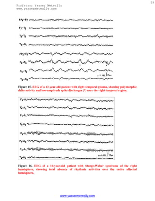
to a focal epileptic process. Static lesions such as infantile hemiplegia or Sturge-Weber
syndrome[64] are associated with marked attenuation and often total absence of rhythmic
activities (alpha or beta activity) over the entire affected hemisphere (Fig. 16). In contrast
to progressive hemispheric lesions, such as cerebral tumor, there is very little, if any, slow
activity over the involved hemisphere in such lateralized static focal processes.
www.yassermetwally.com](https://image.slidesharecdn.com/eeg-120912084228-phpapp02/85/Textbook-of-electroencephalography-61-320.jpg)
![60
Professor Yasser Metwally
www.yassermetwally.com
Certainly, attenuation, disorganization, and slowing of the background activity on the side
of the focal cerebral lesion and presence of PDA are EEG hallmarks of a focal cerebral
process. Less often, the amplitude of the background activity may be higher on the side of
the focal cerebral lesion,[65] which may lead to an erroneous interpretation of the side of
the lesion. Such increase in the amplitude of the background activity is encountered with
cerebral infarcts that have "healed," with skull defect related to previous craniotomy or in
patients with slowly progressive tumors (Fig. 17). Often the enhanced background activity
(such as alpha rhythm) over the side of the focal cerebral process is slightly slower in
frequency as well as less reactive to eye opening,[63] which should alert the interpreter to
the abnormality. Breach rhythms[66] associated with skull defects are focal "mu-like"
rhythms in Rolandic or temporal region with sporadic slow waves and spiky or sharp
transients (Fig. 18). These rhythms are unrelated to epilepsy and do not indicate
recurrence of a tumor. The "spiky" grapho-elements should not be overinterpreted as
epileptogenic discharges. For proper assessment of EEG asymmetries, it is therefore
essential to know if the patient has had a craniotomy or skull defect, which may enhance
background activities on the side of the breach of the skull.
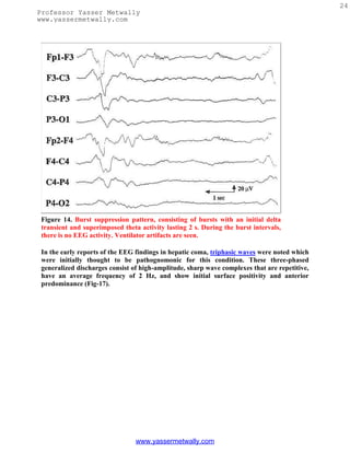
Figure 17. EEG of a 47-year-old patient with a low-grade glioma of the left temporal lobe,
showing slightly slow but higher amplitude alpha on the left side.
www.yassermetwally.com](https://image.slidesharecdn.com/eeg-120912084228-phpapp02/85/Textbook-of-electroencephalography-63-320.jpg)
![61
Professor Yasser Metwally
www.yassermetwally.com
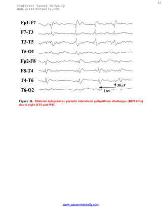
Figure 18. EEG of a 47-year-old patient with history of previous left craniotomy, showing
breach rhythm in the left temporocentral region.
Epileptiform activity, such as focal spikes, sharp waves, or spike wave discharges, also
occur in localized hemispheric lesions usually of an indolent or static nature. With acute
hemispheric lesions, epileptiform discharges are less common but when seen often have a
periodic character. PLEDs consist of sharp waves, repeating more or less regularly at one
per second over a relatively large area of the hemicranium during most of the EEG study
(Fig. 19). This distinctive focal periodic pattern usually occurs in patients with acute
hemispheric strokes, brain abscess, primary (usually glioblastoma) or metastatic
neoplasms, and herpes simplex encephalitis.[44,67]
www.yassermetwally.com](https://image.slidesharecdn.com/eeg-120912084228-phpapp02/85/Textbook-of-electroencephalography-64-320.jpg)
![64
Professor Yasser Metwally
www.yassermetwally.com
Normal components of ongoing background activity: for example, vertex sharp transients
of sleep, POSTs, mu rhythm, lambda waves, drowsy activity during sleep in children that
may often be associated with sharp components, etc.
Epileptiform variants of dubious clinical significance: there are a large number of benign
epileptiform variants that must be recognized, lest they be misinterpreted. Although
morphologically similar, they are nonepileptogenic as they have no established relationship
with the process responsible for generating epileptic seizures. Such sharp transients include
14 to 6 per second positive spikes, small sharp spikes or benign epileptiform transients of
sleep, 6 Hz spike wave or phantom spike wave, wicket spikes, psychomotor variant pattern
or rhythmic midtemporal discharges, breach rhythm, etc. Sleep not only activates
diagnostically useful epileptiform EEG patterns, but also unmasks several types of
nonepileptogenic sharp transients.
It is critical that the EEG interpreter has clear criteria for distinguishing diagnostically
relevant epileptiform discharges from sharply contoured background activity or benign
variants. Useful criteria have been formulated for identification of epileptiform
events[68,69]:
Epileptiform discharges (spikes, sharp waves, and spike wave complexes) should be
unarguably discrete events, not just accentuation of part of an ongoing sequence of waves.
They should be clearly separable from ongoing background activity, not only by their
higher amplitude but also by their morphology and duration.
Most epileptiform discharges have a bi- or triphasic waveform and they have a more
complex morphology than even high-voltage background rhythms.
The epileptiform events are not sinusoidal but rather show asymmetric, rising and falling
phases.
Most spikes and sharp waves are followed by a slow wave.
Finally, they should have a physiological potential field involving more than one electrode
that helps to distinguish them from electrode-related artifacts or muscle potentials.
Specificity of Interictal Epileptiform Abnormalities
Are "hard-core" epileptiform abnormalities encountered in normal children and adults
who do not have a history of epileptic seizures? Different studies, some in children[70,71]
and others in all age groups,[72-74] found an incidence of less than 2 to 4% of epileptiform
abnormalities in the EEG of nonepileptic subjects. In an interesting study on EEG findings
in 13,658 males ages 17 to 25 without a previous history of significant illness who were
medically screened for training in the Royal Air Force of England, 69 (0.5%) had
unequivocal epileptiform discharges.[75] Hence, the incidence of epileptiform
abnormalities in the healthy population was significantly lower than the 2 to 4% noted in
the nonepileptic patients referred to hospital EEG laboratories.
www.yassermetwally.com](https://image.slidesharecdn.com/eeg-120912084228-phpapp02/85/Textbook-of-electroencephalography-67-320.jpg)
![65
Professor Yasser Metwally
www.yassermetwally.com
One can certainly conclude that if an individual has a "blackout spell" or episodic loss of
consciousness, it is very likely to be an epileptic seizure if there are unequivocal
epileptiform discharges recorded in the EEG. To reemphasize, interictal epileptiform
discharges in the EEG are never diagnostic of epilepsy by themselves, but in the
appropriate clinical setting, they provide important circumstantial evidence for the
diagnosis of epilepsy.
Sensitivity of EEG and Techniques to Improve the Yield of Interictal and Ictal EEG
Abnormalities in Patients with Epileptic Disorders
Some patients with unequivocal epilepsy, especially focal epilepsy, may have repeatedly
normal or nonspecific EEG studies. A single routine EEG consisting of half an hour
recording during wakefulness, hyperventilation, and intermittent photic stimulation (IPS)
provides diagnostic findings in approximately half of the patients with epilepsy.[76] The
following describes a few ways to increase the yield of epileptiform abnormalities in an
interictal EEG study.
Serial EEG Studies. EEGs recorded on more than one occasion will increase the chance for
recording a specific epileptiform abnormality. Research[76] has demonstrated that serial
EEG studies increase the yield for epileptiform abnormalities from 50% in the first record
to 84% by the third EEG, and in 92% by the fourth EEG. There was little additional yield
to serial EEGs beyond this point.[76] Thus, four or five EEG studies spread over a few
years provide diagnostic abnormalities in over 90% patients with epilepsy. An opposite
corollary is also true; serial negative EEG studies in a patient with continuing paroxysmal
events should raise suspicion of nonepileptic episodes. It is also well known that interictal
epileptiform discharges markedly increase after a clinical seizure[77]; hence, obtaining an
EEG promptly after a clinical seizure will increase the chances of capturing interictal
epileptiform discharges.
Activating Procedures. Activating procedures (e.g., hyperventilation, IPS), recording
during sleep, are very well known to activate epileptiform discharges not recorded in the
awake tracing. Hyperventilation and IPS are potent activators of generalized spike wave
discharges associated with primary generalized epilepsies. On the other hand, sleep tends
to bring out focal epileptiform abnormalities in patients experiencing focal epileptic
seizures. Sleep activates virtually all focal epileptiform abnormalities; therefore, every
patient suspected of epilepsy should have a sleep recording unless there is an unequivocal
and specific abnormality displayed optimally during wakefulness. One of the best ways to
ensure a sleep EEG is to instruct the patient to come for the EEG test after remaining
awake during the entire or at least a major part of the previous night. Sleep deprivation
appears to have a further activating effect that is additive to natural sleep itself,
particularly in patients with complex partial seizures and in patients with juvenile
myoclonic epilepsy.
Normal response to IPS includes photic driving (photic following) at flash rate or at
harmonics. In about 5% of patients, asymmetric photic driving response (>50% difference
in amplitude) may occur, which by itself (without asymmetric awake and/or sleep
www.yassermetwally.com](https://image.slidesharecdn.com/eeg-120912084228-phpapp02/85/Textbook-of-electroencephalography-68-320.jpg)
![66
Professor Yasser Metwally
www.yassermetwally.com
activities) has no clinical significance. IPS is especially helpful in patients with primary
generalized epilepsy in eliciting abnormal paroxysmal discharges. Photoparoxysmal
response (PPR) has a high correlation with clinical epilepsy. It is characterized by the
occurrence of generalized bilaterally synchronous spike wave or multiple spike wave
discharges occurring with IPS. The most effective frequency is around 15 flashes per
second but other frequencies may be equally effective. Reilly and Peters distinguished two
types of PPR: prolonged (self-sustained), which continues for a short period after the
stimulus has been withdrawn (Fig. 20), and self-limited, which cease before the flashes
stop.[78] There is a much higher incidence of epilepsy in patients with prolonged (93%)
compared with the self-limited (52%) PPR. A 1992 meta-analysis of the studies on PPR
concluded: (1) PPR, prolonged or self-limited, had a significantly higher incidence of
seizures than controls; (2) a prolonged PPR was associated with a much higher incidence of
seizures (85%) than the self-limited group (50%); (3) patients with prolonged PPR more
often had other epileptiform abnormalities in their resting EEG than the self-limited
group; (4) the risk of epilepsy increased if the PPR was associated with epileptiform
abnormalities in the resting EEG; (5) the seizure incidence associated with self-limited PPR
without other epileptiform abnormalities was lower (30%) but still significantly higher
than patients without PPR.[79]
Figure 20. An EEG of a 15-year-old patient with primary generalized epilepsy, showing
prolonged (self-sustained) photoparoxysmal response.
Another photic-induced response that may superficially resemble PPR but has no
significant correlation with epilepsy is a photo-myoclonic response (PMR). It consists of
frontally dominant polyspikes synchronous with the flash rate and accompanied by
rhythmic jerking of the muscles of the forehead, eyelids, and face. The response is blocked
www.yassermetwally.com](https://image.slidesharecdn.com/eeg-120912084228-phpapp02/85/Textbook-of-electroencephalography-69-320.jpg)
![67
Professor Yasser Metwally
www.yassermetwally.com
when the eyes are opened and it promptly stops with withdrawal of photic stimulation.
PMR is generally considered to be a muscular response without cerebral participation but
some regard it to be an expression of cortical response within the spectrum of photic
cortical reflex myoclonus. It is seen in some nervous or tense individuals or in patients with
psychiatric troubles or elderly subjects. Its presence in an individual case has no diagnostic
significance.
Besides photic driving, PPR, and PMR, other less common IPS-induced EEG
responses include:
Posterior hemispheric stimulus-dependent response: an anomalous steady state flash visual
evoked potential (VEP) of unusually sharp waveform or high amplitude. This has no
clinical correlation except when it represents very-high-amplitude and spiky VEPs in
association with Batten's disease or neuronal ceroid lipofuscinosis.
Rarely, IPS may activate a focal epileptiform discharge, usually an occipital spike focus.
Less often IPS may induce a frank seizure with clinical correlates (e.g., an absence, absence
with eyelid myoclonus, single random generalized myoclonic jerks, repeated myoclonic
jerks, occipital onset focal seizure, and rarely even a generalized tonic-clonic seizure).
Remember than 10% of patients with all forms of primary generalized epilepsy show PPR,
with the highest incidence (30 to 35%) in juvenile myoclonic epilepsy.[80] The incidence of
PPR is about 15% in childhood absence epilepsy, <10% in juvenile absence epilepsy, and
10 to 15% in epilepsy with generalized tonic-clonic seizures on awakening. PPR is also
common in infantile and childhood epilepsies with myoclonic seizures such as benign and
severe myoclonic epilepsies of infancy and myoclonic-astatic epilepsy of childhood. PPR is
rare in focal epilepsies and secondary generalized epilepsies (e.g., Lennox-Gastaut
syndrome).
It must be emphasized that PPR can be detected, although rarely, among individuals with
headaches or other complaints, during evaluation for aviation jobs, or as a genetic marker
in a susceptible individual with a family history of idiopathic generalized epilepsy. PPR in
nonepileptic subjects has a prevalence of 1 to 4%; the response then is usually brief and
less prominent.
IPS is not a totally benign activating procedure. One can induce a generalized tonic-clonic
convulsion (often the first one) if photic stimulation is continued over a long duration in a
patient who shows prominent PPR. It is recommended that the photic stimulation be
limited to short periods (1 to 5 seconds) and terminated promptly as soon as generalized
spike wave activity is recorded (Fig. 20).
Special Electrodes. Although the standard 10 to 20 international system of electrode
placement provides reasonable coverage of the whole head, certain areas that have high
epileptogenicity, such as the mesial temporal lobes in patients with mesial temporal
sclerosis, are not fully explored by conventional placement and may require additional
www.yassermetwally.com](https://image.slidesharecdn.com/eeg-120912084228-phpapp02/85/Textbook-of-electroencephalography-70-320.jpg)
![68
Professor Yasser Metwally
www.yassermetwally.com
electrodes. Nasopharyngeal electrodes have been widely used in the past in patients
suspected to have with temporal lobe epilepsy. They are associated with variable degrees of
discomfort and may prevent the patient from attaining sleep during the EEG recording.
They have now been largely replaced by the use of anterior temporal electrodes, which are
placed 1 cm above and one third the distance along the line from the external auditory
meatus to the external canthus of the eye.[81]
In a comparison of the percentages of spikes detected by standard scalp electrodes, anterior
temporal, mini-sphenoidal, surface sphenoidal, and nasopharyngeal electrodes in patients
with suspected complex partial seizures, the anterior temporal electrodes provided
significant improvement in detecting epileptiform abnormalities.[82] Recordings from
standard scalp electrodes detected 58% of the discharges. Anterior temporal electrodes
were the best; they detected 70% of all the discharges by themselves, and 81% in
combination with standard scalp electrodes. It can be concluded that recordings from
anterior temporal electrodes must be done to improve the detection of interictal
epileptiform abnormalities in patients suspected of having temporal lobe epilepsy.
Sphenoidal electrodes are almost invariably employed during video EEG monitoring as a
part of the presurgical evaluation of patients with medically intractable complex partial
seizures. The yield of abnormality from sphenoidal recordings is certainly greater than that
with nasopharyngeal or anterior temporal electrodes, but it is difficult to justify the use of
invasive electrode placement in routine EEG study for paroxysmal disorders.
Activation of an Actual Seizure During Routine EEG Study. All efforts must be made to
capture the patient's habitual episode during a routine EEG. If precipitating factors are
known, these are appropriately exploited. Hyperventilation, a potent precipitator of an
absence seizure in a child with primary generalized epilepsy, must always be utilized for at
least 5 minutes, once in the beginning and again at the end of a routine EEG study. In rare
patients with reflex epilepsy, playing specific music in musicogenic epilepsy, asking a
patient to read from a book in reading epilepsy, bathing the patient in bathing epilepsy,
asking the patient to eat his meals (eating epilepsy), smelling gasoline, and so on, may all be
carried out to promote an ictal event.
In patients with possible pseudoseizures, suggestion protocols are often useful in
precipitating episodes and demonstrating EEG changes.[83] It is important to emphasize
that induced seizures must be clinically typical of a patient's habitual episodes before the
diagnosis of pseudoseizures is strongly considered.
Some Interpretive Challenges of EEG Findings During Paroxysmal Events
Some of the pitfalls regarding ictal EEG changes during an actual seizure need to be
stressed. All epileptic seizures are not associated with distinctive concomitant surface EEG
changes. Seizures that remain very localized, including epilepsia partialis continua and
simple partial seizures (focal seizures with preserved consciousness), may not have changes
in the scalp EEG because the diagnostic discharge may be deep-seated or involve only a
small pool of neuronal tissue. However, epileptic seizures manifested by loss of
consciousness, on the other hand, are accompanied by demonstrable changes in the scalp
www.yassermetwally.com](https://image.slidesharecdn.com/eeg-120912084228-phpapp02/85/Textbook-of-electroencephalography-71-320.jpg)

![70
Professor Yasser Metwally
www.yassermetwally.com
Figure 21. EEG of a
46-year-old patient
with primary
generalized epilepsy,
showing atypical
generalized
bisynchronous spike
wave activity.
Besides the presence of brief (1 to 3 seconds) generalized spike wave discharges, there are
no interictal epileptiform abnormalities that are specific for individual syndromes included
under PGE (childhood absence epilepsy, juvenile absence epilepsy, juvenile myoclonic
epilepsy, epilepsy with myoclonic absences, and generalized tonic-clonic seizures on
awakening). There are a few EEG features that are more common with certain syndromes:
(1) polyspike wave discharges are more common with myoclonic epilepsies; (2) paroxysms
of occipital-dominant rhythmic delta activity in the EEG is a feature most commonly
encountered with childhood absence epilepsy; (3) short paroxysms of spike wave discharges
of higher frequency (4.0 to 4.5 Hz) are more common with PGE manifesting primarily with
generalized tonic-clonic seizures; and (4) PPR is most common with juvenile myoclonic
epilepsy.
In patients with PGE, a routine EEG may capture one or more absence seizures or
epileptic myoclonic jerks. In children an absence seizure may be induced during the EEG
study by hyperventilation with characteristic generalized 3 Hz spike wave discharge, which
is sustained for more than 3 seconds in duration. Epileptic myoclonic jerks are associated
in the EEG with high-amplitude generalized polyspike wave discharges in association with
myoclonic jerks. Not well recognized is the fact that the EEG in patients with PGE may
record focal or lateralized spikes in addition to the overwhelming generalized
bisynchronous spike wave activity.[84,85] Similarly, spike or spike wave activity occurring
bilaterally but restricted to the frontal areas (where the generalized paroxysms are usually
maximum) is also common. Such discharges are often called "abortive" spike wave
complexes. Roughly one quarter of patients with 3 Hz spike wave activity in their EEG may
show such focal or lateralized discharges,[84] which should generally be viewed as isolated
fragments or limited expression of what is fundamentally a generalized epileptic
www.yassermetwally.com](https://image.slidesharecdn.com/eeg-120912084228-phpapp02/85/Textbook-of-electroencephalography-73-320.jpg)
![73
Professor Yasser Metwally
www.yassermetwally.com
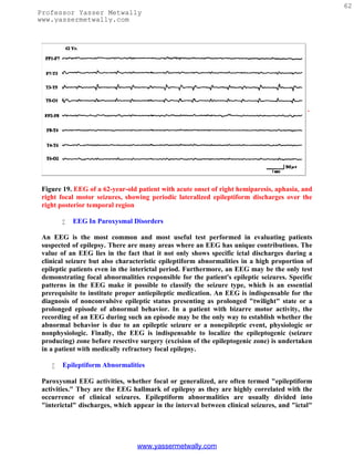
Hypsarrhythmia, which is an EEG pattern, and infantile spasms do not have an absolutely
constant relationship and are not interchangeable terms. Typical and modified
hypsarrhythmia occurs in two thirds of the EEGs of infants with infantile spasms, whereas
the remaining one third show generalized slow spike wave discharges (described
below).[86] Besides various pathologic conditions associated with a severe cortical insult,
hypsarrhythmic pattern is often encountered in infants suffering from tuberous sclerosis or
genetically determined metabolic conditions such as non-ketotic hyperglycenemia.[87]
Children with Aicardi's syndrome (agenesis of corpus callosum, mental retardation,
infantile spasms, choreoretinal lesions) show a markedly asymmetric hypsarrhythmic
pattern with virtually complete interhemispheric asynchrony of a suppression-burst-like
background.[88]
The hypsarrhythmic pattern is a maturational pattern most commonly expressed between
the ages of 4 and 12 months. As the infant grows older, beyond the age of 2 years, it is rare
to encounter typical hypsarrhythmia, although infantile spasms may still continue.
Hypsarrhythmia is replaced by different EEG patterns such as a diffusely slow tracing,
slow spike wave discharges as seen with Lennox-Gastaut syndrome, IMSD, and, rarely, a
normal tracing.
Adrenocorticotrophic hormone therapy often has a dramatic effect on infantile spasms as
well as the hypsarrhythmic EEG pattern, which may virtually disappear in a matter of a
few days to a few weeks after initiation of therapy. However, despite these clinical and EEG
improvements, the long-term neurocognitive development remains subnormal.
Lennox-Gastaut syndrome (childhood epileptic encephalopathy) is another common form
of SGE manifesting in early childhood with developmental delay, neurocognitive deficits,
and frequent generalized seizures including tonic seizures. The EEG shows generalized,
slow spike wave discharges (1.5 to 2.5 Hz) superimposed on abnormally slow background
activity (Fig. 24).[89,90] It is important to distinguish these EEG findings from those seen
with primary generalized epilepsy where the background activity is normal for age and the
generalized spike wave discharges are usually of faster frequency (3 to 5 Hz). Although
appearing widespread and bilaterally synchronous, the slow spike wave activity is usually
higher in amplitude over the anterior head regions (in 90% of patients); less commonly the
amplitude is highest over the occipital areas. The duration of the paroxysms varies widely
from isolated complexes to almost continuous slow spike wave activity, commonly without
an identified behavioral or awareness change. Hence, the slow spike wave activity in
Lennox-Gastaut syndrome is considered an interictal pattern, although it must be
understood that subtle changes of behavior in retarded and uncooperative children are
hard to recognize.
www.yassermetwally.com](https://image.slidesharecdn.com/eeg-120912084228-phpapp02/85/Textbook-of-electroencephalography-76-320.jpg)
![74
Professor Yasser Metwally
www.yassermetwally.com
Figure 24. EEG of a 16-year-old child with mental retardation and tonic seizures, showing
slow spike wave activity superimposed on a slow background.
When one encounters prolonged episodes of slow spike wave activity lasting several seconds
to minutes, the interpretative challenge is to decide if these electrographic events represent
an ictal pattern (atypical absences or nonconvulsive status) or they simply represent more
pronounced interictal pattern. A history of similar long episodes of slow spike wave activity
in one or more previous EEGs would support an interictal finding. Also, giving a small
dose of lorazepam intravenously will have no affect on an interictal pattern but will usually
abort an ictal pattern, at least temporarily.
If a tonic seizure is recorded during the EEG of a patient with Lennox-Gastaut syndrome,
the characteristic finding is an electrodecrement or "flattening" lasting several seconds. In
addition, high-frequency rhythmic activity in the alpha-beta frequency range commonly
occurs during the electrodecrement.
Another distinctive EEG pattern of a symptomatic generalized epilepsy syndrome is IMSD
characterized by the presence of three or more independent and noncontiguous foci of
spike or spike wave activity with at least one focus in one hemisphere (Fig. 25).[91,92] As
expected, the background activity is invariably disorganized and slow in frequency.
www.yassermetwally.com](https://image.slidesharecdn.com/eeg-120912084228-phpapp02/85/Textbook-of-electroencephalography-77-320.jpg)
![75
Professor Yasser Metwally
www.yassermetwally.com
Figure 25. EEG of a 7-month-old child, showing independent multifocal spike discharges.
There is a close correlation between the three EEG patterns of hypsarrhythmia, slow spike
wave, and IMSD associated with SGE. All of them are associated with diffuse or multifocal
cerebral abnormalities and have similar clinical correlates of mental retardation, multiple
and medically intractable seizure types, and a high incidence of neurologic deficits.[91]
Furthermore, serial studies over time may show a change of one pattern to the other in the
same patient. Also, in the same EEG study, more than one of these patterns may coexist
(e.g., IMSD during wakefulness and slow spike wave activity during sleep). It is very well
known that at least 20% of infants with hypsarrhythmia may show slow spike wave usually
by the second to fourth year of life. Both of these patterns may further change to IMSD in
early childhood. Thus, these three EEG patterns have a common physiopathologic basis
and are probably dependent more on cerebral maturation than on a particular kind of
cerebral pathologic process.[91] Hypsarrhythmia is usually seen in the later half of the first
year of life in response to a cerebral insult prenatally, perinatally, or in the immediate
postnatal period. It rarely results from cerebral insults after the second year of life. The
slow spike wave pattern associated with Lennox-Gastaut syndrome is commonly observed
between the ages of 2 and 5 years. The IMSD pattern is seen commonly throughout the first
decade of life.
A unique EEG pattern of GPFA is seen predominantly during sleep consisting of high-
frequency, 12 to 25 Hz repetitive spike discharges occurring synchronously over both
hemispheres (Fig. 26).[93] It is associated most commonly with SGE (usually Lennox-
Gastaut syndrome) but it may rarely occur also with PGE or in patients with focal seizures,
www.yassermetwally.com](https://image.slidesharecdn.com/eeg-120912084228-phpapp02/85/Textbook-of-electroencephalography-78-320.jpg)
![76
Professor Yasser Metwally
www.yassermetwally.com
particularly with a frontal lobe focus. This EEG pattern is usually not associated with an
obvious clinical change, although subtle tonic seizures (opening of eyes and jaw, eye
deviation upward) may be missed. In rare patients with PGE and 3 Hz generalized spike
wave, the awake EEG may appear rather benign but the presence of GPFA during sleep is
a warning that more severe encephalopathy may be present. In such patients, motor
seizures are common and the disorder is likely to persist in adulthood.[94]
Figure 26. EEG of an 11-year-old patient with Lennox-Gastaut syndrome, showing
generalized paroxysmal fast activity (B).
EEG in Focal or Localization-Related Epilepsies
Focal epilepsies are usually categorized into two subgroups: (1) asymptomatic (secondary)
localization-related epilepsies due to acquired focal cortical processes; and (2) idiopathic
(primary) localization-related epilepsies, which are largely age-dependent and genetically
based disorders, the best-known being benign Rolandic epilepsy (BRE).
The hallmark of focal epilepsy in the interictal EEG is the presence of a focus of
epileptiform activity (i.e., spikes, spike waves, or spike wave complexes). The interictal
www.yassermetwally.com](https://image.slidesharecdn.com/eeg-120912084228-phpapp02/85/Textbook-of-electroencephalography-79-320.jpg)
![77
Professor Yasser Metwally
www.yassermetwally.com
paroxysmal activity is characteristically random, occurring at inconstant intervals.
Intuitively, the positive correlation of an abundance of epileptiform discharges with a more
frequent occurrence of clinical seizures would be expected. This is, however, not true.
Abundance and rate of repetition of epileptiform discharges correlate very poorly with the
frequency of clinical seizures. It is not uncommon to see rare interictal spikes in a patient
who has frequent complex partial seizures. The reverse is also true, as in BRE where the
child may have only rare clinical seizures but very abundant epileptiform discharges,
particularly during sleep. An important fact regarding interictal epileptiform discharges is
that they become much more frequent in the EEG immediately after a clinical focal
seizure.[77]
Another characteristic of the interictal EEG is the presence of a focal abnormality of the
background activity. This may take the form of intermittent low-voltage slow waves and
attenuation of faster rhythm with the same localization as that of the epileptiform
discharges. In general, when the focal epileptiform activity occurs along with abnormal
focal organization, the possibility of a structural lesion is more likely and the focal epilepsy
is most likely to be symptomatic. On the other hand, occurrence of random focal spikes
without any accompanying features of focal disorganization of the background activity is
usual with idiopathic or primary localization-related epilepsies such as BRE or benign
occipital epilepsy.
The majority (85%) of patients with complex partial seizures have temporal lobe onset,
whereas a small proportion (15%) have frontal lobe onset partial epilepsy. Temporal lobe
epilepsy, which is the most common type of focal epilepsy in adults, is associated with
interictal epileptiform discharges over one or both anterior temporal regions (Fig. 27). The
peak of the potential field involves inferior frontal (F7 or F8), midtemporal (T3 or T4), and
the ear lobe electrodes (A1 or A2). The F7 and F8 electrodes of the 10 to 20 electrode
placement system more commonly record activity originating in the anterior temporal lobe
rather than in the frontal areas. In suspected patients with temporal lobe epilepsy,
recording from additional TI and T2 electrodes must be performed to optimally elicit
epileptiform abnormalities of temporal lobe origin. In patients undergoing presurgical
evaluation, anterior sphenoidal electrodes are commonly inserted, which markedly
increase the yield of demonstrating temporal epileptiform abnormalities. In frontal lobe
epilepsy, interictal epileptiform discharges are recorded over the frontal region but often
the EEGs, even on several occasions, may remain normal.[95,96] Special attention must be
paid to the midline electrodes (FZ and CZ), which can demonstrate low-amplitude
epileptiform discharges. Also, additional supraorbital and midfrontopolar (FPZ) electrodes
may be helpful in demonstrating epileptiform abnormalities in frontal lobe epilepsy.
www.yassermetwally.com](https://image.slidesharecdn.com/eeg-120912084228-phpapp02/85/Textbook-of-electroencephalography-80-320.jpg)
![78
Professor Yasser Metwally
www.yassermetwally.com
Figure 27. EEG of a 68-year-old patient with a long history of complex partial seizures,
showing a focus of sharp waves and low-amplitude slow activity over the right anterior
temporal region.
BRE constitutes the most common primary (idiopathic) localization-related partial epilepsy
with onset between the ages of 4 and 10 years. Virtually all recover by the age of 15 or 16
years when the patients become seizure-free and their EEGs revert to normal. The
background EEG activity in wakefulness and sleep is normal. There are epileptiform
discharges over the central or centrotemporal region, which are markedly activated during
sleep (Fig. 28).[97] In some children, epileptiform discharges are restricted to sleep
recording only.
www.yassermetwally.com](https://image.slidesharecdn.com/eeg-120912084228-phpapp02/85/Textbook-of-electroencephalography-81-320.jpg)
![79
Professor Yasser Metwally
www.yassermetwally.com
Figure 28. EEG of a 9-year-old child with benign Rolandic epilepsy, showing a focus of
right centrotemporal spike discharges. The right half of the figure shows a spike discharge
with horizontal dipole distribution.
A horizontal dipole field is a highly characteristic feature of the centrotemporal
epileptiform discharges associated with BRE.[98] The negative end of the dipole is located
at the centrotemporal area, and the positive end toward the frontal regions bilaterally (see
right half of Fig. 28). In 10 to 15% of patients with BRE, additional EEG abnormalities are
seen. Independent spike foci may occur over the occipital and less commonly over the
frontal regions. Interestingly, bisynchronous spike wave discharges also occur in
approximately 10% of patients with BRE during routine EEG recordings.[99]
Centrotemporal or Rolandic spikes are not specific for BRE. Children with acquired brain
insults involving fronto-centro-temporal regions (who may also have neurologic deficits,
e.g., cerebral palsy) may show epileptiform activity over one or both central areas. In
contrast to BRE such "lesional" patients usually have abnormal background activity,
either focally or diffusely, and have secondary (symptomatic) partial epilepsy.
Many studies[100-102] have stressed that a small proportion of epileptics have midline
spike foci; the epileptiform abnormalities are localized at the midline electrodes with some
spread to parasagittal electrodes on both sides. They are best demonstrated in the coronal
montage, which includes midline (FZ, CZ, and PZ) electrodes; the amplitude may be
www.yassermetwally.com](https://image.slidesharecdn.com/eeg-120912084228-phpapp02/85/Textbook-of-electroencephalography-82-320.jpg)
![80
Professor Yasser Metwally
www.yassermetwally.com
somewhat asymmetric in parasagittal electrodes. The midline spike foci have highest
amplitude at the CZ electrode in most of the patients. Such foci are common in children,
and at least half of the patients show these discharges only during sleep. Certainly, caution
must be exercised in distinguishing midline CZ spikes from normal sleep potentials such as
vertex sharp transients. Midline spike foci have a high correlation with clinical seizures of
focal or generalized type, and a significant proportion show evidence of cognitive or
neurologic deficits.
Rarely, a focal seizure is recorded in a routine EEG in a patient with focal epilepsy. As
mentioned before, the hallmark of an ictal EEG pattern associated with a focal seizure is
the sudden appearance of focal rhythmic activity that evolves during a short period with a
progressive change in the amplitude and frequency with a topographic spread. Postictally,
there may be a period of "flattening" followed by variable degrees of slow wave activity,
which may be generalized initially but usually becomes lateralized or focal later in the
postictal period before the preictal EEG pattern becomes reestablished. A transient delta
focus after the seizure is a very reliable sign of a localized lesion or at least a focal origin of
the previous epileptic seizure. The postictal focal slowing may persist for a few seconds to a
few hours or even a few days, depending upon such factors as the age of the patient, size of
the lesion, duration, and number of seizures.
The ictal EEG pattern of complex partial seizures of temporal lobe onset usually starts with
a focal rhythmic 4 to 7 Hz theta activity over one temporal region, which then spreads
bilaterally, becoming higher in amplitude, slower in frequency, and often interspersed with
sharp wave components.
Complex partial seizures of frontal lobe onset have a unique semiology compared with the
complex partial seizures of temporal lobe onset.[95,96] The former are brief (less than 1
minute), commonly nocturnal, often occurring in clusters with many per day, have complex
motor automatisms with kicking and thrashing, sexual automatism, vocalization, and with
very short or no postictal period of confusion after the seizure. The frantic and often
bizarre motor thrashing and kicking behavior and rapid return of awareness at the end of
the seizure frequently leads to an erroneous diagnosis of pseudoseizures. Diagnostic
difficulty is further compounded by the fact that routine EEG, even on many occasions,
may be nonrevealing in patients with frontal lobe seizures.[95,96] Interictal abnormalities,
when present, are often seen near the vertex bifrontally so that localization is not easy.
Furthermore, the ictal EEG during a frontal lobe complex partial seizure often fails to
provide diagnostic information because of marked contamination with ongoing movement
artifacts during the seizure. Careful evaluation of the ictal EEG using coronal montage and
high-frequency filtering may reveal rhythmic ictal patterns in frontal/central electrodes
near the midline, spreading bilaterally to the parasagittal region. The frontal lobe complex
partial seizures not only resemble pseudoseizures clinically but also lack diagnostic
interictal and ictal EEG findings. Their brief duration, emergence out of sleep, and
stereotypic semiology are highly characteristic features that help distinguish them from
pseudoseizures.
www.yassermetwally.com](https://image.slidesharecdn.com/eeg-120912084228-phpapp02/85/Textbook-of-electroencephalography-83-320.jpg)
![81
Professor Yasser Metwally
www.yassermetwally.com
Certain EEG Patterns Morphologically Similar to Interictal/ictal Epileptiform
Discharges But Unrelated to Epilepsy
There are some EEG patterns that, by virtue of having spike or sharp wave components or
being rhythmic, closely resemble diagnostically important epileptiform discharges.
Although they share some of the same electrographic features, their presence in the EEG
has no correlation with epileptic seizures. These patterns have been reviewed in detail.[103]
The 14 and 6 Hz positive spikes are brief paroxysms, usually less than 1 second, of positive
spikes with a frequency of 6 Hz at some times and 14 Hz at others. They are distributed
over the posterior hemispheric regions, occurring usually unilaterally. If enough of them
are recorded in a tracing, they are expressed on both sides of the head. They are most
commonly seen in children and adolescents during drowsiness and light sleep. This pattern
has been the subject of many reviews and long-drawn controversy but it is now held to be a
normal sleep activity or a normal variant.
Small sharp spikes or benign epileptiform transients of sleep are spike potentials that are
recorded in adults during drowsiness and light sleep. They are sporadic and shift from one
side to the other, and have widespread distribution over the scalp despite low amplitude.
The spikes are usually low amplitude (less than 50 µV), very short duration (less than 50
milliseconds), and mono- or diphasic potentials with no or minimal slow wave component
following the spikes (Fig. 29). They are never associated with focal slowing or other
abnormalities of the background activities. Their special distribution on the scalp may
suggest a horizontally oriented dipole generator extending across the sagittal midline with
opposite polarity on the two sides of the head. They are especially frequent in the EEG
performed after a period of sleep deprivation and are considered to have no correlation
with epileptic seizure disorder.[104]
The 6 Hz spike wave or phantom spike wave consists of bilaterally synchronous, short (less
than 1 second) paroxysms of 5 to 7 Hz spike wave activity.[105] Although widespread in
distribution, they are usually best developed over the posterior hemispheric region (Fig.
30). The spike component is usually low in amplitude, whereas the slow wave following it is
more prominent; hence, the term "phantom spike wave." It occurs in young adults and is
best expressed during drowsiness. There is still some controversy regarding their clinical
significance, especially the ones that are anteriorly dominant. It has been suggested[106]
that the 6 Hz spike wave activity that occurs with high amplitude and predominance over
the anterior hemisphere and is recorded during wakefulness may have a high correlation
with seizures, whereas predominantly occipital, low-amplitude, spike wave discharges
occurring in drowsiness have no correlation with epileptic seizures.
"Psychomotor variant" or rhythmic midtemporal discharges is a rare EEG finding
observed in less than 1% of the patient population coming to an EEG lab. It is
characterized by long (several seconds to a minute) paroxysms of 5 to 7 Hz rhythmic
activity with an admixture of sharp components occurring over the midtemporal (T3/T4)
regions (Fig. 31).[107] The paroxysms are unilateral or bilateral. Bilateral paroxysms may
appear independently on the two sides or simultaneously with variable asymmetry. The
www.yassermetwally.com](https://image.slidesharecdn.com/eeg-120912084228-phpapp02/85/Textbook-of-electroencephalography-84-320.jpg)
![82
Professor Yasser Metwally
www.yassermetwally.com
pattern is invariant and without evolution from beginning to end. This EEG finding occurs
during drowsiness or light sleep and has no correlation with epileptic seizure disorder.
The wicket temporal pattern is characterized by negative sharp transients that occur in
runs and have a wicket shape. The repetition rate varies between 6 and 11 Hz in different
and even in the same bursts. They are best seen in T3/T4 or F7/F8 electrodes with variable
spread to A1/A2. They are usually bitemporal, occurring independently on the two sides.
The pattern occurs exclusively in adults and is seen in both wakefulness and sleep.[108]
Subclinical rhythmic EEG discharges of adults is a rare EEG pattern[109,110] that closely
resembles an ictal pattern, but there is no evidence that it represents an epileptic seizure. It
occurs mainly in elderly subjects and may extend beyond 1 minute but without any clinical
change. It is predominant over the posterior head regions and usually bilateral but may be
asymmetric. In the most characteristic form the pattern begins with a series of rhythmic
sharp-contoured waves that gradually merge into a sustained theta frequency. It may
subside abruptly or gradually.[103]
There are many normal activities, particularly during sleep in children that, being sharp in
configuration, are often mistaken for epileptiform discharges. These include vertex sharp
transients and positive occipital sharp transients. In children, the vertex sharp transients
may be quite high in amplitude and spiky in configuration, very closely resembling
epileptiform discharges. One has to be very careful in interpreting sharp waves or spikes in
children of Rolandic location expressed only during sleep. Normal mu rhythm in central
leads and breach rhythm recorded in patients with iatrogenic or acquired skull defect may
also consist of sharp components that may be mistaken for epileptiform discharges.
Lambda waves, which are sharply contoured activity in the occipital region seen when a
person has eyes open and scans the surrounding environment, need to be differentiated
from posterior hemispheric epileptiform discharges.
www.yassermetwally.com](https://image.slidesharecdn.com/eeg-120912084228-phpapp02/85/Textbook-of-electroencephalography-85-320.jpg)
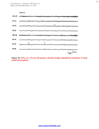
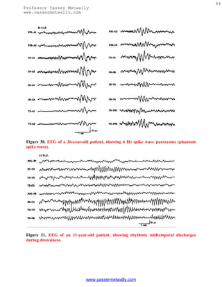
![85
Professor Yasser Metwally
www.yassermetwally.com
EEG in Neonates
In recent years there has been much interest in using EEG to evaluate full-term or
premature neonates[111,112] due to the serious limitations in performing an adequate
neurologic examination. The neonate may be confined to an isolette, may be intubated, or
may be paralyzed for ventilatory control. Under such circumstances, EEG is a very
important tool to assess an encephalopathic process or occurrence of epileptic seizures. In
addition, the background abnormalities have been classified in neonates and used to
predict neurologic outcome.
The EEG of a neonate shows distinctive patterns related to the conceptional age (CA) and
the behavioral state (awake, active sleep, quiet sleep). Space does not permit a description
of EEG patterns associated with different conceptional ages, but this is summarized in
Table 2. It is important to emphasize that the EEG maturation runs parallel in utero and in
incubator; only minimal or no differences have been found between babies of the same CA
born after different periods in utero.
Table 2. EEG Maturation in Preterm Neonates
In a full-term neonate four EEG patterns are observed related to the wakefulness/sleep
cycle: (1) low-voltage irregular (LVI) is characterized by the presence of continuous low-
amplitude (<50 µV), mainly widespread theta activity; (2) mixed pattern is characterized
by continuous moderate amplitude (usually <100 µV) theta and delta activities; (3) high-
voltage slow (HVS) consists of continuous high-amplitude semirhythmic, mostly delta
activity (0.5 to 3.0 cps) in all regions with an amplitude of 50 to 150 µV; and (4) tracé
www.yassermetwally.com](https://image.slidesharecdn.com/eeg-120912084228-phpapp02/85/Textbook-of-electroencephalography-88-320.jpg)
![86
Professor Yasser Metwally
www.yassermetwally.com
alternant (TA) pattern is characterized by the occurrence of 3 to 5 second bursts of high
amplitude (50 to 100 µV) slow activity (0.5 to 3.0 Hz), which occur at intervals of 3 to 10
seconds when the background is relatively low amplitude (10 to 25 µV) consisting of theta
waves. In other words, there is an alteration of bursts of large-amplitude slow activity
separated by quiescent or "flat" periods of low-voltage activity.
These four EEG patterns are recorded in different states. The LVI EEG is usually
recorded in wakefulness and in active sleep. The mixed pattern can also be recorded in
active sleep and relaxed wakefulness. The TA and HVS patterns are characteristic of quiet
sleep. A unique characteristic of neonate sleep is that as the neonate falls asleep, he usually
enters a period of active (REM) sleep. This differs from older infants, children, and adults
who never begin their sleep with a period of active (REM) sleep. It is only by 10 to 12 weeks
postterm that sleep onset changes from active to quiet (NREM) sleep.
A couple of perils and pitfalls need emphasized in interpreting the neonatal EEG. The TA
pattern of quiet sleep in a normal full-term neonate and tracé discontinua pattern in a
normal premature neonate during quiet sleep have a superficial resemblance to the
suppression-burst pattern that carries a poor prognosis. Differentiation between normal
and abnormal discontinuous patterns becomes an important challenge in interpreting the
neonatal EEG. The suppression-burst pattern is invariant and nonreactive to stimulation.
It usually signifies a severe encephalopathy (usually ischemic/hypoxic), although it may
occur transiently due to recent intravenous sedative/hypnotic medication. On the other
hand, the TA and tracé discontinua pattern associated with quiet sleep in full-term and
premature neonates, respectively, are state dependent and react to stimulation. In addition,
long recordings would demonstrate activities characteristic of wakefulness and active sleep
in normal full-term and premature (over 32 weeks CA) neonates. The TA pattern
gradually disappears over 6 weeks postterm when the HVS pattern becomes the sole EEG
accompaniment of quiet sleep.[113]
Another striking feature of the neonatal EEG is the frequent occurrence of multifocal
sharp transients during indeterminate and quiet sleep. These start appearing at 35 weeks
CA and constitute a normal finding in full-term neonates. The clinical significance of these
sharp transients remains controversial. It is difficult to clearly separate abnormal
(pathologic) sharp waves from normal sharp transients of the neonate. In general, the
normal sharp transients are infrequent in occurrence, usually blunt in morphology (rather
than assuming spiky configuration), arise from any scalp location but are common over
frontal and temporal regions, are truly random in occurrence, without persistent focality,
and largely confined to the burst phase of the TA pattern. However, no universal criteria
have been established to separate normal sharp transients of the newborn from the
abnormal sharp wave activity. Unless the sharp wave discharges are repetitive, periodic, or
localized over one region, an epileptogenic significance must not be assigned to them. When
the multifocal sharp transients are very frequent and occur even during active sleep and
wakefulness, the EEG is considered to be abnormal but suggestive merely of a nonspecific
encephalopathic process.
www.yassermetwally.com](https://image.slidesharecdn.com/eeg-120912084228-phpapp02/85/Textbook-of-electroencephalography-89-320.jpg)
![87
Professor Yasser Metwally
www.yassermetwally.com
A unique EEG pattern of pathologic significance is the presence of positive Rolandic sharp
waves (PRS).[114] PRS may be confined to one hemisphere; if they are bilateral they may
be consistently more abundant over one hemisphere. They are usually maximum at CZ but
may be lateralized to C3 or C4 electrodes (Fig. 32). Their positive polarity is a distinctive
feature in addition to their localization. Although initially described as the EEG correlates
of the intraventricular hemorrhage, PRS waves may be seen in other conditions, including
periventricular leukomalacia, parenchymal hemorrhage, hydrocephalus, hypoxic/ischemic
insult, and other conditions.[112,115-117] Presently, the view is that PRS waves represent a
marker of different white matter lesions rather than being specific for
periventricular/intraventricular hemorrhage. Positive polarity sharp waves at other
locations (especially in the temporal region) have no distinctive significance and may just
be a part of multifocal sharp transients in the neonates.
Figure 32. EEG of a neonate of
35 weeks conceptional age,
showing low-amplitude
positive Rolandic sharp wave
discharges at the vertex (CZ)
electrode. Patient had grade
IV intraventricular
hemorrhage and was
unresponsive.
Abnormalities of the background activity in full-term neonates are usually classified as
either severe or mild[118-120] and are summarized in Table 3. The severe EEG
abnormalities indicate severe impairment of brain function and carry a poor prognosis for
survival and/or neurologic development. Severely abnormal EEG patterns consist of: (1)
isoelectric EEG showing activity consistently below 5 µV; (2) persistent low-voltage tracing,
EEG showing activity between 5 and 15 µV with very little variability or sleep/wake
www.yassermetwally.com](https://image.slidesharecdn.com/eeg-120912084228-phpapp02/85/Textbook-of-electroencephalography-90-320.jpg)
![88
Professor Yasser Metwally
www.yassermetwally.com
differentiation; (3) paroxysmal tracing or suppression-burst pattern (discontinuous,
invariant, and nonreactive pattern characterized by 1 to 10 second paroxysms of
polymorphic activities such as sharp waves, spikes, and theta-delta activities interspersed
with long quiescent periods as long as > 20 seconds); (4) invariant high-amplitude delta
activity (persistent and nonchanging high-amplitude 0.5 to 3.0 Hz generalized activity); and
(5) the presence of gross asynchrony and asymmetry of the EEG activity over the two sides
of the head. Studies have established that the presence of these EEG abnormalities in a full-
term neonate, particularly if the abnormalities have been persistent in serial EEGs, carry a
very poor prognosis for survival or future neurologic development; over 90% of neonates
with such severe abnormalities have an unfavorable outcome.[118,119]
Table 3. Classification of EEG Abnormalities in Neonates
Mild abnormalities of the background activities include more than the usual asynchrony
and/or asymmetry; EEG being immature for the conceptional age; lack of recognizable
sleep states; excessive discontinuity ("flat" periods longer than 30 seconds); abnormal
monorhythmic activities; and excessive multifocal sharp transients. The presence of more
than one mild abnormality may suggest an underlying encephalopathic process of varying
severity, particularly if the EEG shows persistent abnormality on serial studies. Several of
the above mild abnormalities do occur in neonates who are heavily sedated; hence,
iatrogenic causes need to be excluded before ascribing them to permanent neurologic
insult.
Neonatal Seizures
One of the major reasons an EEG is performed is if a neonate is suspected of having
epileptic seizures. In neonates, epileptic seizures are often characterized clinically by subtle
www.yassermetwally.com](https://image.slidesharecdn.com/eeg-120912084228-phpapp02/85/Textbook-of-electroencephalography-91-320.jpg)
![89
Professor Yasser Metwally
www.yassermetwally.com
motor behavior such as elevation of a limb, eye deviation, eyelid flutter, tonic posturing,
bicycling movements of the legs, apnea, and so on. The EEG is indispensable in establishing
the epileptic nature of the motor activity by demonstrating an associated ictal pattern.
There are many unique features of neonatal seizures that are different from the seizures
encountered in older children and adults. The International Classification of Epileptic
Seizures is obviously inappropriate for neonates. The immature brain at this age is unable
to initiate and sustain generalized epileptic discharges as in older children; hence, typical
tonic-clonic seizures do not occur. Many of the neonatal seizures are subtle seizures as
described above. At least some of these do not show a close relationship to an EEG change.
The significance of such stereotypic motor events with no concomitant EEG changes
becomes a controversial issue regarding diagnosis and management. Whether these events
represent "epileptic" dysfunction (not "picked up" by scalp electrodes) or whether these
stereotypic behaviors signify episodes of brain stem release phenomena has yet to be
resolved.
The opposite situation is also common. An electrographic ictal pattern may occur without
an obvious clinical change. Such "subclinical" seizures are common with a pharmacologic
neuromuscular blockade, stupor and coma following severe hypoxic/ischemic
encephalopathy, multifocal status epilepticus, and following apparently successful
treatment of status epilepticus using antiepileptic drugs.
The EEG ictal pattern is highly variable but consists of rhythmic activity of some sort,
which is well localized to a relatively small area of the brain. With rare exception, the ictal
pattern in neonates is focal, unifocal, or multifocal. When multifocal, the ictal pattern may
occur over two different regions at the same time but the discharges have different
waveforms and different repetition rates. Multifocal seizures simultaneously occurring on
the two sides is a unique feature of neonatal epileptogenesis.
Interictal epileptiform abnormalities are rarely present to aid in the diagnosis of neonatal
seizures. Multifocal sharp transients over the frontal and temporal regions are common
even in healthy neonates and, as mentioned above, do not correlate with present or future
occurrence of epileptic seizures. It appears that in neonates the epileptic process exhibits
"all or none" features: either a seizure manifests overtly with appropriate electrographic
features or has no interictal epileptiform markers.
Ictal EEG patterns associated with neonatal seizures are of four basic types[111]:
Focal spikes or spike wave discharges superimposed on a more or less normal background
is an ictal pattern most commonly located over the Rolandic region (C3 or C4), and the
frequency of the ictal discharge is usually over two per second (Fig. 33). In neonates, focal
EEG discharges and clinical seizures do not necessarily imply focal brain lesions. Common
etiologies include metabolic disturbances, such as hypocalcemia or hypoglycemia, and
subarachnoid hemorrhage. Such an ictal pattern, when associated with normal a EEG
background, is prognostically favorable.
www.yassermetwally.com](https://image.slidesharecdn.com/eeg-120912084228-phpapp02/85/Textbook-of-electroencephalography-92-320.jpg)
![90
Professor Yasser Metwally
www.yassermetwally.com
Another focal ictal pattern consists of slow-frequency sharp waves or complex waveforms
repeating approximately one per second, never recruiting at a faster rate (Fig. 34). This
pattern is similar to PLEDs. The background activity is almost always low in amplitude.
This pattern, called "depressed" brain discharges,[121] is associated with a severe cerebral
insult (e.g., hypoxic/ischemic encephalopathy, encephalitis, cerebrovascular accident, etc.).
The accompanying clinical seizures are usually subtle or the EEG discharges are entirely
subclinical.
Focal monorhythmic pattern in the beta, theta, and delta frequency is a unique ictal
pattern in neonates. It may start with low-amplitude focal 8 to 14 Hz activity that slows
down to 4 to 7 Hz and then to 0.5 to 3.0 Hz rhythmic pattern. All types of combinations of
the frequency band are possible but some ictal discharges may remain essentially
monorhythmic ("alpha band" seizures) during a given seizure (Fig. 35). The background is
always abnormal and usually low in amplitude. This type of pattern has been referred to as
pseudo-beta-alpha-theta-delta ictal pattern[111] and is usually associated with subtle
seizures, tonic or myoclonic seizures, or no behavioral clinical change. This ictal pattern is
associated with severe CNS dysfunction and correlates with a poor outcome.
Multifocal ictal pattern is characterized by an abnormal background activity and the
development of an ictal pattern independently or, rarely, simultaneously over two or more
areas of one or both hemispheres. Two or more focal seizures may appear concomitantly in
the same or, more commonly, the opposite hemisphere and appear to progress
independently. This ictal EEG pattern is usually associated with subtle seizures; the
underlying pathology consists of severe encephalopathy due to infection, congenital
anomalies, birth trauma, or anoxia. This pattern carries a poor prognosis for normal
neurologic development.
www.yassermetwally.com](https://image.slidesharecdn.com/eeg-120912084228-phpapp02/85/Textbook-of-electroencephalography-93-320.jpg)
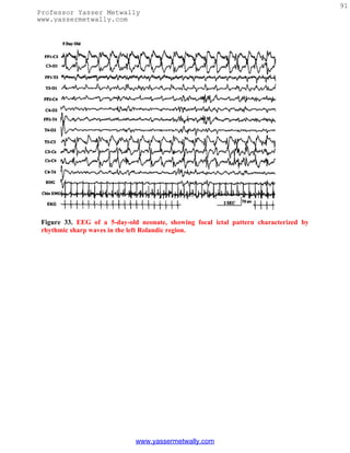
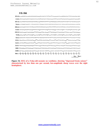
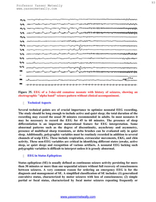
![94
Professor Yasser Metwally
www.yassermetwally.com
epilepsia partialis continua with the patient remaining fully conscious; and (3)
nonconvulsive status (NCSE) characterized by a variable alteration of consciousness with
minimal or no motor activity.
NCSE poses many challenging nosologic, diagnostic, and therapeutic problems. NCSE
includes: (1) absence status, occurring in the setting of generalized epilepsy (idiopathic or
symptomatic) and (2) complex partial status associated with focal or partial epilepsy of
frontal or temporal onset. In both types, the patient may present with mental status
alteration (e.g., slowness in behavior and mentation, confusion, and, rarely, stupor or
coma). Then, there are patients who after treatment of generalized convulsive status
continue to be obtunded or comatose and show epileptiform discharges in their EEG.
These patients are often designated as having "subtle" SE or lumped under NCSE.
It is relatively easy to diagnose NCSE associated with focal epilepsy when there are
frequent electrographic focal seizures with an ictal EEG pattern that evolves over time
with change in the amplitude, frequency, and spatial distribution. However, it is quite
common for the ictal EEG pattern associated with complex partial status associated with
focal epilepsy to be generalized spikes or sharp waves repeating at 1 to 6 Hz frequency.
Such a generalized EEG pattern is similar to that seen in typical absence status associated
with idiopathic generalized epilepsy (absence epilepsy) and atypical absence status in
children with secondary generalized epilepsy of the Lennox-Gastaut type. To complicate
the situation even further, patients with Lennox-Gastaut syndrome interictally have
generalized 1.0 to 3.0 cps spike wave discharges that may be very frequent, and one needs
to decide if they represent an ictal pattern (hence atypical absence status) or simply
represent a prominent interictal pattern. Some waxing or waning of such generalized
epileptiform discharges may not help in the distinction because this may be simply related
to state changes.
Some helpful criteria are proposed by Young et al[122] in patients who show almost
continuously occurring generalized, nonevolving epileptiform discharges in their EEGs,
including repetitive generalized or focal epileptiform discharges (spikes, sharp waves, and
spike waves) that repeat at a rate faster than three per second, very likely represent an ictal
pattern. Such repetitive discharges at a frequency slower than three per second are likely
to be ictal if significant clinical and/or EEG improvement is demonstrated following small
doses of intravenous lorazepam or diazepam. Rhythmic sinusoidal waves of any frequency
(ranging from to frequency) may represent an ictal pattern if there is an evolving pattern
at the onset (increasing amplitude and/or decreasing frequency) or a decrement pattern at
the termination (decremental amplitude or frequency) or postdischarge slowing or voltage
attenuation.
In a patient with obtundation or mental status change of recent onset, an EEG is indicated
to rule out NCSE. If repetitive generalized epileptiform discharges are recorded in the
EEG, 1 to 2 mg of lorazepam or 5 to 10 mg of diazepam are injected intravenously while
the EEG is running. A marked clinical improvement of obtundation and disappearance of
generalized paroxysmal activity in the EEG would strongly support the diagnosis of NCSE
www.yassermetwally.com](https://image.slidesharecdn.com/eeg-120912084228-phpapp02/85/Textbook-of-electroencephalography-97-320.jpg)
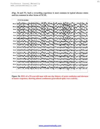
![96
Professor Yasser Metwally
www.yassermetwally.com
Figure 37. EEG of the same patient seen in Figure 36 following 4 mg of intravenous
lorazepam, showing disappearance of all paroxysmal activity and mental clearing, highly
suggestive of nonconvulsive status epilepticus.
Reviewing the previous EEG and obtaining follow-up EEG studies also provide a helpful
distinction between ictal and interictal basis for the repetitive generalized spike wave
discharges seen in children with Lennox-Gastaut syndrome. A period of frequent repetitive
generalized spike wave discharges associated with clinical deterioration of mental status is
more likely an episode of atypical absence status, particularly if the previous EEGs or
follow-up EEGs display dramatically fewer epileptiform abnormalities.
"Subtle" SE commonly includes patients who had a known episode of convulsive or
generalized tonic-clonic status, brought under control by intravenous antiepileptic therapy
(e.g., phenytoin, lorazepam, and barbiturates), but continue to remain obtunded or
comatose without significant motor activity. EEG of such patients often show repetitive
discharges, which may include lateralized periodic discharges (e.g., PLEDs or BiIPLEDS)
or generalized periodic discharges (PEDs). Some epileptologists[123] are of the opinion that
progressive sequential EEG changes occur during generalized convulsive SE with an
www.yassermetwally.com](https://image.slidesharecdn.com/eeg-120912084228-phpapp02/85/Textbook-of-electroencephalography-99-320.jpg)
![97
Professor Yasser Metwally
www.yassermetwally.com
"intermediary" pattern of PEDs and PLEDs (unilateral or bilateral) before disappearance
of all paroxysmal EEG activities, and that the presence of these "intermediary" EEG
patterns necessitate further aggressive therapeutic measures (e.g., inducing pentobarbital
coma, etc.). Such views are not universal. Many, on the other hand, consider PED and
PLED patterns observed during the course of convulsive status not an ictal pattern but
suggestive of a severe epileptic encephalopathy reflective of a neuronal dysfunction from
underlying brain damage.[124]
Refractory SE is usually treated by continuous intravenous anesthesia maintained by
pentobarbital, propofol, or medazolam. The dose is regulated such as to control all clear-
cut clinical or electrographic seizures and to maintain a suppression-burst pattern in the
EEG. Therefore, continuous bedside EEG is monitored. There is no consensus as to the
duration of "burst" and "flat" periods for optimal dosing. Most consider that establishing
and maintaining any degree of suppression-burst pattern is adequate.
One final note of caution is that focal motor seizures including epilepsia partialis continua
may not show ictal changes in the EEG because of the limited size of neuronal tissue
involved during the epileptic seizure or because the ictal pattern in the EEG may be
obscured by artifacts. Careful review of the EEG using different montages (especially a
transverse bipolar montage going through the midline electrodes) and use of appropriate
muscle filters may reveal a low-amplitude ictal pattern.
References
1. Markand ON. Alpha rhythms. J Clin Neurophysiol 1990; 7:163-189
2. Adams A. Studies on the flat electroencephalogram in man. Electroencephalogr Clin
Neurophysiol 1959;11:35-41
3. Obrist WD. The electroencephalogram of normal aged adults. Electroencephalogr
Clin Neurophysiol 1954;6:235- 244
4. Busse EM, Obrist WD. Significance of focal electroencephalographic changes in the
elderly. Postgrad Med 1963;34: 179-182
5. Orbrist WD. Problems of aging. In: Remond A, ed. Handbook of
Electroencephalography and Clinical Neurophysiology. Vol. 6A. Amsterdam:
Elsevier; 1976:275-292
6. Hubbard O, Sunde D, Goldenshon ES. The EEG changes in centenarians.
Electroencephalogr Clin Neurophysiol 1976;40:407-417
7. Torres F, Faovo A, Loewenson R, et al. The electroencephalogram of elderly
subjects revisited. Electroencephalogr Clin Neurophysiol 1983;56:391-398
8. Kooi KA, Gurener AM, Tupper CJ, et al. Electroencephalographic patterns of the
temporal region in normal adults. Neurology 1964;14:1029-1035
9. Katz RI, Horowitz GR. Electroencephalogram in septuagenarian: studies in a
normal geriatric population. J Am Geriatr Soc 1982;3:273-275
10. Markand ON. Electroencephalography in diffuse encephalopathies. J Clin
Neurophysiol 1984;1:357-407
www.yassermetwally.com](https://image.slidesharecdn.com/eeg-120912084228-phpapp02/85/Textbook-of-electroencephalography-100-320.jpg)
![98
Professor Yasser Metwally
www.yassermetwally.com
11. Hockaday JM, Potts F, Epstein E, et al. Electroencephalographic changes in acute
cerebral anoxia from cardiac or respiratory arrest. Electroencephalogr Clin
Neurophysiol 1965; 18:575-586
12. Prior PF. The EEG in Acute Cerebral Anoxia. Amsterdam: Excerpta Medica; 1973
13. Cordeau JP. Monorhythmic frontal delta activity in human electroencephalogram:
a study of 100 cases. Electroencephalogr Clin Neurophysiol 1959;11:733-746
14. Schaul N, Lüders H, Sachdev K. Generalized, bilaterally synchronous bursts of slow
waves in the EEG. Arch Neurol 1981;38:690-692
15. Bickford RG, Butt HR. Hepatic coma: the electroencephalographic pattern. J Clin
Invest 1955;34:790-799
16. Simsarian JP, Harner RN. Diagnosis of metabolic encephalopathy: significance of
triphasic waves in the electroencephalogram [abstract]. Neurology 1972;22:456
17. Karnze DS, Bickford RG. Triphasic waves: a reassessment of their significance.
Electroencephalogr Clin Neurophysiol 1984;57:193-198
18. Fisch BJ, Klass DW. The diagnostic specificity of triphasic wave patterns.
Electroencephalogr Clin Neurophysiol 1988; 70:1-8
19. Reiher J. The electroencephalogram in the investigation of metabolic comas.
Electroencephalogr Clin Neurophysiol 1970;28:104
20. Bahmon-Dussan JE, Celesia GG, Grigg-Damberger MM. Prognostic significance of
EEG triphasic waves in patients with altered states of consciousness. J Clin
Neurophysiol 1989;6:313-319
21. MacGillivray BB, Kennedy J. The "triphasic waves" of hepatic encephalopathy
[abstract]. Electroencephalogr Clin Neurophysiol 1970;28:428P
22. Aoki Y, Lombroso CT. Prognostic value of electroencephalography in Reye's
syndrome. Neurology 1973;23: 333-343
23. Yamada T, Young S, Kimura J. Significance of positive spike bursts in Reye's
syndrome. Arch Neurol 1977;34:376-380
24. Drury I. 14-and-6 positive bursts in childhood encephalopathies.
Electroencephalogr Clin Neurophysiol 1989;72: 479-485
25. Carroll WM, Mastaglia FL. Alpha and beta coma in drug intoxication
uncomplicated by cerebral hypoxia. Electroencephalogr Clin Neurophysiol
1979;46:95-105
26. Kuroiwa Y, Furukawa T, Inaki K. Recovery from drug-induced alpha-coma.
Neurology 1981;31:1359-1361
27. Stockard TJ, Werner SS, Albers JA, et al. EEG findings in phencyclidine
intoxication. Arch Neurol 1976;33:200-203
28. Kuroiwa Y, Celesia GG, Chung HD. Periodic EEG discharges and status spongiosus
of the cerebral cortex in anoxic encephalopathy: a necropsy case report. J Neurol
Neurosurg Psychiatry 1982;45:740-742
29. Paz D, Brenner RD. Bilateral independent periodic lateralized epileptiform
discharges, clinical significance. Arch Neurol 1981;38:713-715
30. McCarty GE, Marshall DW. Transient eyelid opening associated with postanoxic
EEG suppression-burst pattern. Arch Neurol 1981;38:754-756
31. Reeves AL, Westmoreland BF, Klass DW. Clinical accompaniments of the burst-
suppression EEG pattern. J Clin Neurophysiol 1997;14:150-153
www.yassermetwally.com](https://image.slidesharecdn.com/eeg-120912084228-phpapp02/85/Textbook-of-electroencephalography-101-320.jpg)
![99
Professor Yasser Metwally
www.yassermetwally.com
32. Chokroverty S. "Alpha-like" rhythms in electroencephalograms in coma after
cardiac arrest. Neurology 1975;25:655- 663
33. Westmoreland BF, Klass DW, Sharbrough FW, et al. "Alpha coma:"
electroencephalographic, clinical, pathological and etiological correlation. Arch
Neurol 1975;32:713-718
34. Alving J, Miller M, Sindrup E, et al. Alpha-pattern coma following cerebral anoxia.
Electroencephalogr Clin Neurophysiol 1979;47:95-101
35. Kuroiwa Y, Furukawa T. EEG prognostication in drug related alpha coma [letter].
Arch Neurol 1981;38:200
36. Chatrian GE, White LE Jr, Shaw CM. EEG pattern resembling wakefulness in
unresponsive decerebrate state following traumatic brainstem infarct.
Electroenceph Clin Neurophysiol 1964;16:285-289
37. American EEG Society. Guideline three: minimum technical standards for EEG
recording in suspected cerebral death. J Clin Neurophysiol 1994;11:10-13
38. Report of Special Task Force. Guidelines for the determination of brain death in
children. Pediatrics 1987;80:298-300
39. Markand ON. EEG in the diagnosis of CNS infections. In: Roos KL, ed. Central
Nervous System Infectious Diseases and Therapy. New York: Marcel Dekker;
1997:667-689
40. Illis LS, Taylor RM. The electroencephalogram in herpes simplex encephalitis.
Lancet 1972;1:718-721
41. Gupta PC, Seth P. Periodic complex in herpes simplex encephalitis: a clinical and
experimental study. Electroencephalogr Clin Neurophysiol 1973;35:67-74
42. Chien LT, Boehm RM, Robinson H, et al. Characteristic early
electroencephalographic changes in herpes simplex encephalitis: clinical and
virologic studies. Arch Neurol 1977; 34:361-364
43. Upton A, Gumpert J. Electroencephalography in the diagnosis of herpes simplex
encephalitis. Lancet 1970;1:650-652
44. Markand ON, Daly DD. Pseudoperiodic lateralized paroxysmal discharges in
electroencephalogram. Neurology 1971; 21:975-981
45. Cobb W, Hill D. Electroencephalogram in subacute progressive encephalitis. Brain
1950;73:392-404
46. Cobb W. The periodic events of subacute sclerosing leukoencephalitis.
Electroenceph Clin Neurophysiol 1966; 21:278-294
47. Markand ON, Panszi JG. The electroencephalogram in subacute sclerosing
panencephalitis. Arch Neurol 1975;32:719- 726
48. Westmoreland BF, Gomex MR, Blume WT. Activation of periodic complexes of
subacute sclerosing panencephalitis by sleep. Ann Neurol 1977;1:185-187
49. Burger LJ, Rowan J, Goldenshon E. Creutzfeldt-Jakob disease. Arch Neurol
1972;26:428-433
50. Au WJ, Gabor AJ, Viyan N, Markand ON. Periodic lateralized epileptiform
complexes (PLEDs) in Creutzfeldt-Jakob disease. Neurology 1980;30:611-617
51. Furlan A, Henry CE, Sweeny PI, et al. Focal EEG abnormalities in Heidenhain's
variant of Jakob-Creutzfeldt disease. Arch Neurol 1981;38:312-314
52. Chiofalo N, Fuentes A, Galvez S. Serial EEG findings in 27 cases of Creutzfeldt-
Jakob disease. Arch Neurol 1980;37: 143-145
www.yassermetwally.com](https://image.slidesharecdn.com/eeg-120912084228-phpapp02/85/Textbook-of-electroencephalography-102-320.jpg)
![100
Professor Yasser Metwally
www.yassermetwally.com
53. Chiappa KH, Burke CJ, Young RR. Creutzfeldt-Jakob disease: periodic sharp
waves in the EEG as an invariant element of the clinical syndrome. Presented at the
American Academy of Neurology Meeting, Chicago, 1979
54. Gloor P, Kalaby O, Giard N. The electroencephalogram in diffuse
encephalopathies: electroencephalographic correlates of gray and white matter
lesion. Brain 1968;91:779-802
55. Pampiglione G, Harden A. Neurophysiological identification of a late infantile form
of "neuronal lipidosis." J Neurol Neurosurg Psychiatry 1973;36:68-74
56. Letemendia F, Pampiglione G. Clinical and EEG observation in Alzheimer's
disease. J Neurol Neurosurg Psychiatry 1958;21:167-172
57. Liddell DW. Investigation of EEG findings in presenile dementia. J Neurol
Neurosurg Psychiatry 1958;21:173-176
58. Gordon EB, Sim M. The EEG in presenile dementia. J Neurol Neurosurg Psychiatry
1967;30:285-291
59. Muller HF, Kral VA. The electroencephalogram in advanced senile dementia. J Am
Geriatr Soc 1967;15:415-426
60. Margerison JH, Scott DF. Huntington's chorea: clinical EEG and neuropathological
findings [abstract]. Electroencephalogr Clin Neurophysiol 1965;19:314
61. Daly DD, Markand ON: Focal brain lesions. In: Daly DD, Pedley TA, eds. Current
Practice of Clinical Electroencephalography. 2nd ed. New York: Raven Press;
1990:335- 370
62. Walter WG. The location of cerebral tumours by electroencephalography. Lancet
1936;11:305-308
63. Arfel G, Fischgold H. EEG-signs in tumours of the brain. Electroencephalogr Clin
Neurophysiol 1961;19(suppl):36- 50
64. Brenner R, Sharbrough F. Electroencephalographic evaluation in Sturge-Weber
syndrome. Neurology 1976;26:629-632
65. Kershmann J, Conde A, Gibson WC. Electroencephalography in differential
diagnosis of supratentorial tumors. Arch Neurol Psychiatry (Chicago) 1949;62:255-
268
66. Cobb WA, Guillof RJ, Cast J. Breach rhythm: the EEG related to skull defects.
Electroencephalogr Clin Neurophysiol 1979;47:251-271
67. Chatrian GE, Shaw CM, Leftman H. The significance of periodic lateralized
epileptiform discharges in EEG: an electrographic, clinical and pathological study.
Electroencephalogr Clin Neurophysiol 1964;17:177-193
68. Maulsby RL. Some guidelines for assessment of spikes and sharp waves in EEG
tracings. Am J EEG Technol 1971; 11:3-16
69. Gloor P. The EEG and differential diagnosis of epilepsy. In: Van Duijn H, Donker
DNJ, Van Huffelen AC, eds. Current Concepts in Clinical Neurophysiology. The
Netherlands: NV Drukkerij Trio; 1977:9-21
70. Petersen I, Eeg-Olofsson O, Sellden U. Paroxysmal activity in EEG of normal
children. In: Kellaway P, Petersen I, eds. Clinical Electroencephalography of
Children. New York: Grune & Stratton; 1968:167-188
71. Eeg-Olofsson O, Petersen I, Sellden U. The development of the
electroencephalogram in normal children from the age of one through fifteen years:
paroxysmal activity. Neuropediatrie 1971;4:375-404
www.yassermetwally.com](https://image.slidesharecdn.com/eeg-120912084228-phpapp02/85/Textbook-of-electroencephalography-103-320.jpg)
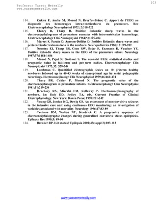

![105
Professor Yasser Metwally
www.yassermetwally.com
electroencephalography, which, after artifacts are ruled out, can only show facts. These
disturbing facts are in need of a reasonable interpretation. Let us contemplate the
indubitable fact that a complete medical evaluation will yield certain physical shortcomings
and organic abnormalities in practically every healthy individual; even acne pimples are
cutaneous lesions and hence abnormalities. What the electroencephalographer needs in
such cases is a common sense philosophy as a basis for a wise interpretation. General
medicine is full of examples of seemingly irrelevant and yet unmistakably present
abnormalities which the prudent, seasoned physician will integrate into a holistic view of
the individual. Seen from this angle, electroencephalography does not differ from the rest
of medicine.
Figure 1. Examples of sharp
waves [left] and spike [right]
Table 1. Electroclinical criteria of spike/ sharp wave discharge
A spike is a transient, clearly distinguished from the background activity, with
pointed peak at conventional paper speeds and a duration from 20 to under 70
msec; the main component is generally negative. Amplitude is variable. Spikes
represent the basic element of paroxysmal activity in the EEG
A sharp wave is a transient, clearly distinguished from background activity,
with pointed peak at conventional paper speeds and duration of 70 to 200 msec.
The main component is generally negative relative to other areas.
Both spikes and sharp waves have multiphasic characters, being composed of a
sequence of a minor positive, a major negative, and a second minor positive
component is typical in most instances. The long duration of a sharp wave
permits better insight into the multiphasic character of this potential.
The spike/sharp wave potentials are reliable indicators of a potential seizure
focus because they result from the characteristic neurophysiological event "the
paroxysmal depolarization shift" (PDS). This phenomenon consists of thousands
of neurons simultaneously undergoing large depolarization with superimposed
action potentials. Both synaptic events and intrinsic cellular currents have been
implicated in this process. EEG spikes/sharp waves are due to the slow
depolarization currents in the PDS. Neurons surrounding the focus are inhibited
during the paroxysmal depolarization shift, and within the focus the the
paroxysmal depolarization shift is followed by a hyperpolarization potential.
Both an increase in depolarizing events and a loss of inhibitory mechanisms can
lead to persistence and propagation of the discharge as a seizure.
www.yassermetwally.com](https://image.slidesharecdn.com/eeg-120912084228-phpapp02/85/Textbook-of-electroencephalography-108-320.jpg)
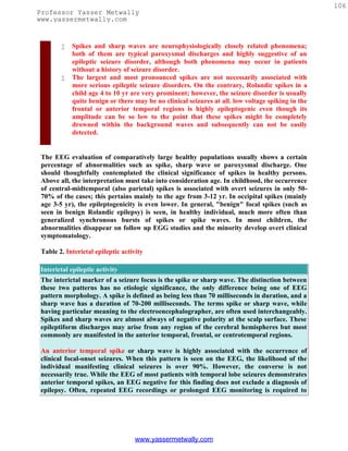
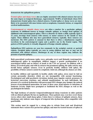
![109
Professor Yasser Metwally
www.yassermetwally.com
types, previously called primary and secondary, respectively. Idiopathic epilepsies are
thought to be genetically heritable, are associated with normal intelligence, and occur
during specific age periods. The symptomatic epilepsies are likely the result of a CNS
injury, which in a symptomatic partial epilepsy consists of a focal lesion and in a
symptomatic generalized epilepsy consists of diffuse cerebral abnormality. Symptomatic
epilepsies are typically lifelong conditions.
It cannot be overemphasized that the diagnosis of epilepsy is based primarily on the clinical
history. As noted above, a clinical seizure rarely occurs during an EEG, and thus the EEG
is rarely diagnostic of a seizure disorder or epilepsy. In a large, populationbased EEG
study by Zivin and Ajmone-Marsan [1] involving subjects without a history of seizures,
approximately 2 percent of the subjects had EEGs with epileptiform discharges. Of the
individuals in this subgroup, only 15 percent subsequently developed a seizure disorder.
Therefore, epileptiform discharges seen on an EEG should not be referred to as interictal
discharges unless it is known that the patient has a clinically defined seizure disorder. Focal
or generalized epileptiform discharges should be noted as consistent with the interictal
expression of either a partial or a generalized epilepsy, respectively. When applied in the
appropriate clinical setting, the EEG is useful in classifying the seizure type, predicting the
long-term outcome, and choosing the appropriate antiepileptic medication.
Overall, symptomatic partial seizure disorders are the most common type of epilepsy. The
clinical semiology of the partial seizure generally depends on the site of onset. In children,
focal epileptiform discharges arising from the temporal region have the greatest incidence
of clinical seizures, ranging from 85 to 95 percent. The next highest incidence (70 to 75
percent) is associated with frontal discharges. The central, parietal and occipital regions
have the lowest incidence of seizures related to epileptiform discharges. estimated at 40 to
70 percent. In addition to the characteristics of recorded epileptiform activity, the age of
the patient and the presence or absence of neurological deficits on examination are
important factors that are helpful in determining the clinical significance of epileptiform
discharges and in classifying the partial seizure disorder as either symptomatic or
idiopathic. The occurrence of a clinical seizure with a focal electrographic correlate is
diagnostic of a partial epilepsy. Blume and colleagues [2] presented several types of scalp
EEG correlates for partial seizures, most of which began with rhythmic sinusoidal activity
or repetitive sharp wave activity that subsequently evolved in frequency. Most patients
with complex partial seizures were noted to have a scalp correlate on the EEG. Patients
with simple partial seizures were less likely to have a scalp correlate.
The best-defined idiopathic partial epilepsy is benign rolandic epilepsy. The classic EEG
finding in this childhood seizure disorder is a characteristic monomorphic centrotemporal
sharp wave. The sharp waves are often seen independently in the centrotemporal and
adjacent regions, and they are accentuated by light sleep. The waking background rhythm
is generally normal.
Of the idiopathic generalized epilepsies, the absence seizure is the most common type. The
interictal EEG feature of this type of seizure disorder consists of generalized, high-
amplitude, anteriorly predominant 3-Hz spike and wave discharges, called typical 3-Hz
www.yassermetwally.com](https://image.slidesharecdn.com/eeg-120912084228-phpapp02/85/Textbook-of-electroencephalography-112-320.jpg)
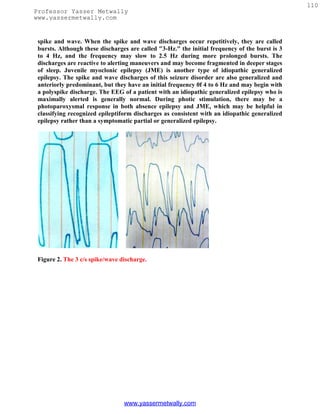
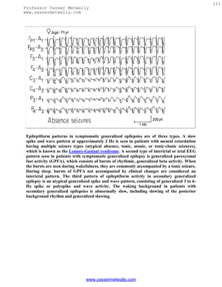
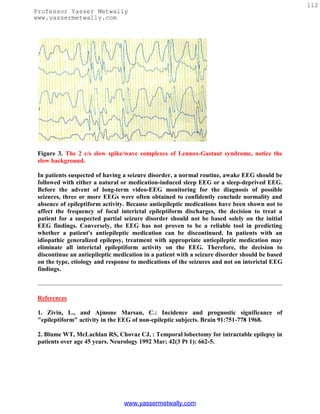
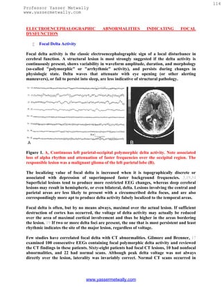
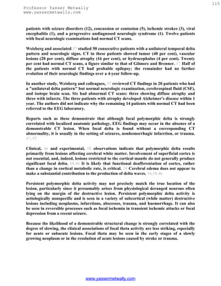
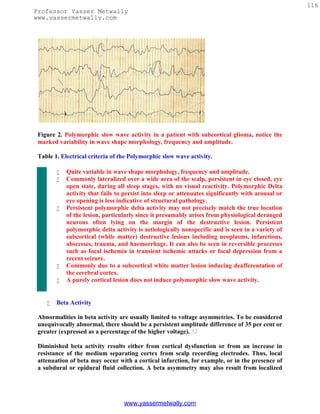
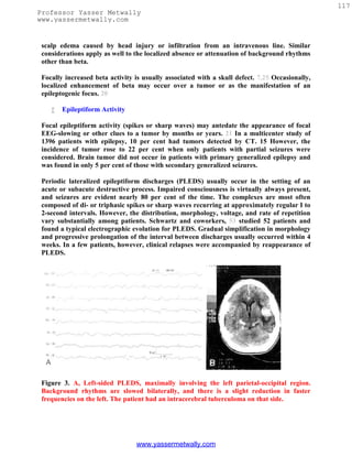
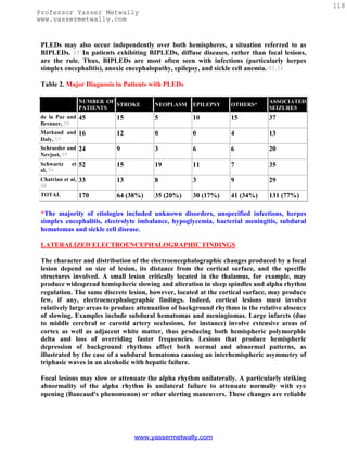
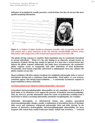
![120
Professor Yasser Metwally
www.yassermetwally.com
the EEGs of 154 patients with diencephalic or posterior fossa lesions. Only 12 per cent of
patients with diencephalic lesions had normal EEGS, whereas 60 per cent and 73 per cent
of patients with lower brain-stem or cerebellar pathology respectively had normal EEGS.
If the EEG abnormality was clearly lateralized, an infratentorial lesion was unlikely.
Paroxysmal bursts of rhythmic delta waves with frontal or occipital predominance (the
latter especially common in children) have been associated with subfrontal, deep midline,
or posterior fossa lesions. In fact, however, intermittent rhythmic delta activity (IRDA) is
nonspecific and is seen much more often in the setting of metabolic disorders or other
encephalopathies affecting the brain diffusely than with focal lesions, regardless of location.
IRDA may appear against an otherwise normal background. In contrast to polymorphic
delta, IRDA is usually reactive to alerting maneuvers, disappears in sleep, and is
augmented by hyperventilation or drowsiness. Correlative studies using CT and PET
16,29,41,46 have failed to demonstrate a particular anatomic structure responsible for
generating IRDA. Physiologic investigations, 18,46 implicate dysfunction of
thalamocortical interactions.
Rhythmic delta activity consists of sinusoidal waveforms of approximately 2.5 Hz that
occur intermittently in the EEG recording. It is most often symmetric but can be
lateralized. In adults, the delta activity has a frontal predominance (frontal intermittent
rhythmic delta activity [FIRDA]). In children, it is maximal posteriorly (occipital
intermittent rhythmic delta activity [OIRDA]). Intermittent rhythmic delta activity is
associated with structural lesions, most commonly diencephalic, infratentorial or
intraventricular tumors, or with diffuse encephalopathies. FIRDA occurring in patients
with a normal EEG background suggests that the pattern is due to a structural lesion;
when associated with EEG background abnormalities, it is likely to be due to
encephalopathy. In cases of encephalopathy with FIRDA, the pathophysiologic processes
are believed to involve cortical and subcortical gray matter. OIRDA is associated with
absence epilepsy in children aged 6-10 years.
Figure 6. The intermittent rhythmic delta activity [left image] and the the polymorphic
slow wave activity [right image]
www.yassermetwally.com](https://image.slidesharecdn.com/eeg-120912084228-phpapp02/85/Textbook-of-electroencephalography-123-320.jpg)
![121
Professor Yasser Metwally
www.yassermetwally.com
Table 3. Electrical criteria of The intermittent rhythmic delta activity.
Consists of sinusoidal waveforms of approximately 2.5 Hz that occur
intermittently in the EEG recording. It is most often symmetric but can be
lateralized.
In adults, the delta activity has a frontal predominance (frontal intermittent
rhythmic delta activity [FIRDA]). In children, it is maximal posteriorly
(occipital intermittent rhythmic delta activity [OIRDA])
The intermittent rhythmic delta activity shows visual reactivity and is commonly
suppressed in the eye open state unless the patient is comatose.
Intermittent rhythmic delta activity is associated with structural lesions, most
commonly diencephalic, infratentorial or intraventricular tumors, or with
diffuse encephalopathies.
FIRDA occurring in patients with a normal EEG background suggests that the
pattern is due to a structural lesion; when associated with EEG background
abnormalities, it is likely to be due to encephalopathy.
OIRDA is associated with absence epilepsy in children aged 6-10 years.
CLINICAL APPLICATIONS
The EEG as a Screen for Further Investigation
Patients with focal findings on neurologic examination require CT or MRI. When the
clinical data do not strongly implicate a localized disease process, however, some clinicians
have advocated using EEG as a cost-effective screen for more extensive, especially
radiologic, investigation. Rosenberg and associated, 49 retrospectively reviewed 136
patients with abnormal neurologic examinations but whose findings and histories did not
suggest focal pathology. Clinically, the patients could be divided into six groups: headache,
first seizure, recurrent seizure, confusion or dementia, transient ischemic attacks, and
miscellaneous disorders. Electroencephalographic findings could be assigned to one of
three groups. Twenty-one patients had focal abnormalities (slowing, spikes, or
attenuation). Four of these (20 per cent of this group) had CT scans showing stroke, tumor,
or porencephaly; the remainder had normal CT scans. Sixty-four patients had generalized
electroencephalographic abnormalities. in this group only two patients had abnormal CT
scans: one showing multiple metastases and the other cerebral hemiatrophy. Fifty-one
patients with normal EEGs had normal CT scans. Rosenberg and coworkers, 49 concluded
that careful neurologic examination and EEG may obviate CT in many patients.
Weisberg and colleagues reviewed the results of CT in several hundred patients with
chronic recurrent headaches, normal physical examinations, and normal EEGS. CT
abnormalities occurred in under 0.5 per cent, and some of these were considered incidental.
On the other hand, in patients with chronic headache and abnormal EEGs (9 per cent of
their headache group), a focal EEG abnormality correctly predicted focal CT lesions. CT
abnormalities did not occur in patients with diffuse or bilateral electroencephalographic
www.yassermetwally.com](https://image.slidesharecdn.com/eeg-120912084228-phpapp02/85/Textbook-of-electroencephalography-124-320.jpg)
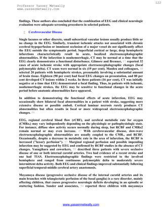
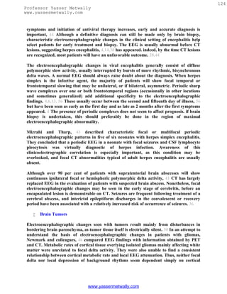
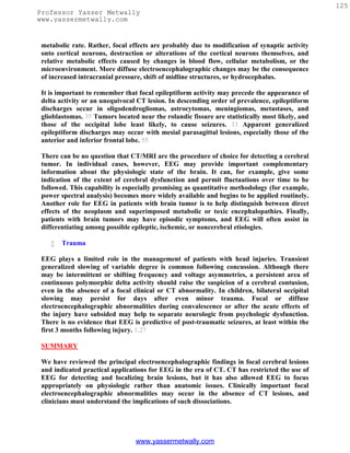
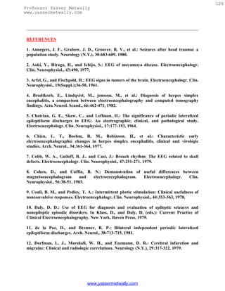
![127
Professor Yasser Metwally
www.yassermetwally.com
13. Elian, M.: Herpes simplex encephalitis: Prognosis and long-term follow-up. Arch.
Neurol., 32:39-43, 1975.
14. Enzmann, D. R., Ransom, B., Norman, D., et al.: Computed tomography of herpes
simplex encephalitis. Radiology, 129:419-425, 1978.
15. Gastaut, H.: Conclusions: Computerized transverse axial tomography in epilepsy.
Epilepsia, 17:337-338, 1976.
16. Gastaut, J. L., Michel, B., Hassan, S., et al.: Electroencephalography in brain edema
(127 cases of brain tumor investigated by cranial computerized tomography).
Electroencephalogr. Clin. Neurophysiol., 46:239-255, 1979.
17. Gilmore, P. C., and Brenner, R. P.: Correlation of EEG, computerized tomography,
and clinical findings: Study of 100 patients with focal delta activity. Arch. Neurol., 38:371-
372, 1981.
18. Gloor, P., Ball, G., and Schaul, N.: Brain lesions that produce delta waves in the EEG.
Neurology (Minneap.), 27:326-333, 1977.
19. Goldensohn, E. S.: Use of the EEG for evaluation of focal intracranial lesions. In Klass,
D., and Daly, D. (eds.): Current Practice of Clinical Electroencephalography. New York,
Raven Press, 1979.
20. Green, R. L. and Wilson, W. P.: Asymmetries of beta activity in epilepsy, brain tumor,
and cerebrovascular disease. Electroencephalogr. Clin. Neurophysiol., 13:75-78, 1961.
21. Hess, R.: Brain tumors and other space occupying processes. In Remorid, A., and Hess,
R. (eds): Handbook of Electroencephalography and Clinical Neurophysiology. Vol. 14, Part
C. Amsterdam, Elsevier Scientific Publishing Co., 1975.
22. Hockaday, J. M., and Whitty, C. W. M.: Factors determining the electroencephalogram
in migraine: A study of 560 patients according to clinical type of migraine. Brain, 92:769-
788, 1969.
23. Hungerford, G. D., du Boulay, G. H., and Zilka, K. J.: Computerized axial tomography
in patients with severe migraine: A preliminary report. J. Neurol. Neurosurg. Psychiatry,
39:990-994, 1976.
24. jabbari, B., Maulsby, R. L., Holtzapple, P. A., et a].; Prognostic value of EEG in acute
vascular aphasia: A long-term clinical-EEG study of 53 patients. Clin. Electroencephalogr.,
10:190-197, 1979.
25. Jaffe, R. and Jacobs, L.: The beta focus: Its nature and significance. Acta Neurol.
Scand., 48:191 203, 1972.
www.yassermetwally.com](https://image.slidesharecdn.com/eeg-120912084228-phpapp02/85/Textbook-of-electroencephalography-130-320.jpg)
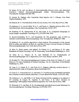
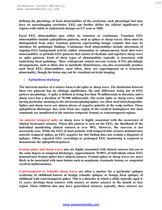
![133
Professor Yasser Metwally
www.yassermetwally.com
nocturnal. The EEG pattern is unusual in that there is often a simultaneous negative
waveform in the centrotemporal region and a positive one in the frontal region. This
pattern of EEG polarity is virtually diagnostic of benign rolandic epilepsy.
Epileptiform EEG patterns are seen less commonly in the occipital, central, or parietal
regions. Occipital spikes typically are seen in young children and may or may not be
associated with clinical seizures. Discharges in any of these regions may indicate the
presence of partial epilepsy.
Figure 1. Examples of sharp
waves [left] and spike [right]
Table 1. Electroclinical criteria of spike/ sharp wave discharge
A spike is a transient, clearly distinguished from the background activity, with
pointed peak at conventional paper speeds and a duration from 20 to under 70
msec; the main component is generally negative. Amplitude is variable. Spikes
represent the basic element of paroxysmal activity in the EEG
A sharp wave is a transient, clearly distinguished from background activity,
with pointed peak at conventional paper speeds and duration of 70 to 200 msec.
The main component is generally negative relative to other areas.
Both spikes and sharp waves have multiphasic characters, being composed of a
sequence of a minor positive, a major negative, and a second minor positive
component is typical in most instances. The long duration of a sharp wave
permits better insight into the multiphasic character of this potential.
The spike/sharp wave potentials are reliable indicators of a potential seizure
focus because they result from the characteristic neurophysiological event "the
paroxysmal depolarization shift" (PDS). This phenomenon consists of thousands
of neurons simultaneously undergoing large depolarization with superimposed
action potentials. Both synaptic events and intrinsic cellular currents have been
implicated in this process. EEG spikes/sharp waves are due to the slow
depolarization currents in the PDS. Neurons surrounding the focus are inhibited
during the paroxysmal depolarization shift, and within the focus the the
paroxysmal depolarization shift is followed by a hyperpolarization potential.
Both an increase in depolarizing events and a loss of inhibitory mechanisms can
lead to persistence and propagation of the discharge as a seizure.
Spikes and sharp waves are neurophysiologically closely related phenomena;
both of them are typical paroxysmal discharges and highly suggestive of an
www.yassermetwally.com](https://image.slidesharecdn.com/eeg-120912084228-phpapp02/85/Textbook-of-electroencephalography-136-320.jpg)
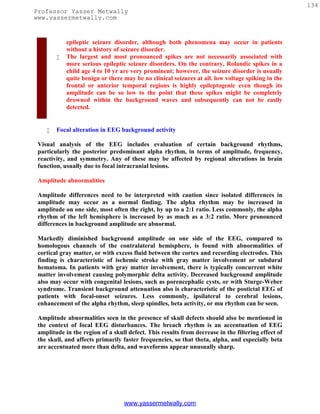
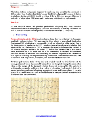
![136
Professor Yasser Metwally
www.yassermetwally.com
Figure 2. Polymorphic slow wave activity in a patient with subcortical glioma, notice the
marked variability in wave shape morphology, frequency and amplitude.
Table 2. Electrical criteria of the Polymorphic slow wave activity.
Quite variable in wave shape morphology, frequency and amplitude.
Commonly lateralized over a wide area of the scalp, persistent in eye closed, eye
open state, during all sleep stages, with no visual reactivity. Polymorphic Delta
activity that fails to persist into sleep or attenuates significantly with arousal or
eye opening is less indicative of structural pathology.
Persistent polymorphic delta activity may not precisely match the true location
of the lesion, particularly since it presumably arises from physiological deranged
neurons often lying on the margin of the destructive lesion. Persistent
polymorphic delta activity is aetiologically nonspecific and is seen in a variety of
subcortical (while matter) destructive lesions including neoplasms, infarctions,
abscesses, trauma, and haemorrhage. It can also be seen in reversible processes
such as focal ischemia in transient ischemic attacks or focal depression from a
recent seizure.
Commonly due to a subcortical white matter lesion inducing deafferentation of
the cerebral cortex.
A purely cortical lesion does not induce polymorphic slow wave activity.
Rhythmic delta activity consists of sinusoidal waveforms of approximately 2.5 Hz that
occur intermittently in the EEG recording. It is most often symmetric but can be
lateralized. In adults, the delta activity has a frontal predominance (frontal intermittent
rhythmic delta activity [FIRDA]). In children, it is maximal posteriorly (occipital
intermittent rhythmic delta activity [OIRDA]). Intermittent rhythmic delta activity is
associated with structural lesions, most commonly diencephalic, infratentorial or
intraventricular tumors, or with diffuse encephalopathies. FIRDA occurring in patients
with a normal EEG background suggests that the pattern is due to a structural lesion;
when associated with EEG background abnormalities, it is likely to be due to
encephalopathy. In cases of encephalopathy with FIRDA, the pathophysiologic processes
are believed to involve cortical and subcortical gray matter. OIRDA is associated with
absence epilepsy in children aged 6-10 years.
www.yassermetwally.com](https://image.slidesharecdn.com/eeg-120912084228-phpapp02/85/Textbook-of-electroencephalography-139-320.jpg)
![137
Professor Yasser Metwally
www.yassermetwally.com
Figure 3. The intermittent rhythmic delta activity [left image] and the the polymorphic
slow wave activity [right image]
Table 3. Electrical criteria of The intermittent rhythmic delta activity.
Consists of sinusoidal waveforms of approximately 2.5 Hz that occur
intermittently in the EEG recording. It is most often symmetric but can be
lateralized.
In adults, the delta activity has a frontal predominance (frontal intermittent
rhythmic delta activity [FIRDA]). In children, it is maximal posteriorly
(occipital intermittent rhythmic delta activity [OIRDA])
The intermittent rhythmic delta activity shows visual reactivity and is commonly
suppressed in the eye open state unless the patient is comatose.
Intermittent rhythmic delta activity is associated with structural lesions, most
commonly diencephalic, infratentorial or intraventricular tumors, or with
diffuse encephalopathies.
FIRDA occurring in patients with a normal EEG background suggests that the
pattern is due to a structural lesion; when associated with EEG background
abnormalities, it is likely to be due to encephalopathy.
OIRDA is associated with absence epilepsy in children aged 6-10 years.
Focal theta activity is less likely to reflect a macroscopic structural lesion than is focal
delta. Theta is commonly, however, associated with a functional disturbance, such as
epileptogenic cortex, especially postictally, after amplitude suppression and focal delta have
resolved. In addition, localized theta is usually superimposed on focal delta to some degree;
the relative proportion of delta and theta reflects the size and/or severity of the underlying
structural or functional cerebral abnormality.
Periodic EEG patterns
Periodic lateralized epileptiform discharges (PLEDs) are EEG abnormalities consisting of
repetitive spike or sharp wave discharges, which are focal or lateralized over one
hemisphere, recur at intervals of 0.5-5 seconds, and continue through most of the duration
www.yassermetwally.com](https://image.slidesharecdn.com/eeg-120912084228-phpapp02/85/Textbook-of-electroencephalography-140-320.jpg)
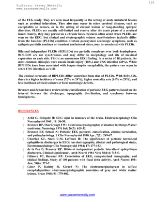
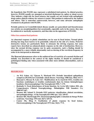
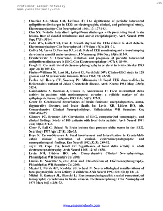
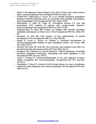

![149
Professor Yasser Metwally
www.yassermetwally.com
to the lesion. Thus IRDA is a nonspecific nonlocalizing EEG pattern, unless associated with
other focal findings on the EEG. Although the mechanisms for production of IRDA are
understood incompletely, studies correlating with pathologic specimens suggest that IRDA
is associated primarily with diffuse gray matter disease.
The degree of encephalopathy manifested appears to correspond to the proportion of IRDA
on the EEG. This pattern must be distinguished from the frequently encountered frontally
maximal intermittent delta that can be seen in drowsy elderly patients.
Figure 1. The intermittent rhythmic delta activity [left image] and the the polymorphic
slow wave activity [right image]
Table 1. Electrical criteria of The intermittent rhythmic delta activity.
Consists of sinusoidal waveforms of approximately 2.5 Hz that occur
intermittently in the EEG recording. It is most often symmetric but can be
lateralized.
In adults, the delta activity has a frontal predominance (frontal intermittent
rhythmic delta activity [FIRDA]). In children, it is maximal posteriorly
(occipital intermittent rhythmic delta activity [OIRDA])
The intermittent rhythmic delta activity shows visual reactivity and is commonly
suppressed in the eye open state unless the patient is comatose.
Intermittent rhythmic delta activity is associated with structural lesions, most
commonly diencephalic, infratentorial or intraventricular tumors, or with
diffuse encephalopathies.
FIRDA occurring in patients with a normal EEG background suggests that the
pattern is due to a structural lesion; when associated with EEG background
abnormalities, it is likely to be due to encephalopathy.
OIRDA is associated with absence epilepsy in children aged 6-10 years.
www.yassermetwally.com](https://image.slidesharecdn.com/eeg-120912084228-phpapp02/85/Textbook-of-electroencephalography-152-320.jpg)
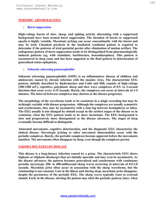
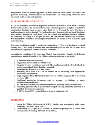
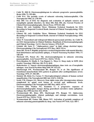
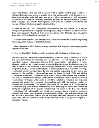
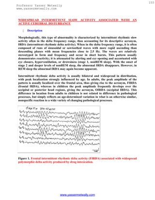
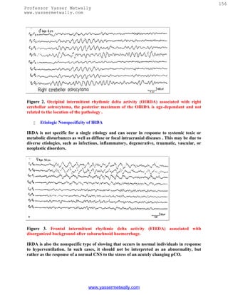
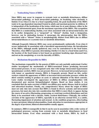
![158
Professor Yasser Metwally
www.yassermetwally.com
nonlocalizing. The common denominator in the wide variety of pathological processes
producing IRDA is that, when such an abnormality appears, it is likely to be associated
with the development of widespread CNS dysfunction; the earliest clinical correlates are
fluctuating levels of alertness and attention. With focal lesions, one mechanism may be an
increased transventricular pressure with secondary disturbances at both the subcortical
and cortical levels. With primary intracranial encephalopathies, it appears to be due to
widespread involvement of the gray matter at subcortical and cortical levels.
Figure 4. The intermittent rhythmic delta activity [left image] and the the polymorphic
slow wave activity [right image]
Table 1. Electrical criteria of The intermittent rhythmic delta activity.
Consists of sinusoidal waveforms of approximately 2.5 Hz that occur
intermittently in the EEG recording. It is most often symmetric but can be
lateralized.
In adults, the delta activity has a frontal predominance (frontal intermittent
rhythmic delta activity [FIRDA]). In children, it is maximal posteriorly
(occipital intermittent rhythmic delta activity [OIRDA])
The intermittent rhythmic delta activity shows visual reactivity and is commonly
suppressed in the eye open state unless the patient is comatose.
Intermittent rhythmic delta activity is associated with structural lesions, most
commonly diencephalic, infratentorial or intraventricular tumors, or with
diffuse encephalopathies.
FIRDA occurring in patients with a normal EEG background suggests that the
pattern is due to a structural lesion; when associated with EEG background
abnormalities, it is likely to be due to encephalopathy.
OIRDA is associated with absence epilepsy in children aged 6-10 years.
www.yassermetwally.com](https://image.slidesharecdn.com/eeg-120912084228-phpapp02/85/Textbook-of-electroencephalography-161-320.jpg)
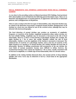
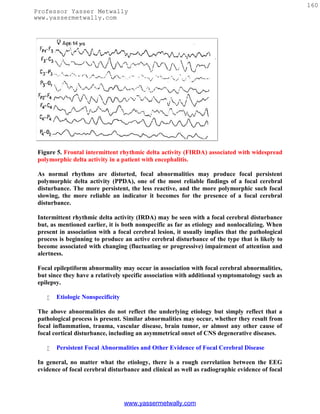
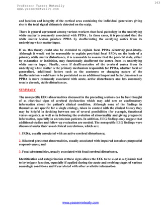
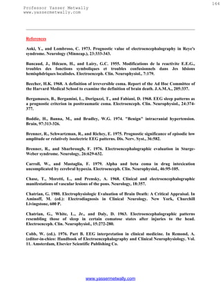
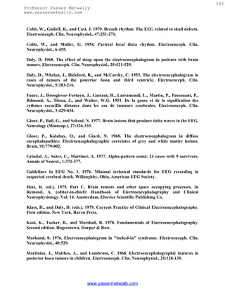
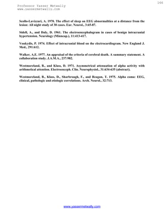

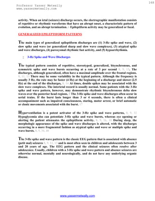
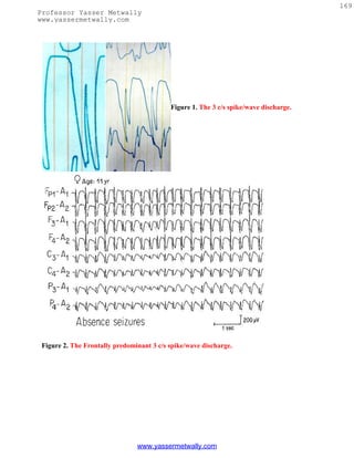
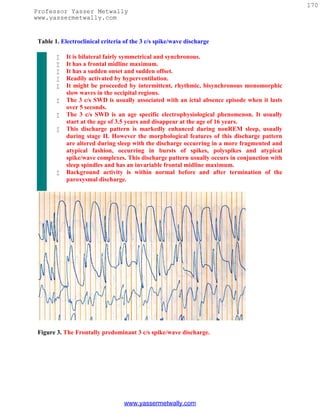
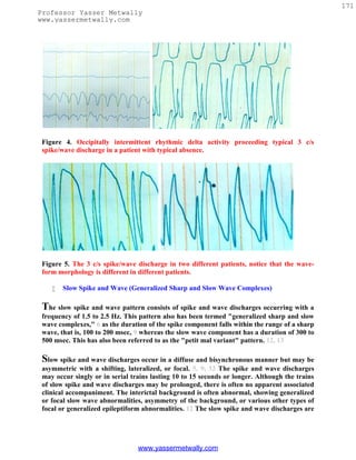
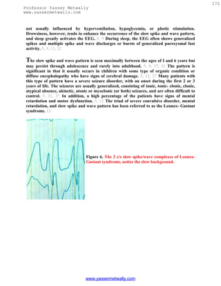
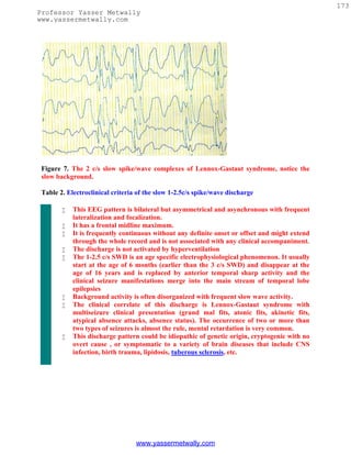
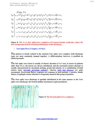
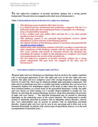
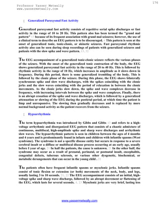
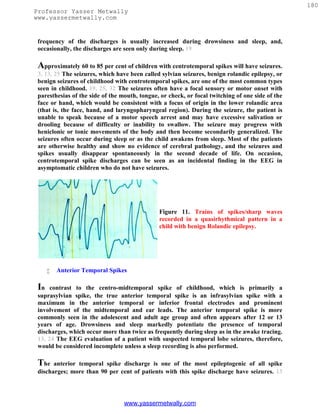
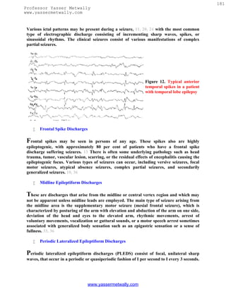
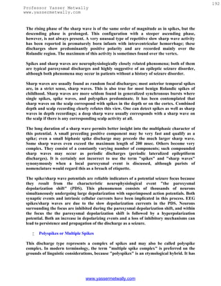
![193
Professor Yasser Metwally
www.yassermetwally.com
been defined as a complex paroxysmal EEG pattern with close association of two or more
diphasic spikes occurring more or less rhythmically in bursts of variable duration,
generally with large amplitudes.
Polyspike bursts are readily elicited by electrical stimulation of single depth leads,
especially in limbic regions. On the scalp, however, polyspikes occur mostly as bilateral or
generalized synchronous discharges. Exceptional focal polyspikes are occasionally
encountered; these usually have a frontal maximum, except for occipital accentuation in
hypsarrhythmia. Polyspikes and also polyspike-wave complexes are sometimes associated
with concomitant myoclonus, especially in primary generalized epilepsy and in
photosensitive individuals with this type of seizure disorder. Children with Lennox-Gastaut
syndrome may also show the association of polyspikes and myoclonus.
Runs of Rapid Spikes
This pattern has been described as "grand mal discharge", "fast paroxysmal rhythms",
and "rhythmic spikes". It is seen only in sleep and occurs in older children, adolescents,
and younger adults. It consists of bursts of spike discharges at a rate from 10-25/sec,
usually generalized but with a well defined maximum over frontal regions; it may even be
confined to the frontal leads. The voltage is in the medium to high range, often exceeding
100 or even 200 iLv. The discharge rate is in most cases somewhat irregular. The bursts
last for about 2 to 10 sec; bursts of more than 5 sec duration are usually associated with
tonic seizures and thus represent an ictal pattern.
The obvious misnomer, grand mal discharges, is based on certain similarities with the ictal
EEG of a grand mal seizure. In a patient population with grand mal seizures, runs of rapid
spikes as an interictal pattern are a very rare finding. This pattern occurs in patients with
secondary generalized epilepsy who suffer from more than one type of seizure and
especially from akinetic seizures. This discharge is very typical in patients with Lennox-
Gastaut syndrome; it is hardly ever found outside this clinical entity.
Figure 1. Examples of sharp
waves [left] and spike [right]
www.yassermetwally.com](https://image.slidesharecdn.com/eeg-120912084228-phpapp02/85/Textbook-of-electroencephalography-196-320.jpg)
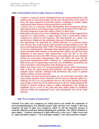
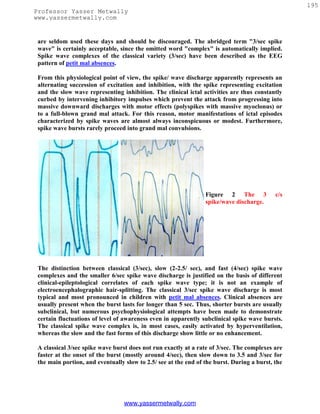
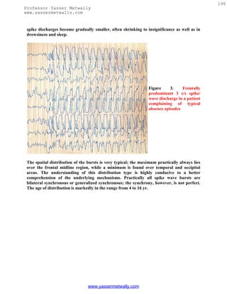
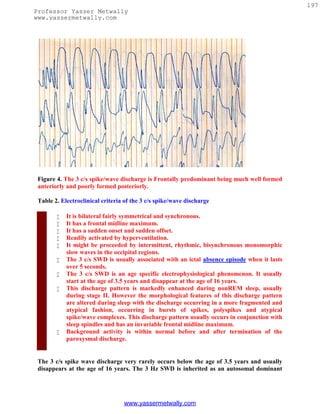
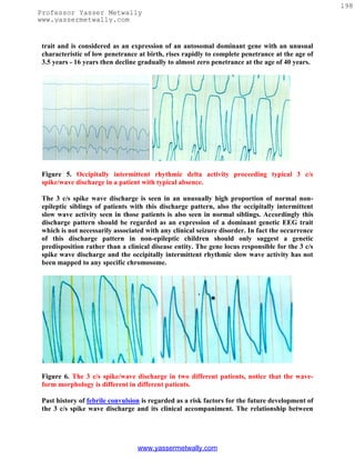
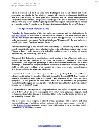
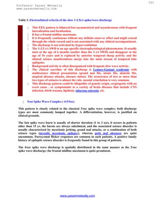
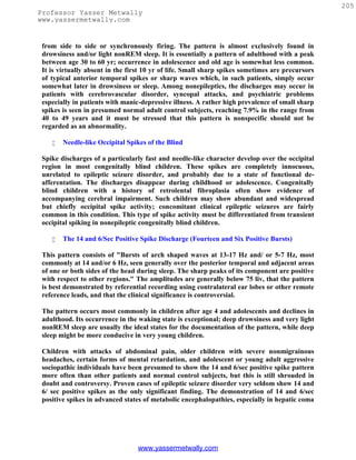
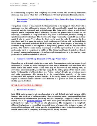
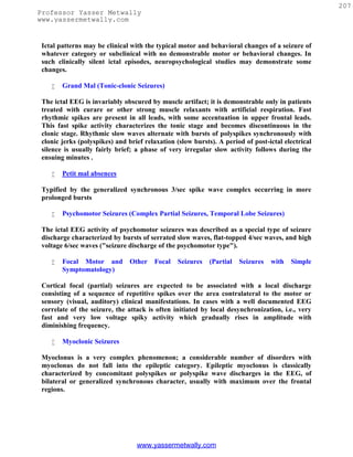
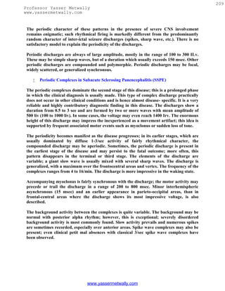
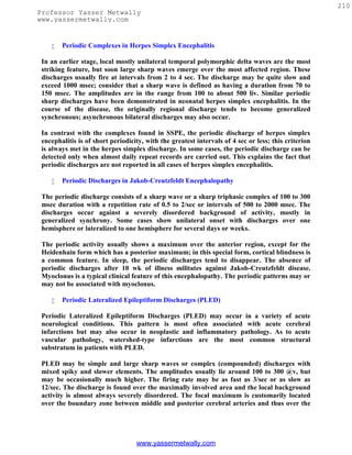
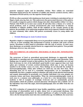
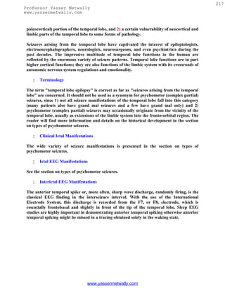
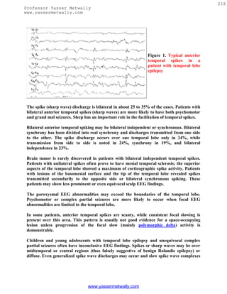
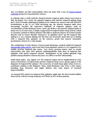
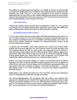
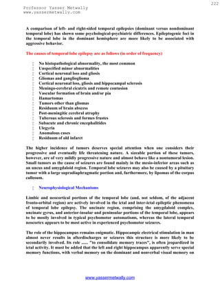
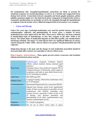
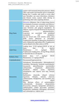
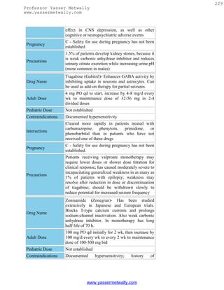
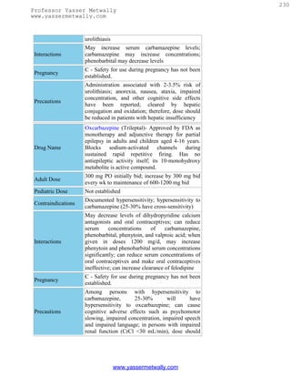
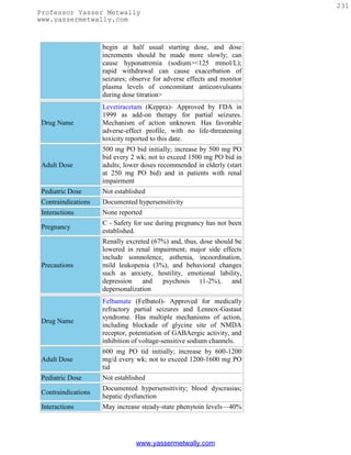
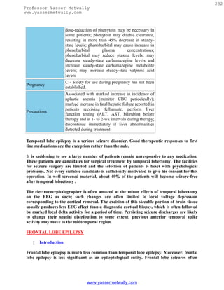
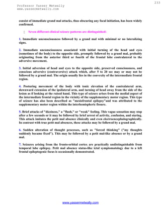
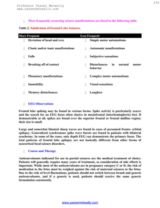
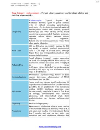
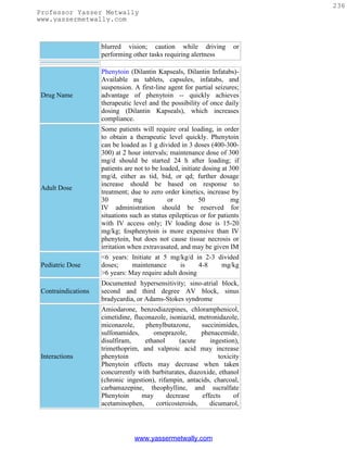

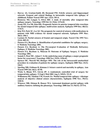

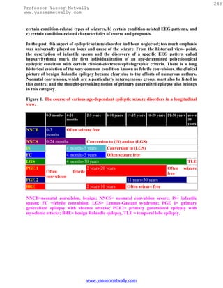
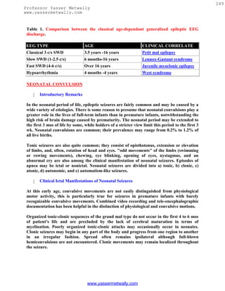
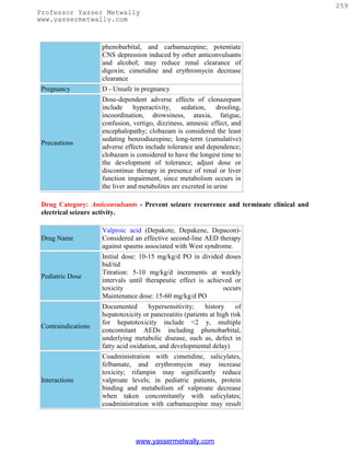
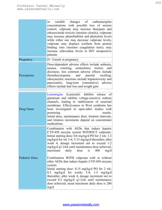
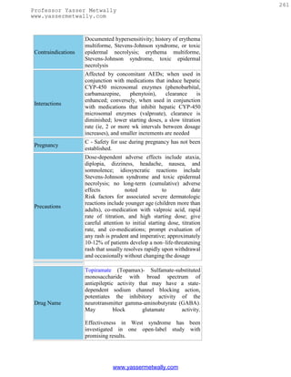
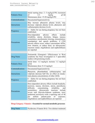

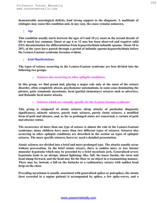
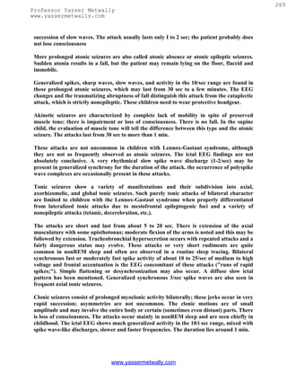
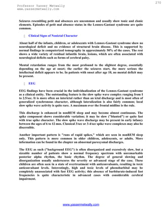
![271
Professor Yasser Metwally
www.yassermetwally.com
Etiological and Neuropathological Considerations
The aforementioned normal computed tomography (CT) scan findings in 50% of the cases
further support the view that about half of the cases are idiopathic and hence without
structural cerebral changes. The nature of idiopathic forms is completely unclear. Genetic
predisposition is certainly more than just a hypothesis.
Acquired pathology of residual character is present in a considerable number of cases;
with CNS infection and birth trauma/asphyxia as the leading problems. One could argue
that such pathology alone can hardly account for this severe form of epileptic seizure
disorder and that a genetic predisposition is a prerequisite. Even progressive pathology
such as intracranial tumors may be associated occasionally with Lennox-Gastaut
syndrome. Phenylketonuria, forms of lipidosis, tuberous sclerosis, lead encephalopathy,
and toxoplasmosis have been specifically mentioned as etiological factors. In essence, we
are dealing with the same dichotomy of idiopathic and symptomatic forms as in infantile
spasms-hypsarrhythmia.
Pathogenetic Concepts
The debatable concept of secondary generalized epilepsy was discussed earlier in the
section on infantile spasms. All that was said there also applies to the Lennox-Gastaut
syndrome.
Therapy and Prognosis
Medical Care:
The goals of treatment for patients with LGS are the same as for all epilepsy
patients: the best quality of life with the fewest seizures (hopefully none), the fewest
treatment side effects, and the least number of medications.
Antiepileptic medications (AEDs) are the mainstay of therapy for patients with
LGS. Unfortunately, no one medical treatment gives satisfactory relief for all or
even most patients with LGS. A combination of medical treatment modalities
frequently is required.
The various medical treatment options for patients with LGS can be divided into the
following 3 major groups:
o First-line treatments based on clinical experience or conventional wisdom
(eg, valproic acid, benzodiazepines [specifically clonazepam, nitrazepam,
clobazam])
o Suspected effective treatments based on open-label uncontrolled studies (eg,
vigabatrin, zonisamide)
o Proven effective treatments based on double-blind placebo-controlled studies
(eg, lamotrigine, topiramate, felbamate)
www.yassermetwally.com](https://image.slidesharecdn.com/eeg-120912084228-phpapp02/85/Textbook-of-electroencephalography-274-320.jpg)
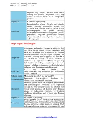
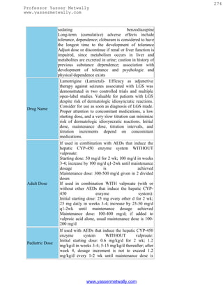
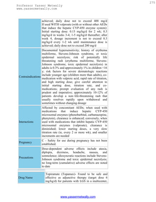
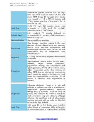


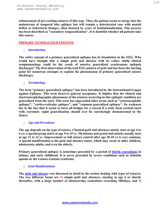
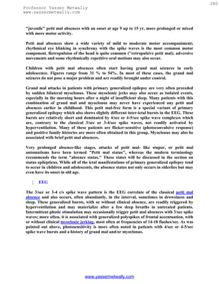
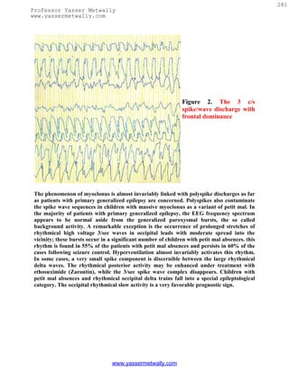
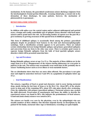
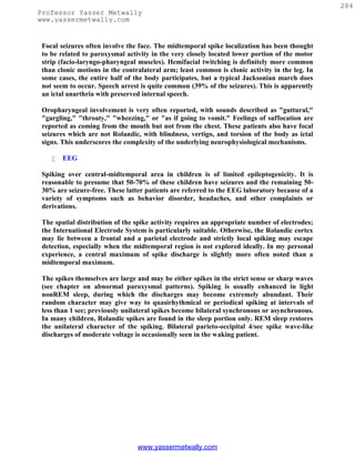
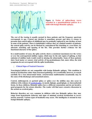


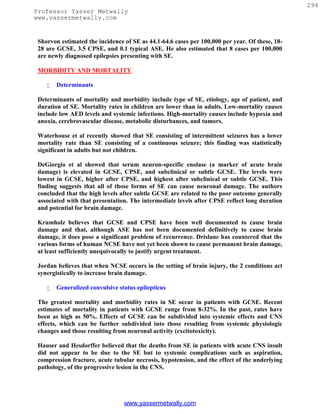
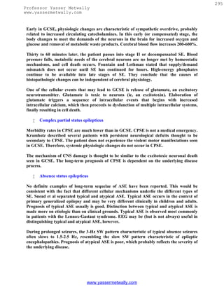
![296
Professor Yasser Metwally
www.yassermetwally.com
ELECTROENCEPHALOGRAPHY (EEG)
EEG frequently is obscured by muscle and movement artifact during an episode of GCSE,
and thus its usefulness may be limited. In diagnosis, EEG can distinguish SE from other
causes of altered mental status. EEG also is useful in classifying SE into SW and non-SW
forms, in choosing and monitoring therapy, and in formulating prognosis.
Animals with experimentally induced GCSE progress through a sequence of EEG stages
according to Treiman's classification. These include discrete generalized seizures with
interictal slowing, merged discrete seizures with waxing and waning of frequency and
amplitude, continuous seizure discharge, almost continuous seizure discharge interrupted
by flat periods, and periodic epileptiform discharges (PEDs) on a flat background (ie,
periodic lateralized epileptiform discharges [PLEDs]).
Disagreement exists regarding the number of stages and the descriptions of individual
stages, but enough independent descriptions indicate some sort of progression, at least in
experimental animals, to support this proposal. Treiman believes that most cases of human
GCSE are actually secondarily GCSE. Whether a similar sequence of changes occurs in
primarily GCSE is uncertain. The full sequence has not been reported in humans.
Although all stages have been reported in various individuals (sometimes several in a single
individual), some investigators believe that humans, unlike experimental animals, do not
always progress through a predictable sequence of EEG changes during SE. Whether
PEDs comprise an ictal pattern or a marker of severe cortical dysfunction is still a subject
of disagreement.
Nowack and Shaikh have reported fragments of Treiman's sequence in human patients
with CPSE. Such a sequence may occur in cases of partial-onset (probably non-SW) human
SE. In partial status epilepticus, Grand'Maison et al. have described a somewhat different
set of stages than Treiman.Granner and Lee studied EEGs from 85 episodes of NCSE.
They speculate that many cases with generalized-appearing ictal discharge (especially those
with focal predominance) may actually have focal onset of the SE and that diazepam can be
a useful tool for distiguishing between generalized and focal onset in this heterogenous
group.
A patient who displays the violent motor manifestations of GCSE usually presents no
diagnostic difficulty. However, Walker et al reported that almost half of patients referred
to a specialized center for "refractory SE" had nonepileptic conditions that mimic SE.
Howell et al. discussed the difficulties of differentiating SE from pseudostatus epilepticus.
Kaplan described 25 patients with NCSE in whom diagnosis was delayed up to 5 days
because of presentations that suggested other entities. EEG can be helpful in making the
correct diagnosis. Privitera et al reported that 74 of 198 patients with altered levels of
consciousness but no clinical convulsions had EEG evidence of definite or probable non–
tonic-clonic SE. They believed that correct diagnosis could not be made on clinical signs
alone. Their conclusion was that an emergency EEG should be obtained in all patients with
altered levels of consciousness.
www.yassermetwally.com](https://image.slidesharecdn.com/eeg-120912084228-phpapp02/85/Textbook-of-electroencephalography-299-320.jpg)
![301
Professor Yasser Metwally
www.yassermetwally.com
25. Krumholz A: Epidemiology and evidence for morbidity of nonconvulsive status
epilepticus. J Clin Neurophysiol 1999 Jul; 16(4): 314-22; discussion 353.
26. Litt B et al: Nonconvulsive status epilepticus in the critically ill elderly. Epilepsia
1998; 39: 1194-1202.
27. Lowenstein DH, Bleck T, Macdonald RL: It's time to revise the definition of status
epilepticus. Epilepsia 1999 Jan; 40(1): 120-2.
28. Nei M, Lee JM, Shanker VL: The EEG and prognosis in status epilepticus.
Epilepsia 1999 Feb; 40(2): 157-63.
29. Nowack WJ, Shaikh IA: Progression of electroclinical changes in complex partial
status epilepticus: filling in the blanks. Clin Electroencephalogr 1999 Jan; 30(1): 5-
8.
30. Otahara S, Otsuka Y: Myoclonic status epilepticus. In: Engel P, eds. Epilepsy: A
Comprehensive Textbook. Lippincott-Raven; 1997:725-729.
31. Privitera M, Hoffman M, Moore JL: EEG detection of nontonic-clonic status
epilepticus in patients with altered consciousness. Epilepsy Res 1994 Jun; 18(2):
155-66.
32. Shinnar S, Berg AT, Moshe SL: How long do new-onset seizures in children last?
Ann Neurol 2001 May; 49(5): 659-64.
33. Shorvon S: Status Epilepticus: Its Clinical Features and Treatment in Children and
Adults. Cambridge University Press; 1994.
34. Simon R, et al: Acute morbidity and mortality of status epilepticus. In: Engel J,
Pedley TA, eds. Epilepsy: A Comprehensive Textbook. Philadelphia: Lippincott-
Raven; 1997:741-753.
35. Snead OC III: Absence status epilepticus. In: Engel J, Pedley TA, eds. Epilepsy: A
Comprehensive Textbook. Philadelphia: Lippincott-Raven; 1997:701-707.
36. Thomas P, Beaumanoir A, Genton P: 'De novo' absence status of late onset: report
of 11 cases [see comments]. Neurology 1992 Jan; 42(1): 104-10.
37. Towne AR, Waterhouse EJ, Boggs JG: Prevalence of nonconvulsive status
epilepticus in comatose patients. Neurology 2000 Jan 25; 54(2): 340-5.
38. Treiman DM: Electroclinical features of status epilepticus. J Clin Neurophysiol
1995 Jul; 12(4): 343-62.
39. Treiman DM, Meyers PD, Walton NY: A comparison of four treatments for
generalized convulsive status epilepticus. Veterans Affairs Status Epilepticus
Cooperative Study Group [see comments]. N Engl J Med 1998 Sep 17; 339(12): 792-
8.
40. Walker MC, Shorvon SD: Treatment of status epilepticus and serial seizures. In:
Shorvon S, Dreifuss F, Fish D, Thomas D (eds): The Treatment of Epilepsy 1996;
Blackwell Science: Oxford: 269-285.
41. Walker MC, Howard RS, Smith SJ: Diagnosis and treatment of status epilepticus
on a neurological intensive care unit. QJM 1996 Dec; 89(12): 913-20.
42. Walton NY, Treiman DM: Response of status epilepticus induced by lithium and
pilocarpine to treatment with diazepam. Exp Neurol 1988 Aug; 101(2): 267-75.
43. Waterhouse EJ, Garnett LK, Towne AR: Prospective population-based study of
intermittent and continuous convulsive status epilepticus in Richmond, Virginia.
Epilepsia 1999 Jun; 40(6): 752-8.
www.yassermetwally.com](https://image.slidesharecdn.com/eeg-120912084228-phpapp02/85/Textbook-of-electroencephalography-304-320.jpg)


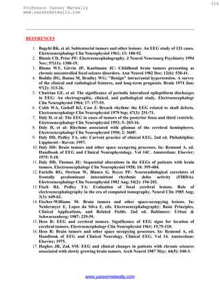
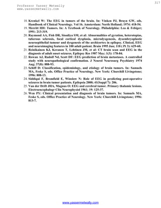
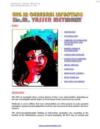
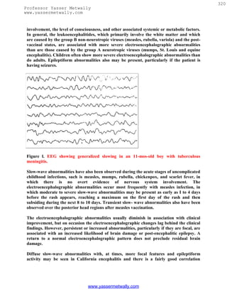
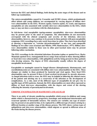
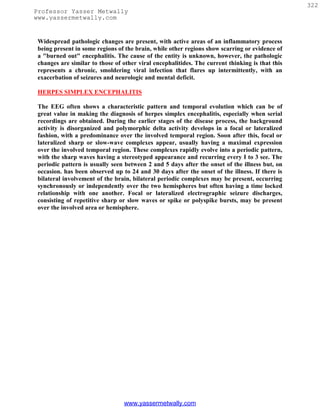
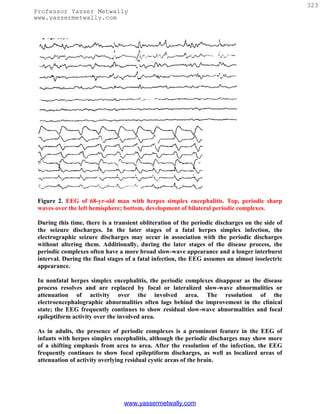

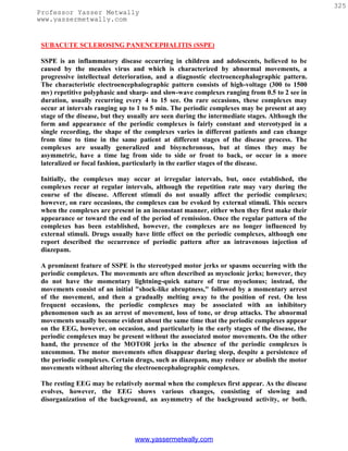
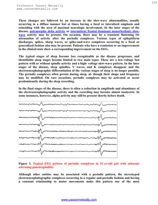

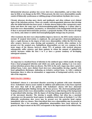


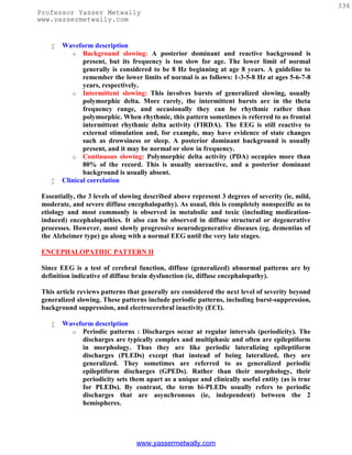
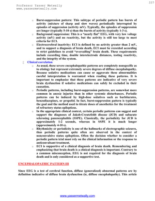
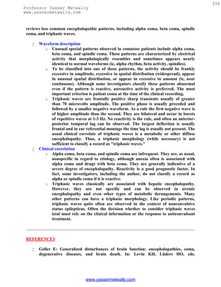
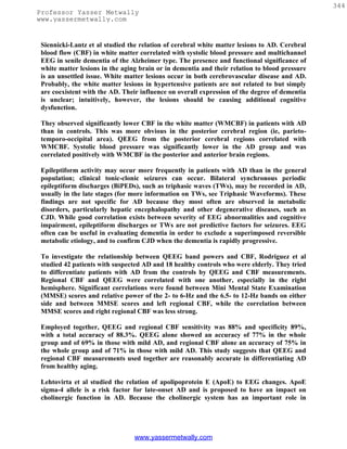
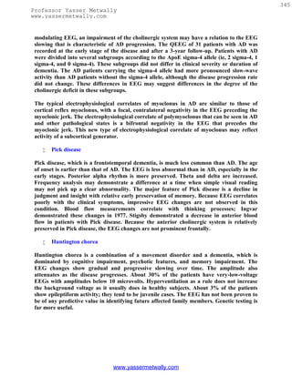
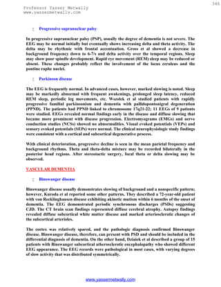
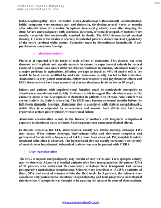
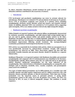
![358
Professor Yasser Metwally
www.yassermetwally.com
TRIPHASIC WAVEFORMS
TWs initially were described by Foley et al in hepatic encephalopathy. They later were
described in other metabolic states and brain tumors. Most electroencephalographers now
agree that TWs are a relatively nonspecific pattern observed in a number of metabolic
conditions, degenerative dementias, and anoxia. In a bipolar montage, TWs usually
comprise a high-voltage, positive wave followed by a smaller negative deflection; they
usually are bilaterally synchronous and maximal frontally. A fronto-occipital
(anteroposterior) phase lag varies from 25-140 ms. This is expressed less in referential
montages.
Figure 1. Triphasic waves
TWs have not been reported in children. Generally, the TW pattern carries a poor
prognosis with a high mortality rate if it occurs in association with rapid neurological and
clinical deterioration. However, TWs in a psychiatric population described by Blatt and
Brenner carried a different prognosis. In a large retrospective study consisting of 15,326
EEGs performed from 1983-1992 in a psychiatric institute, 83 EEGs (62 patients—13 men
and 49 women aged 59-90 years, with a mean age of 74 years) had TWs. All 62 patients
were awake, though they often were confused. Most (n=56) had dementia, usually severe;
15 also had delirium. Six patients had no dementia. Infrequent etiologies included
neuroleptic malignant syndrome (n=1) and hepatic encephalopathy (n=1); in 4, the cause
was uncertain, although all were receiving lithium.
EEG features analyzed included frequency of background rhythms, distribution of the
TWs, periodicity, and epileptiform abnormalities. Background rhythms were slow in all
but 7 patients (mean 6.2 +1-1.7 [SD] Hz). TWs were maximal posteriorly in 47 patients and
anteriorly in 6 patients and were diffuse in 9 patients. Neuroimaging studies demonstrated
prominent posterior abnormalities in only 1 individual. Periodicity was prominent in 4
patients; in 2, the TWs were maximal anteriorly. Interictal epileptiform activity was
present in 6 patients, a history of seizures in 8, and myoclonus in 4. TWs are uncommon in
psychiatric populations; they occur primarily in elderly and severely demented patients.
Agugila et al discussed nonmetabolic causes of TWs and described 2 patients with TWs on
their EEGs in the absence of metabolic disturbances. One patient had coma associated with
cerebellar hematoma, the other had mild dementia associated with idiopathic calcifications
www.yassermetwally.com](https://image.slidesharecdn.com/eeg-120912084228-phpapp02/85/Textbook-of-electroencephalography-361-320.jpg)
![359
Professor Yasser Metwally
www.yassermetwally.com
of the basal ganglia and healthy auditory brainstem responses and subcortical and cortical
SEPs. Neurologic examination revealed no asterixis in either patient.
The literature on nonmetabolic causes of TWs also was reviewed, and the clinical and
anatomic reports of 10 patients were analyzed. Seven patients had focal brainstem-
diencephalic lesions (craniopharyngioma [2], thalamic gliomas [3], pontine stroke [2]).
Three patients suffered from diffuse subcortical or multifocal encephalopathies
(Binswanger encephalopathy [1], cerebral carcinomatosis [1], multifocal cerebral
lymphoma [1]).
From the clinical point of view, patients with nonmetabolic diseases causing TWs presented
with either disturbance of higher cerebral functions with no asterixis or sudden onset of
coma. Agugila et al concluded that TWs may result from focal brainstem/diencephalic
lesions or from diffuse subcortical or multifocal encephalopathies in the absence of
concomitant metabolic abnormalities. Nonmetabolic causes of TW should be suspected in
patients presenting with neurological disturbances not associated with asterixis.
TWs also were evaluated by Sundaram et al, and their clinical correlates and morphology
were assessed. Twenty-six (41%) of 63 consecutive patients with TWs had various types of
metabolic encephalopathies, while 37 patients (59%) had nonmetabolic encephalopathies,
usually senile dementia. TWs were not found to be specific for any single type of metabolic
encephalopathy. Etiology was linked more closely to level of consciousness at recording
than any morphologic or distributional feature of the TWs themselves. Thus, all 31 alert
patients had nonmetabolic encephalopathies, while all 13 comatose patients had metabolic
encephalopathies.
The second, positive component (wave II) most often had the highest voltage, while equally
maximal waves I and II occurred next most commonly. In these patients, TWs most often
were expressed maximally anteriorly. Among patients with metabolic encephalopathies, a
posterior-anterior delay or lag of the wave II peak occurred more commonly than did the
better known anterior-posterior lag. Lags occurred with both metabolic and nonmetabolic
conditions but were more common with the former. No difference in quantity or mode of
appearance existed between the metabolic and nonmetabolic groups when matched for
consciousness level.
Prognosis for patients with either metabolic or nonmetabolic encephalopathies was
unfavorable. Only 4 of 24 patients with metabolic encephalopathy and 1 of 35 patients with
nonmetabolic encephalopathy were well at follow-up more than 2 years later. Forty percent
of EEGs with sharp and slow-wave complexes (slow spike waves) had sporadically
appearing TWs. The relative amplitudes of the 3 components differed from those of TWs in
other conditions; equally maximal waves II and III were the most usual form.
DIGITAL EEG
As stated in the assessment report of the American Academy of Neurology and the
American Clinical Neurophysiology Society, digital EEG is an established substitute for
www.yassermetwally.com](https://image.slidesharecdn.com/eeg-120912084228-phpapp02/85/Textbook-of-electroencephalography-362-320.jpg)
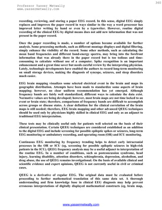
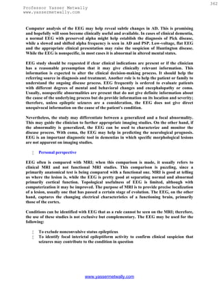
![363
Professor Yasser Metwally
www.yassermetwally.com
To attempt to record functional disturbance in individuals whose brain MRI is
"normal" but brain dysfunction is evident clinically (eg, metabolic
encephalopathies)
To attempt to record disease-specific patterns in the proper clinical setting, such as
progressive myoclonic epilepsies, CJD, SSPE
To help a psychiatrist with the multitude of complex disorders masking as potential
epilepsy or encephalopathy (eg, lithium intoxication may present with BiPEDs)
To identify focal or lateralized changes that suggest a structural cause to the
encephalopathy
The truth often is stated that EEG is nonspecific and cannot diagnose etiology or
localization well (eg, the cause of coma). However, nonspecificity is often not the question in
general medical practice because most of the referrals in general neurology are individuals
in whom the cause is pretty well clear, or reasonably suspected, on the basis of clinical
history and laboratory chemistry. The question from the clinician is whether the brain is
involved and the extent of brain damage, if any. To answer these questions, presently no
clinical tool is more useful than the EEG.
REFERENCES
1. Adams DH, Ponsford S, Gunson B: Neurological complications following liver
transplantation . Lancet 1987 Apr 25; 1(8539): 949-51.
2. Aguglia U, Gambardella A, Oliveri RL, et al: Nonmetabolic causes of triphasic
waves: a reappraisal . Clin Electroencephalogr 1990 Jul; 21(3): 120-5.
3. Aguglia U, Farnarier G, Tinuper P: Subacute spongiform encephalopathy with
periodic paroxysmal activities: clinical evolution and serial EEG findings in 20 cases
. Clin Electroencephalogr 1987 Jul; 18(3): 147-58.
4. Al-Mateen M, Philippart M, Shields WD: Rett syndrome. A commonly overlooked
progressive encephalopathy in girls . Am J Dis Child 1986 Aug; 140(8): 761-5.
5. Aoki T, Kobayashi K, Jibiki I: An autopsied case of Creutzfeldt-Jakob disease with
the lateral geniculate body lesion showing antagonizing correlation between periodic
synchronous discharges and photically induced giant evoked responses. Psychiatry
Clin Neurosci 1998 Jun; 52(3): 333-7.
6. Bittenbender JB, Quadfasel FA: Rigid and akinetic forms of Huntington’s chorea.
Arch Neurol (Chicago) 1962; 7: 275-288.
7. Blatt I, Brenner RP: Triphasic waves in a psychiatric population: a retrospective
study. J Clin Neurophysiol 1996 Jul; 13(4): 324-9.
8. Boyd SG, Harden A, Egger J: Progressive neuronal degeneration of childhood with
liver disease ("Alpers' disease"): characteristic neurophysiological features .
Neuropediatrics 1986 May; 17(2): 75-80.
9. Dzialek E, Szymanska R, Bogucki A: [The electroencephalographic image in
Binswanger's subcortical arteriosclerotic encephalopathy]. Neurol Neurochir Pol
1989 May-Jun; 23(3): 214-9.
www.yassermetwally.com](https://image.slidesharecdn.com/eeg-120912084228-phpapp02/85/Textbook-of-electroencephalography-366-320.jpg)
![364
Professor Yasser Metwally
www.yassermetwally.com
10. Edman A, Matousek M, Sjogren M: Longitudinal EEG findings in dementia related
to the parietal brain syndrome and the degree of dementia. Dement Geriatr Cogn
Disord 1998 Jul-Aug; 9(4): 199-204.
11. Flaherty MP, O'Flaherty SJ: Neurological deterioration following head injury: the
eyes had it. J Paediatr Child Health 1998 Apr; 34(2): 202-5.
12. Flaten TP, Alfrey AC, Birchall JD, et al: Status and future concerns of clinical and
environmental aluminum toxicology . J Toxicol Environ Health 1996 Aug 30; 48(6):
527-41.
13. Foley JM, Watson CW, Adams RD: Significance of electroencephalographic
changes in hepatic coma. Trans Am Neurol Assoc 1950; 75: 161-165.
14. Ghika-Schmid F, Ghika J, Regli F: Hashimoto's myoclonic encephalopathy: an
underdiagnosed treatable condition? Mov Disord 1996 Sep; 11(5): 555-62.
15. Gross RA, Spehlmann R, Daniels JC: Sleep disturbances in progressive
supranuclear palsy . Electroencephalogr Clin Neurophysiol 1978 Jul; 45(1): 16-25.
16. Hallett M, Wilkins DE: Myoclonus in Alzheimer's disease and minipolymyoclonus.
Adv Neurol 1986; 43: 399-405.
17. Hansen HC, Zschocke S, Sturenburg HJ: Clinical changes and EEG patterns
preceding the onset of periodic sharp wave complexes in Creutzfeldt-Jakob disease.
Acta Neurol Scand 1998 Feb; 97(2): 99-106.
18. Hietaharju A, Jaaskelainen S, Hietarinta M: Central nervous system involvement
and psychiatric manifestations in systemic sclerosis (scleroderma): clinical and
neurophysiological evaluation. Acta Neurol Scand 1993 May; 87(5): 382-7.
19. Hill D: The EEG in organic disease. Proc R Soc Med 1948; 41: 242-248.
20. Ingvar DH: History of brain imaging in psychiatry. Dement Geriatr Cogn Disord
1997 Mar-Apr; 8(2): 66-72.
21. Ingvar DH: [The cerebral ideogram] . Encephale 1977; 3(1): 5-33.
22. Ingvar DH, Philipson L: Distribution of cerebral blood flow in the dominant
hemisphere during motor ideation and motor performance. Ann Neurol 1977; 2:
23~237.
23. Isozumi K, Fukuuchi Y, Tanaka K, et al: A MELAS (mitochondrial myopathy,
encephalopathy, lactic acidosis, and stroke-like episodes) mtDNA mutation that
induces subacute dementia which mimics Creutzfeldt-Jakob disease . Intern Med
1994 Sep; 33(9): 543-6.
24. Iznak AF, Gavrilova SI, Zhigul'skaia SE: [Quantitative EEG in early and
differential diagnosis of mild dementia of different genesis]. Vestn Ross Akad Med
Nauk 1999; (1): 11-6.
25. Jelic V, Blomberg M, Dierks T: EEG slowing and cerebrospinal fluid tau levels in
patients with cognitive decline. Neuroreport 1998 Jan 5; 9(1): 157-60.
26. Juvarra G, Bettoni L, Olivieri MF, et al: Hypercalcemic encephalopathy in the
course of hyperthyroidism . Eur Neurol 1985; 24(2): 121-7.
27. Kameda K, Itoh N, Nakayama H: Frontal intermittent rhythmic delta activity
(FIRDA) in pituitary adenoma. Clin Electroencephalogr 1995 Jul; 26(3): 173-9.
28. Koppel BS, Poon TP, Khandji A, et al: Subacute sclerosing panencephalitis and
acquired immunodeficiency syndrome: role of electroencephalography and
magnetic resonance imaging . J Neuroimaging 1996 Apr; 6(2): 122-5.
www.yassermetwally.com](https://image.slidesharecdn.com/eeg-120912084228-phpapp02/85/Textbook-of-electroencephalography-367-320.jpg)
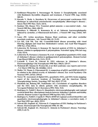
![366
Professor Yasser Metwally
www.yassermetwally.com
46. Siennicki-Lantz A, Lilja B, Rosen I: Cerebral blood flow in white matter is
correlated with systolic blood pressure and EEG in senile dementia of the Alzheimer
type. Dement Geriatr Cogn Disord 1998 Jan-Feb; 9(1): 29-38.
47. Steinhoff BJ, Kropp S, Riedemann C: [Elecroencephalographic charactistics of
Creutzfeldt-Jakob disease and its differential diagnosis]. Fortschr Neurol Psychiatr
1998 Aug; 66(8): 357-65.
48. Stevens A, Kircher T: Cognitive decline unlike normal aging is associated with
alterations of EEG temporo-spatial characteristics. Eur Arch Psychiatry Clin
Neurosci 1998; 248(5): 259-66.
49. Stigsby B, Johannesson G, Ingvar DH: Regional EEG analysis and regional cerebral
blood flow in Alzheimer's and Pick's diseases . Electroencephalogr Clin
Neurophysiol 1981 May; 51(5): 537-47.
50. Strik WK, Chiaramonti R, Muscas GC: Decreased EEG microstate duration and
anteriorisation of the brain electrical fields in mild and moderate dementia of the
Alzheimer type. Psychiatry Res 1997 Oct 31; 75(3): 183-91.
51. Su PC, Goldensohn ES: Progressive supranuclear palsy. Electroencephalographic
studies. Arch Neurol (Chicago) 1973; 29: 18~186.
52. Sundaram MB, Blume WT: Triphasic waves: clinical correlates and morphology .
Can J Neurol Sci 1987 May; 14(2): 136-40.
53. Takahashi M, Kubota F, Nishi Y: Persistent synchronous periodic discharges
caused by anoxic encephalopathy due to cardiopulmonary arrest. Clin
Electroencephalogr 1993 Oct; 24(4): 166-72.
54. Tatum WO 4th, Ross J, Cole AJ: Epileptic pseudodementia . Neurology 1998 May;
50(5): 1472-5.
55. Thomas P, Borg M: Reversible myoclonic encephalopathy revealing the AIDS-
dementia complex. Electroencephalogr Clin Neurophysiol 1994 Feb; 90(2): 166-9.
56. Wada Y, Nanbu Y, Koshino Y: Reduced interhemispheric EEG coherence in
Alzheimer disease: analysis during rest and photic stimulation. Alzheimer Dis Assoc
Disord 1998 Sep; 12(3): 175-81.
57. Widagdo MM, Pierson JM, Helme RD: Age-related changes in qEEG during
cognitive tasks. Int J Neurosci 1998 Jul; 95(1-2): 63-75.
58. Wszolek ZK, Lagerlund TD, Steg RE: Clinical neurophysiologic findings in patients
with rapidly progressive familial parkinsonism and dementia with pallido-ponto-
nigral degeneration. Electroencephalogr Clin Neurophysiol 1998 Sep; 107(3): 213-
22.
59. Xu CY: [An EEG evaluation in diagnosing subacute spongiform encephalopathy].
Chung Hua Shen Ching Ching Shen Ko Tsa Chih 1989 Feb; 22(1): 37-8, 63.
www.yassermetwally.com](https://image.slidesharecdn.com/eeg-120912084228-phpapp02/85/Textbook-of-electroencephalography-369-320.jpg)
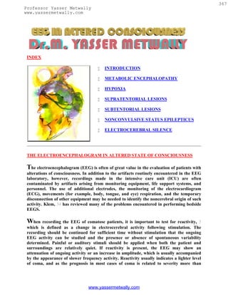
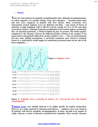
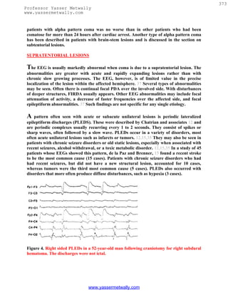
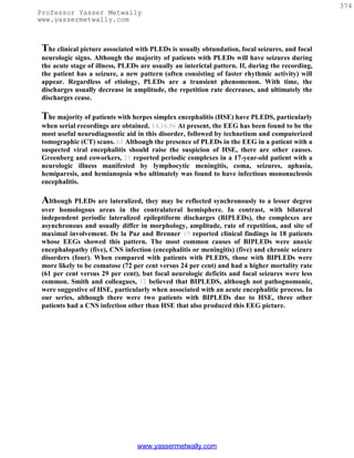
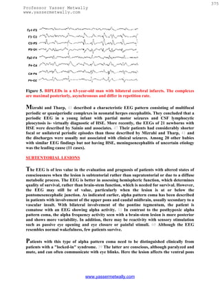
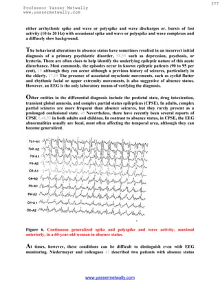
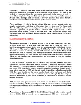
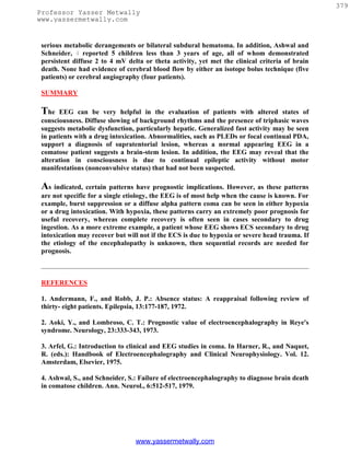
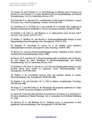
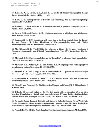
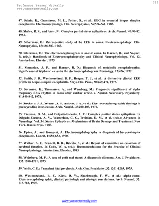


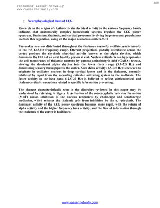
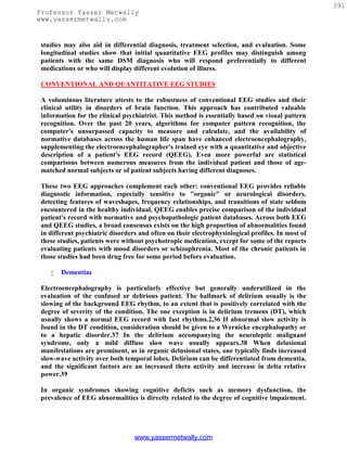
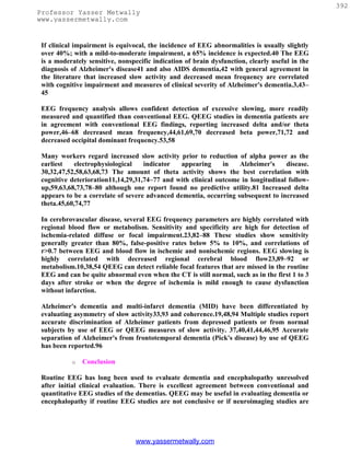
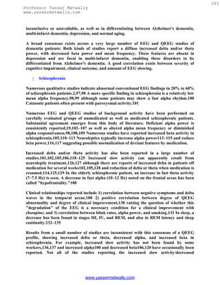
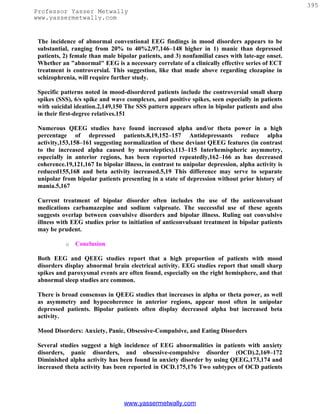
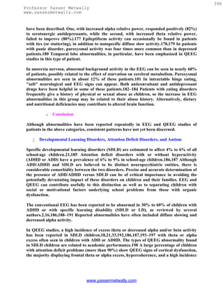
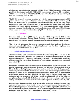
![398
Professor Yasser Metwally
www.yassermetwally.com
o Conclusion
There is a broad consensus that both EEG and QEEG reveal marked abnormalities in
alcoholics and substance abusers. The effects vary depending on the drug. Either increased
slow activity with lower alpha and beta or the converse have been reported; this reflects the
diversity of substances or states focused upon.
However, among numerous QEEG studies, there is a consensus of increased beta relative
power in alcoholism and increased alpha in cannabis or crack cocaine users.
Mild Head Injury or Postconcussion Syndrome
Patients with complaints of cognitive memory or attentional deficit after mild head injury
without loss of consciousness frequently present for psychiatric evaluation for worker's
compensation and disability benefits. Objective evidence of brain dysfunction in such cases
is critical in the endeavor to separate the truly dysfunctional patient from the malingerer.
EEG/QEEG evidence can play a critical role in such cases. Although the absence of
abnormal brain electrical activity cannot definitively exclude the possibility of brain
dysfunction, the presence of abnormalities, especially those most frequently associated with
unequivocal traumatic brain insult, must be considered supportive of such claims.
There is a high incidence of diffuse axonal injury, about 50%, in the 50,000 patients per
year with head injury who recover; these patients characteristically do not display
structural lesions on CT or even on MRI scans early after injury.217,218 Among those who
recover after moderate head injury, 90% have cognitive or neuropsychological deficits.219
Among such patients, studies involving many hundreds of cases have reported normal
neurologic examinations but abnormal EEG.220–226 Numerous EEG and QEEG studies of
severe head injury (Glasgow Coma Scale [GCS] 4–8) and moderate injury (GCS 9–12),
using samples of 50 to 200 patients, have agreed that increased theta and decreased alpha
power and/or decreased coherence and asymmetry often characterize such patients.
Changes in these measures provide the best predictors of long-term outcome.219,227–229
The studies cited above generally concur that the most characteristic QEEG or EEG
abnormalities persisting after mild or moderate head injury are similar in type to those
found after severe head injury, namely increased power in the theta band, decreased alpha,
low coherence, and increased asymmetry. It is noteworthy that similar EEG abnormalities
have been reported in boxers,230 correlated with the numbers of bouts or knockouts, and
in professional soccer players who were "headers."231 These observations lend further
credibility to the multiple reports of discriminant functions based on QEEG variables that
successfully separated normal individuals from patients with a history of mild to moderate
head injury years after apparent clinical recovery.232,233
www.yassermetwally.com](https://image.slidesharecdn.com/eeg-120912084228-phpapp02/85/Textbook-of-electroencephalography-401-320.jpg)
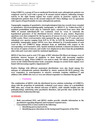
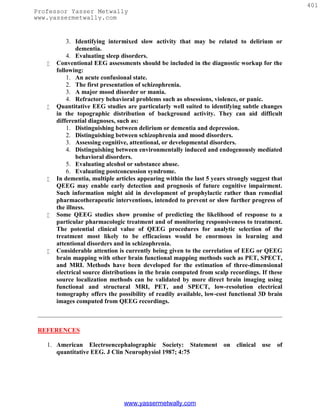
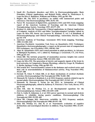
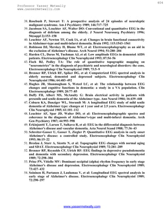
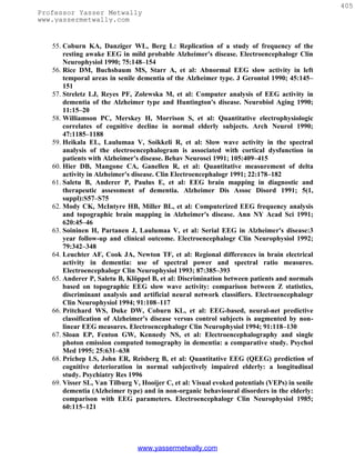
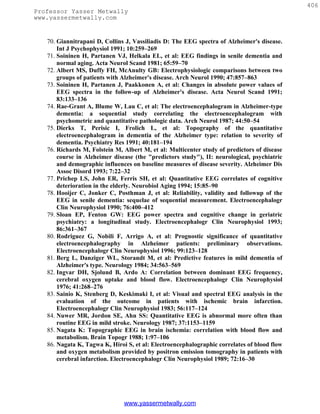
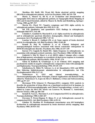
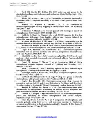
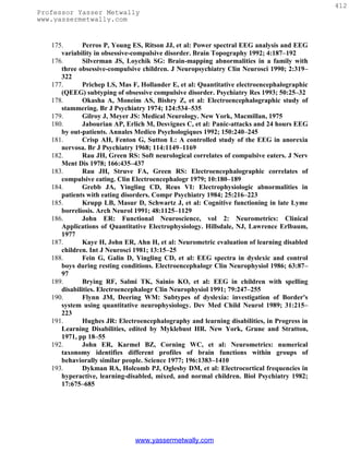
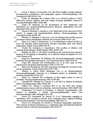
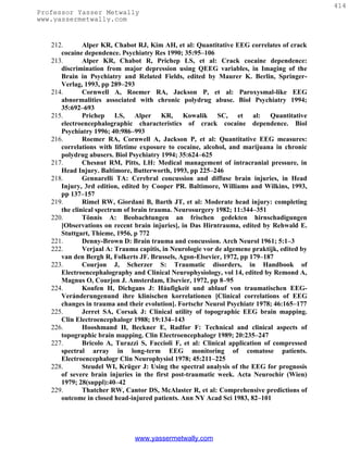

The document summarizes the history and technical aspects of conventional EEG. It discusses how EEG works to detect and amplify the brain's electrical activity, which is measured using electrodes placed on the scalp. Different electrode placements and montages are used to view brain activity from various regions and perspectives. While imaging techniques now provide anatomical details, EEG remains clinically useful for evaluating brain function in various neurological disorders.