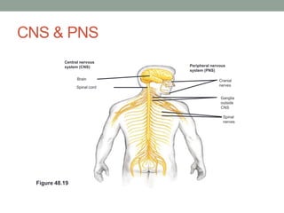
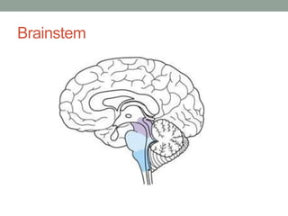
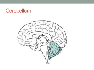
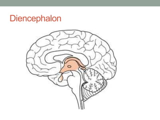
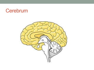
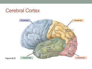
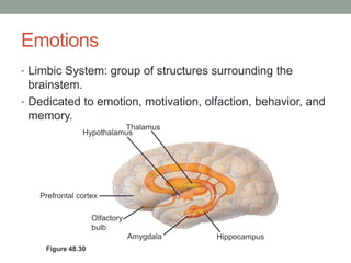
THE NERVOUS
SYSTEM
Introduction
• Basic to the functioning of any animal.
• In order to survive and reproduce, an animal must
  respond to environmental stimuli both external and
  internal.
• Provides both immediate and long-term responses.
Circuits of neurons and supporting cells
• Most animals with nervous systems have clusters of
  neurons that perform specialized functions.
• Such clustering is absent in the cnidarians.
• Most cnidarians have radially symmetrical bodies.
• Most cnidarians have nerve nets.
Nerves vs. Nerve Nets
• Simple animals, ex. Cnidarians, have nerve nets.
• More complex animals: axons of multiple nerve cells are
 bundled together to form nerves.
  • Better suited to controlling elaborate movements than a
    single diffuse nerve net.
Cephalization
• Animals with elongated, bilaterally symmetrical bodies
  have even more specialized nervous systems.
• Such animals exhibit cephalization.
Nervous System Organization
• Look at the picture on page 1065
CNS & PNS in Vertebrates
• CNS= Central Nervous System
  • Brain and spinal cord.
• PNS= Peripheral Nervous System
  • Nerves and ganglia.
• Regional specialization is a hallmark of both systems.
• The PNS connects to the CNS
CNS & PNS
            Central nervous
            system (CNS)        Peripheral nervous
                                system (PNS)

                  Brain                       Cranial
                  Spinal cord                 nerves


                                               Ganglia
                                               outside
                                               CNS

                                                Spinal
                                                nerves


  Figure 48.19
Spinal cord
• The spinal cord runs lengthwise inside the spine.
• It conveys information to and from the brain and
  generates basic patterns of locomotion.
• It acts independently of the brain.
Reflexes
• The spinal cord acts as part of the simple nerve circuits
  that produce reflexes.
• A reflex protects the body by triggering a rapid,
  involuntary response to a particular stimulus.
• Ganglia run along the outside of the spinal cord
Origin of the brain & spinal cord
• Derived from the dorsal embryonic
  nerve cord.
• During development it is transformed
  into the narrow central canal of the
  spinal cord and the ventricles of the
  brain.
Fluid & Matter
• Both the central canal and the brain are filled with
  cerebrospinal fluid and Gray/White matter
• Cerebrospinal fluid: supplies the brain with essential
  nutrients and hormones.
• Gray Matter- neuron cell bodies and dendrites
• White Matter- bundled axons
Glia in the CNS
• Ependymal cells- circulation of cerebrospinal fluid
• Microglia- protect from invading microorganisms
• Oligodendrocytes- function in axon myelination
• Astrocytes-structural support for neurons
PNS
Overall importance of PNS
• Transmits information to and from the CNS and plays a
  large part in regulating an animal’s movement and internal
  environment.
• Sensory information reaches the CNS along afferent (to
  bring toward) PNS neurons.
• Instructions from the CNS reach the PNS along efferent
  (to carry off) PNS neurons
Nerves
• Cranial Nerves:
  • connect the brain with locations mostly in organs of the head and
    upper body.
• Spinal Nerves:
  • Run between the spinal cord and parts of the body below the head.
Systems of the PNS
• Motor Systems: neurons that carry information to
  muscles.
• Autonomic Nervous System: regulates the internal
  environment. This control is generally involuntary and is
  split into three divisions: Sympathetic, Parasympathetic,
  Enteric.
Sympathetic Division
• Corresponds to arousal and energy generation (fight-or-
 flight).
  • Inhibits saliva production
  • Relaxes bronchi in the lungs
  • Accelerates heart rate
  • Secretion of epinipherene
Parasympathetic Division
• Promote calming and return to self- maintenance.
• Rest-and-digest
  • Slows heart rate
  • Promotes emptying of bladder
  • Constricts bronchi in lungs
Enteric Division
• Networks of neurons in the digestive tract, pancreas, and
  gallbladder.
• Also control peristalsis.
CNS
Brainstem
The Brain Stem
• Adult brainstem midbrain, the pons, and the
  medulla oblongata.
• Sits at the bottom of the brain, on top of the spinal
  cord.
• It relays the information between the spinal cord
  and the brain.
Arousal and Sleep
• Controlled by the brainstem and cerebrum.
• Arousal= you are aware of surroundings.
• Sleep= external stimuli are received but not consciously
  perceived.
• Reticular formation
• Sleep is an active process.
• Melatonin plays an important part in these processes.
Cerebellum
The Cerebellum
• Posterior
• Coordinates movements and balance, specifically helps
  when you have to learn something new.
• Hand eye coordination
Diencephalon
The Diencephalon
• 3 parts: Thalamus, Hypothalamus, and Epithalamus.
Epithalamus
• Location of the pineal gland, origin of melatonin.
• Also contains capillaries that generate cerebrospinal fluid
  from the blood.
• Cerebrospinal fluid: supplies the brain with essential
  nutrients and hormones
Thalamus
• Main input center for center for sensory information going
 to the cerebrum.
Hypothalamus
• Controls homeostasis
  • Ex: body’s thermostat, regulate hunger & thirst.
• Source of posterior pituitary hormones and releasing
  hormones that act on the anterior pituitary.
• Plays a role in sexual and mating behaviors, fight-or-flight,
  and pleasure.
• Four F’s
Biological Clock Regulation
• Specialized nerve cells in the hypothalamus regulate
  circadian rhythms, daily cycles of biological activities.
• As stated previously: regulates sleep, body temperature,
  hunger, and hormone release.
• Molecular mechanisms that direct periodic gene
  expression and cellular activity.
• Typically synchronized with the day and night.
Biological Clock Regulation cont.
• In mammals circadian rhythms are coordinated by a
  group of neurons in the hypothalamus called the
  superchiasmatic nucleus (SCN).
• The SCN determines the circadian rhythm of the whole
  animal.
Cerebrum
The Cerebrum
• Divided into right and left cerebral hemispheres.
• Each hemisphere is made up of an outer covering of gray
 matter, the cerebra cortex; internal white matter; and
 groups of neurons called basal nuclei.
Basal Nuclei
• Basal Nuclei are important centers for planning and
  learning movement sequences.
• Damage in this brain region during embryonic
  development can result in cerebral palsy, a defect
  disrupting how motor commands are issued to the
  muscles.
Cerebral Cortex
               Frontal lobe                                                             Parietal lobe


                                                                        Somatosensory
                                                               Speech
                                                                        association
                      Frontal
                                                                        area
                      association
                      area                                 Taste

                                                                          Reading
                                    Speech
                                                       Hearing

                                                                              Visual
                                         Smell                                association
                                                                              area
                                                 Auditory
                                                 association
                                                 area

                                                                           Vision


                 Temporal lobe                                                              Occipital lobe
Figure 48.27
The Cerebral Cortex
• 4 different lobes: frontal, temporal, occipital, and parietal.
• Frontal lobe: plans actions and controls your movement.
  • Size fits muscles required. Leg- Knee- Hip.
• Parietal lobe: Sensory.
  • Size fits sensory info.
• Temporal lobe: Auditory & Memory Formation
• Occipital lobe: Visual information
Cerebral Cortex
• Accounts for 80% of total brain mass.
• Highly convoluted, allows for large surface area.
• The corpus callosum is a thick band of axons that
 enables communication between the two sides of the
 cerebral cortex.
Language and speech
• Damage to particular regions of the cortex by injuries,
 strokes, or tumors can change in a person’s behavior.
                                                      Max


                        Hearing      Seeing
                         words       words


                                                      Min
                          Speaking
                                       Generating
         Figure 48.29
                           words
                                         words
Broca’s Area
• Pierre Broca studied people who could understand
  language, but not speak it.
• Located in the front part of the primary motor cortex, in the
  frontal lobe.
• Controls the speaking and the generating of the brain.
Wernicke's Area
• Karl Wernicke studied damage to the back of the left
  temporal lobe.
• People could speak, but not comprehend speech.
Lateralization
• Both hemisphere’s have different functions.
• Generally:
  • Left side= language/speech, math, logic
  • Right side= patterns, facial recognition (creative side)
Emotions
• Limbic System: group of structures surrounding the
  brainstem.
• Dedicated to emotion, motivation, olfaction, behavior, and
  memory.
                         Thalamus
               Hypothalamus


   Prefrontal cortex

                   Olfactory
                   bulb
                               Amygdala   Hippocampus
    Figure 48.30
Memory & Learning
• Hippocampus- Forms new memories
• Amygdala- Fear & anxiety
• Short term memories are made by links made in the
 hippocampus. When memories need to be made long
 term, the links are replaced by links in the cerebral cortex
Long Term Potentiation
• LTP: Involves an increase in the strength of
 synaptic transmission
Evolution of Cognition in Vertebrates
• In humans, the outermost part of the cerebral cortex is
  called the neocortex.
• The outer part of the avian cerebral cortex is called the
  pallium.
• The common ancestor of birds and humans probably had
  a pallium as it still present in birds.
Schizophrenia
• 1% of the population has it.
• Distorted perception of reality, hallucinations.
• Two lines of evidence suggest that schizophrenia affects
 neurons that use dopamine:
  • Speed- produces dopamine and produces same effects as schiz.
  • Many drugs used for treatment block dopamine
Depression
• Two types:
• Major depressive disorder- Extreme Sadness
• Bipolar Disorder-
  • Manic phase extremely energetic
  • Depressive phase, same as m.d.d
Drug Addiction
• Altered activity of the brain’s reward system, which
 normally provides motivation for actions.
Alzheimer’s
• Mental deterioration where plaques form in the brain.
• Confusion and memory loss.
Parkinson's
• Motor disease
• Caused by the death of dopamine-secreting neurons
• No cure at the time
Stem Cell Based Therapy
• The adult human brain contains stem cells that can
 differentiate into mature neurons, this will be helpful for
 replacing neurons lost to a disease or trauma.
Conclusion
• Your nervous system has several different parts that play
  their own special parts, but at the same time they work
  together.
• Only 10% of your brain is made up of neurons, the other
  90% is glia.
• There are several neurological diseases.

The nervous system

  • 1.
  • 2.
    Introduction • Basic tothe functioning of any animal. • In order to survive and reproduce, an animal must respond to environmental stimuli both external and internal. • Provides both immediate and long-term responses.
  • 3.
    Circuits of neuronsand supporting cells • Most animals with nervous systems have clusters of neurons that perform specialized functions. • Such clustering is absent in the cnidarians. • Most cnidarians have radially symmetrical bodies. • Most cnidarians have nerve nets.
  • 4.
    Nerves vs. NerveNets • Simple animals, ex. Cnidarians, have nerve nets. • More complex animals: axons of multiple nerve cells are bundled together to form nerves. • Better suited to controlling elaborate movements than a single diffuse nerve net.
  • 5.
    Cephalization • Animals withelongated, bilaterally symmetrical bodies have even more specialized nervous systems. • Such animals exhibit cephalization.
  • 6.
    Nervous System Organization •Look at the picture on page 1065
  • 7.
    CNS & PNSin Vertebrates • CNS= Central Nervous System • Brain and spinal cord. • PNS= Peripheral Nervous System • Nerves and ganglia. • Regional specialization is a hallmark of both systems. • The PNS connects to the CNS
  • 8.
    CNS & PNS Central nervous system (CNS) Peripheral nervous system (PNS) Brain Cranial Spinal cord nerves Ganglia outside CNS Spinal nerves Figure 48.19
  • 9.
    Spinal cord • Thespinal cord runs lengthwise inside the spine. • It conveys information to and from the brain and generates basic patterns of locomotion. • It acts independently of the brain.
  • 10.
    Reflexes • The spinalcord acts as part of the simple nerve circuits that produce reflexes. • A reflex protects the body by triggering a rapid, involuntary response to a particular stimulus. • Ganglia run along the outside of the spinal cord
  • 11.
    Origin of thebrain & spinal cord • Derived from the dorsal embryonic nerve cord. • During development it is transformed into the narrow central canal of the spinal cord and the ventricles of the brain.
  • 12.
    Fluid & Matter •Both the central canal and the brain are filled with cerebrospinal fluid and Gray/White matter • Cerebrospinal fluid: supplies the brain with essential nutrients and hormones. • Gray Matter- neuron cell bodies and dendrites • White Matter- bundled axons
  • 13.
    Glia in theCNS • Ependymal cells- circulation of cerebrospinal fluid • Microglia- protect from invading microorganisms • Oligodendrocytes- function in axon myelination • Astrocytes-structural support for neurons
  • 14.
  • 15.
    Overall importance ofPNS • Transmits information to and from the CNS and plays a large part in regulating an animal’s movement and internal environment. • Sensory information reaches the CNS along afferent (to bring toward) PNS neurons. • Instructions from the CNS reach the PNS along efferent (to carry off) PNS neurons
  • 16.
    Nerves • Cranial Nerves: • connect the brain with locations mostly in organs of the head and upper body. • Spinal Nerves: • Run between the spinal cord and parts of the body below the head.
  • 17.
    Systems of thePNS • Motor Systems: neurons that carry information to muscles. • Autonomic Nervous System: regulates the internal environment. This control is generally involuntary and is split into three divisions: Sympathetic, Parasympathetic, Enteric.
  • 18.
    Sympathetic Division • Correspondsto arousal and energy generation (fight-or- flight). • Inhibits saliva production • Relaxes bronchi in the lungs • Accelerates heart rate • Secretion of epinipherene
  • 19.
    Parasympathetic Division • Promotecalming and return to self- maintenance. • Rest-and-digest • Slows heart rate • Promotes emptying of bladder • Constricts bronchi in lungs
  • 20.
    Enteric Division • Networksof neurons in the digestive tract, pancreas, and gallbladder. • Also control peristalsis.
  • 21.
  • 22.
  • 23.
    The Brain Stem •Adult brainstem midbrain, the pons, and the medulla oblongata. • Sits at the bottom of the brain, on top of the spinal cord. • It relays the information between the spinal cord and the brain.
  • 24.
    Arousal and Sleep •Controlled by the brainstem and cerebrum. • Arousal= you are aware of surroundings. • Sleep= external stimuli are received but not consciously perceived. • Reticular formation • Sleep is an active process. • Melatonin plays an important part in these processes.
  • 25.
  • 26.
    The Cerebellum • Posterior •Coordinates movements and balance, specifically helps when you have to learn something new. • Hand eye coordination
  • 27.
  • 28.
    The Diencephalon • 3parts: Thalamus, Hypothalamus, and Epithalamus.
  • 29.
    Epithalamus • Location ofthe pineal gland, origin of melatonin. • Also contains capillaries that generate cerebrospinal fluid from the blood. • Cerebrospinal fluid: supplies the brain with essential nutrients and hormones
  • 30.
    Thalamus • Main inputcenter for center for sensory information going to the cerebrum.
  • 31.
    Hypothalamus • Controls homeostasis • Ex: body’s thermostat, regulate hunger & thirst. • Source of posterior pituitary hormones and releasing hormones that act on the anterior pituitary. • Plays a role in sexual and mating behaviors, fight-or-flight, and pleasure. • Four F’s
  • 32.
    Biological Clock Regulation •Specialized nerve cells in the hypothalamus regulate circadian rhythms, daily cycles of biological activities. • As stated previously: regulates sleep, body temperature, hunger, and hormone release. • Molecular mechanisms that direct periodic gene expression and cellular activity. • Typically synchronized with the day and night.
  • 33.
    Biological Clock Regulationcont. • In mammals circadian rhythms are coordinated by a group of neurons in the hypothalamus called the superchiasmatic nucleus (SCN). • The SCN determines the circadian rhythm of the whole animal.
  • 34.
  • 35.
    The Cerebrum • Dividedinto right and left cerebral hemispheres. • Each hemisphere is made up of an outer covering of gray matter, the cerebra cortex; internal white matter; and groups of neurons called basal nuclei.
  • 36.
    Basal Nuclei • BasalNuclei are important centers for planning and learning movement sequences. • Damage in this brain region during embryonic development can result in cerebral palsy, a defect disrupting how motor commands are issued to the muscles.
  • 37.
    Cerebral Cortex Frontal lobe Parietal lobe Somatosensory Speech association Frontal area association area Taste Reading Speech Hearing Visual Smell association area Auditory association area Vision Temporal lobe Occipital lobe Figure 48.27
  • 38.
    The Cerebral Cortex •4 different lobes: frontal, temporal, occipital, and parietal. • Frontal lobe: plans actions and controls your movement. • Size fits muscles required. Leg- Knee- Hip. • Parietal lobe: Sensory. • Size fits sensory info. • Temporal lobe: Auditory & Memory Formation • Occipital lobe: Visual information
  • 39.
    Cerebral Cortex • Accountsfor 80% of total brain mass. • Highly convoluted, allows for large surface area. • The corpus callosum is a thick band of axons that enables communication between the two sides of the cerebral cortex.
  • 40.
    Language and speech •Damage to particular regions of the cortex by injuries, strokes, or tumors can change in a person’s behavior. Max Hearing Seeing words words Min Speaking Generating Figure 48.29 words words
  • 41.
    Broca’s Area • PierreBroca studied people who could understand language, but not speak it. • Located in the front part of the primary motor cortex, in the frontal lobe. • Controls the speaking and the generating of the brain.
  • 42.
    Wernicke's Area • KarlWernicke studied damage to the back of the left temporal lobe. • People could speak, but not comprehend speech.
  • 43.
    Lateralization • Both hemisphere’shave different functions. • Generally: • Left side= language/speech, math, logic • Right side= patterns, facial recognition (creative side)
  • 44.
    Emotions • Limbic System:group of structures surrounding the brainstem. • Dedicated to emotion, motivation, olfaction, behavior, and memory. Thalamus Hypothalamus Prefrontal cortex Olfactory bulb Amygdala Hippocampus Figure 48.30
  • 45.
    Memory & Learning •Hippocampus- Forms new memories • Amygdala- Fear & anxiety • Short term memories are made by links made in the hippocampus. When memories need to be made long term, the links are replaced by links in the cerebral cortex
  • 46.
    Long Term Potentiation •LTP: Involves an increase in the strength of synaptic transmission
  • 47.
    Evolution of Cognitionin Vertebrates • In humans, the outermost part of the cerebral cortex is called the neocortex. • The outer part of the avian cerebral cortex is called the pallium. • The common ancestor of birds and humans probably had a pallium as it still present in birds.
  • 48.
    Schizophrenia • 1% ofthe population has it. • Distorted perception of reality, hallucinations. • Two lines of evidence suggest that schizophrenia affects neurons that use dopamine: • Speed- produces dopamine and produces same effects as schiz. • Many drugs used for treatment block dopamine
  • 49.
    Depression • Two types: •Major depressive disorder- Extreme Sadness • Bipolar Disorder- • Manic phase extremely energetic • Depressive phase, same as m.d.d
  • 50.
    Drug Addiction • Alteredactivity of the brain’s reward system, which normally provides motivation for actions.
  • 51.
    Alzheimer’s • Mental deteriorationwhere plaques form in the brain. • Confusion and memory loss.
  • 52.
    Parkinson's • Motor disease •Caused by the death of dopamine-secreting neurons • No cure at the time
  • 53.
    Stem Cell BasedTherapy • The adult human brain contains stem cells that can differentiate into mature neurons, this will be helpful for replacing neurons lost to a disease or trauma.
  • 54.
    Conclusion • Your nervoussystem has several different parts that play their own special parts, but at the same time they work together. • Only 10% of your brain is made up of neurons, the other 90% is glia. • There are several neurological diseases.

Editor's Notes

  • #42 Kind of like mutes
  • #43 Random speech
  • #45 In early development, emotional memory.
  • #55 4 quadrillion synapses.