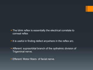
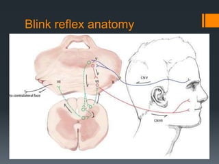
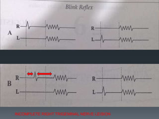
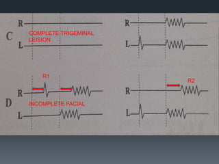
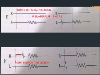
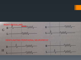
The blink reflex is a disynaptic or multisynaptic reflex that involves the trigeminal and facial nerves. It has two responses - an early ipsilateral R1 response and a late bilateral R2 response. The blink reflex test stimulates the supraorbital nerve branch to evaluate conduction along the trigeminal and facial nerve pathways. Abnormalities in the R1 and R2 responses can localize lesions in different parts of the brainstem or peripheral nerves. The test involves recording electromyography of the orbicularis oculi muscle in response to supraorbital nerve stimulation.
2. The blink reflex is essentially the electrical correlate to
corneal reflex
It is useful in finding defect anywhere in the reflex arc.
Afferent: supraorbital branch of the opthalmic division of
Trigeminal nerve.
Efferent: Motor fibers of facial nerve.
4. Two response: Early R1(ipsilateral) and
Late R2 response(bilateral).
R1: disynaptic- Biphasic
R2: multisynaptic- Polyphasic
5. Procedure
Patient lie relaxed
Two channel recording
Electrodes: Inferior
orbicularis oculi
Active electrode G1-lateral
and inferior to pupil.
Reference electrode G2
placed lateral to lateral
canthus bilaterally.
Ground electrode: mid
forehead
Sweep speed: 5-10ms/division
Sensitivity:100 or 200 micro volt/div
Motor filter: 10 Hz-10Khz
G1
G2
6. Electrode is placed on the supra orbital fissure over
medial supra orbital ridge (depression over bony ridge
over eye brow) to stimulate supra orbital nerve branch of
opthalmic disvision of trigeminal nerve.
Electrical stimulus of 100 micro seconds duration used.
7. The current is increased in small increments 3-5 milli ampere
Until supra maximal stimulation is reached.
Typically no more than 15-25 milli ampere
Four to six trials are obtained on rastered tracing and
superimposed
9. Blink reflex is elicited by stimulation of supraorbital nerve or
infra orbital nerve or by glabellar tap using special reflex
hammer that automatically triggers the oscilloscope sweep
In normal response electrical stimulation elicits R1 response
on the side ipsilateral to stimulation and R2 responses
bilaterally.
10. R 2 latency is the measure of conduction time along the
fastest fibers of afferent pathway of ipsilateral trigeminal
nerve to nucleus of the spinal tract of fifth across multiple
synapses in pons and lateral medulla to both ipsilateral and
contralateral facial n nuclei.
15. Blink reflex R2 changes and localisation of lesions
in the lower brainstem (Wallenberg’s syndrome):
an electrophysiological and MRI study
J Neurol Neurosurg Psychiatry 1999;67:630–636
S Fitzek, C Fitzek, J Marx, H Speckter, P P Urban, F
Thömke, P Stoeter, H C Hopf
16.
17. Modified Rankin scale
0=no symptoms at all;
1=no significant disability despite symptoms;
2=slight disability;
unable to carry out all previous activities but able to look after own
affairs without assistance;
3=moderate disability; requiring some help; but able to walk without
assistance;
4=moderately severe disability; unable to walk without assistance; and
unable to attend to own bodily needs without assistance;
5=severe disability;
bedridden; incontinent; and requiring constant nursing care and
attention;
6=death;
x=no acute infarction