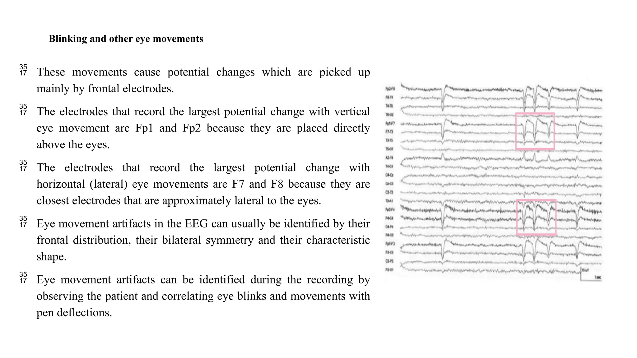
KONGUNADU COLLEGE OF ENGINEERING AND TECHNOLOGY
(AUTONOMOUS)
NAMAKKAL- TRICHY MAIN ROAD, THOTTIAM
DEPARTMENT OF BIOMEDICAL ENGINEERING
Ms. M. Thendral,
Assistant Professor / BME
KNCET
20BM708PE – NEURAL ENGINEERING
(REGULATION-KNCET - UGR2020)
UNIT II – ELECTROENCEPHALOGRAPHY
UNIT II ELECTROENCEPHALOGRAPHY
• Electroencephalography (EEG): General Principles and
Clinical Applications, Neonatal and Paediatric EEG, EEG
Artefacts and Benign Variants, Video EEG monitoring for
epilepsy, Invasive Clinical Neurophysiology in Epilepsy and
movement disorders, Topographic mapping. Frequency
analysis and other quantitative techniques in EEG,
Intraoperative EEG monitoring during carotid endarterectomy
and cardiac surgery, magnetoencephalography
Electroencephalogram
• An electroencephalogram (EEG) is a test that measures electrical
activity in the brain using small, metal discs (electrodes) attached to
the scalp.
• Brain cells communicate via electrical impulses and are active all the
time, even during asleep.
• An EEG can find changes in brain activity that might be useful in
diagnosing brain disorders, especially epilepsy or another seizure
disorder.
BRAIN LOBE
Frontal Lobe
• The frontal lobe is generally where higher executive functions
including emotional regulation, planning, reasoning and problem
solving occur.
Parietal Lobe
• Areas in the parietal lobe are responsible for integrating sensory
information, including touch, temperature, pressure and pain.
Temporal Lobe
• The temporal lobe contains the primary auditory cortex, which
receives auditory information from the ears.
Occipital Lobe
• The occipital lobe is the major visual processing centre in the
brain
BRAIN WAVES
EEG RECORDING SETUP
 Patient cable consists of 21 electrodes and is connected to the 8-channel selector.
 The electrodes are attached to the channel selector in groups of 8 Called Montage
of electrodes.
 The right ear electrodes acts reference electrode for a right brain electrodes and left
ear act as reference electrode for left brain electrode.
 Output from 8 channel connecter goes to differential Amplifier. The output voltage
from the amplifier may either be applied directly to time eight channel display
through filter bank or it may be stored on data on a tape recorder or computer
memory for further processing.
 The system helps to record the potentials generated from Sensory parts of the brain.
To achieve this, output unit is connected with audio, visual and touch stimulus.
EEG RECORDING SETUP
EEG RECORDING SETUP
 It can also measure the time delay between stimulus and response from brain. In addition,
we have a filter bank- Consist of LP, HP and BP filters, they help to removenoise from the
brain waves.
 For the output recording, we can see either pen recorder of CRO.
 Three modes rarely unipolar, bipolar and wilson mode or average mode recording are
used to measure EEG.
 In bipolar technique, the difference in potential between two adjacent electrodes is
measured.
 In the monopolar technique, the potential of each electrode is measured with respect to
a reference electrode attached to ear lobe.
 In the Wilson technique (or) average mode recording techniques the potential is
measured between one of the electrodes (exploring electrode) and the central terminal
which is formed by connecting all electrodes through high, equal resistors to a common
point.
EEG – 10-20 LEAD SYSTEM
 The 10/20 system or International 10/20 system is an
internationally recognized method to describe the location of
scalp electrodes.
 The system is based on the relationship between the location
of an electrode and the underlying area of cerebral cortex.
 The numbers ‘10’ and ‘20’ refer to the fact that the distances
between adjacent electrodes are either 10% or 20% of the total
front- back or right-left distance of the skull.
 Each site has a letter to identify the lobe and a number to
identify the hemisphere location.
Placement of electrodes
• In EEG, electrodes are placed in standard positions on the skull in an
arrangement called 10-20 system, a placement scheme devised by the
International Federation of Societies of EEG. The electrodes in this
arrangement are placed as follows:
• Draw a line on the skull from the nasion, the root of the nose, to the inion,
ossification center (bump) on the occipital lobe.
• Draw a similar line from the left preauricular (ear) point to the right preauricular
point.
• Mark the intersection of these two lines as Cz which is the mid point of the
distance between the nasion and inion (or) the distance between the auricular
points.
• Mark points at 10, 20, 20, 20 and 10% of the total nasion-inion distance. These points
are Fpz, Fz, Cz, Pz and Oz.
Placement of electrodes
 Mark points at 10, 20, 20, 20, 20 and 10% of the total distance between the preauricular
points. These points are T3, C3, Cz, C4 and T4. In these odd numbered points T3 and C3
are on the left and even numbered points C4 and T4 are on the right.
 Measure the distance between Fpz and Oz along the great circle passing through T3 and
mark points at 10, 20, 20, 20, 20 and 10% of this distance. These are the positions of Fp1,
F7, T3, T5 and O1.
 Repeat this procedure on the right side and mark the positions of Fp2, F8, T4, T6 and O2.
 Measure the distance between Fp1 and O1 along the great circle passing through C3 and
mark points at 25% intervals. These points give the positions of F3, C3 and P3. The
ground reference electrode is a metal clip on the earlobe,
 Repeat this procedure on the right side and mark the positions of F4, C4 and P4.
 Check that F7, F3, Fz, F4 and F8 are equidistant along the transverse circle passing
through F7, Fz and F8 and check that T5, P3, Pz, P4 and T6 are equidistant along the
transverse circle passingT5, Pz and T6. In the figure, the positions of the scalp electrodes
are indicated. Further there are nasopharyngeal electrodes Pg1 and Pg2 and ear electrodes
A1 and A2.
APPLICATION OF EEG
EEG helps physicians to diagnose
• The level of consciousness
• Sleep disorders
• Brain death
• Brain tumors
• Epilepsy
• Multiple sclerosis
Level of consciousness
• EEG changes with the level of consciousness.
• Diminished mental activity usually results in a lower frequency and
large amplitude EEG wave.
• EEG has made valuable contribution to the study of sleep physiology.
• The variation of EEG with respect to sleep or the level of
consciousness.,
• REM means rapid eye movement. REM sleep coincides with the
periods of dreaming.
Epilepsy
• Epilepsy is a symptom for brain damage.
• This may due to defects in the birth delivery or head injury during accident
or boxing.
• It may also be due to brain tumor.
• Epilepsy is a disease and is characterised by synchronous discharge of large
groups of neurons, often including the whole brain.
• Epilepsy is divided into two types, grandmal and peritmal.
• The grandmal seizure extends from few seconds to several minutes.
• In the peritmal attack spike type waves are produced with a frequency 3
Hz. and its seizure lasts for 1-20 seconds.
Cerebral death
• EEG displays characteristic features during the application of
anaesthesia.
• As anaesthesia is applied, the brain wave frequency decreases and
the amplitude increases.
• The theta and delta waves appear. In the case of cerebral death (brain
death), EEG shows a permanent absence of brain wave even though
respiration and circulation are maintained.
Brain Tumors
• If the tumor displaces the cortex and if it is large enough, the
electrical activity will be absent in that part of hemisphere.
• since no electric potentials originate in the tumor itself.
• Thus an extinguished or damped EEG over a certain part of cortex can
thus be a sign of a tumor.
EEG Waves for different level of
consciousness
NEONATALAND PAEDIATRIC EEG
 Neonatal and pediatric EEG (Electroencephalography) are specialized forms of brainwave monitoring used to assess the
electrical activity of the brain in infants and children.
 EEG is a non-invasive technique that involves placing electrodes on the scalp to record the electrical signals produced by the
brain's neurons. These signals are then amplified, processed, and displayed as waveforms on a computer screen or paper.
Neonatal EEG:
 Neonatal EEG is performed on newborns and infants, typically up to the age of one month.
 It is used to monitor the brain activity of premature infants or those who are at risk of neurological problems.
 Neonatal EEG can provide valuable information about brain development, detect abnormalities, and aid in diagnosing
conditions such as seizures and other neurological disorders.
 Interpreting neonatal EEG requires specialized knowledge due to the unique patterns of brain activity in newborns.
 Normal sleep patterns, wakefulness, and seizures in neonates differ from those in older children and adults.
Purposes of Neonatal EEG:
• Detection of Seizures: One of the primary purposes of neonatal EEG is to detect and monitor seizures in
newborns. Seizures in neonates can have various causes, such as birth trauma, oxygen deprivation, or
infections. EEG monitoring can help healthcare providers identify seizure activity even if the outward signs
are not obvious.
• Assessment of Brain Health: Neonatal EEG provides information about the overall health and development
of the newborn's brain. Abnormal patterns in the EEG may indicate brain injury, developmental delays, or
other neurological problems.
• Evaluation of Treatment: For newborns who require medical interventions, such as therapeutic hypothermia
(cooling treatment for hypoxic-ischemic encephalopathy), neonatal EEG can help assess the effectiveness of
the treatment and guide ongoing care.
• Prognosis: EEG findings in neonates can offer insights into the long-term prognosis of certain neurological
conditions and guide decisions about treatment and interventions.
Pediatric EEG
 Pediatric EEG (Electroencephalography) is a specialized branch of neurodiagnostics that involves
monitoring and recording the electrical activity of the brain in infants, children, and adolescents.
 This type of EEG is essential for assessing brain function, diagnosing neurological disorders, and guiding
treatment in the pediatric population.
 Pediatric EEG is performed on children, typically from infancy to around 18 years of age.
 It is used to evaluate a wide range of neurological conditions, including epilepsy, developmental delays,
attention disorders, sleep disorders, and other brain-related issues.
 Pediatric EEGs are often used to diagnose and manage epilepsy.
 Epileptic seizures are characterized by abnormal electrical discharges in the brain, which can be detected
and analyzed through EEG recordings.
 EEG can help determine the type of seizures, their frequency, and their origin in the brain, aiding in
treatment planning and medication management.
Purposes of Pediatric EEG:
• Epilepsy Diagnosis and Management: One of the primary uses of pediatric EEG is in the diagnosis and management of
epilepsy. EEG can help identify abnormal electrical discharges in the brain that are characteristic of seizures. It can also
assist in classifying the type of seizures and determining the optimal treatment strategy.
• Evaluation of Neurological Disorders: Pediatric EEG is employed to evaluate various neurological conditions such as
developmental delays, neurodevelopmental disorders (e.g., autism), attention disorders (e.g., ADHD), and movement
disorders. It can provide valuable insights into brain function and abnormalities.
• Sleep Disorders: EEG is utilized to diagnose sleep disorders in children, such as sleep apnea, parasomnias, and disorders
affecting sleep architecture. It helps monitor brain activity during different stages of sleep.
• Monitoring Brain Injury and Recovery: Pediatric EEG is used to monitor brain activity in children who have suffered
head trauma, concussions, or other brain injuries. It can aid in assessing the extent of damage and tracking recovery progress.
• Surgical Planning: For children with certain types of epilepsy that are resistant to medication, EEG can help pinpoint the
specific area of the brain where seizures originate. This information is crucial for planning surgical interventions to remove
or treat the affected brain tissue.
EEG ARTIFACTS
• Physiological artifacts arise from a variety of
body activities that are due to either:
 Movement of head, body or scalp (e.g.
pulsations of the scalp arteries)- that affect the
electrode scalp interface • Bioelectrical
potentials generated within the body from
moving sources (such as eye, tongue, or
pharyngeal muscle movement),
 stationary sources such as the scalp muscles,
heart or sweat glands
 Altered volume conduction due to changes in the
conductance of tissues (scalp, bone, muscle) and
fluids (CSF, blood) between the cerebral cortex
and the recording electrodes.
• Non-physiological artifacts arise
from two main sources:
 external electrical interference from
other power sources such as power
lines or electrical equipment,
 internal electrical malfunctioning of
the recording system, arising from
recording electrodes (electrode
integrity, positioning and
application), cables, amplifiers.
Blinking and other eye movements
 These movements cause potential changes which are picked up
mainly by frontal electrodes.
 The electrodes that record the largest potential change with vertical
eye movement are Fp1 and Fp2 because they are placed directly
above the eyes.
 The electrodes that record the largest potential change with
horizontal (lateral) eye movements are F7 and F8 because they are
closest electrodes that are approximately lateral to the eyes.
 Eye movement artifacts in the EEG can usually be identified by their
frontal distribution, their bilateral symmetry and their characteristic
shape.
 Eye movement artifacts can be identified during the recording by
observing the patient and correlating eye blinks and movements with
pen deflections.
Muscle artifact
 Muscle activity causes very short potentials which
usually recur.
 Muscle artifacts from scalp and face muscles occur
mainly in the frontal and temporal regions.
 Easily identified by its shape and repetition.
 It can be reduced and often eliminated by asking the
patient to relax the jaw or open the mouth slightly, or
change position.
 Distribution that reflects the locations of the muscles
generating it.
 Typical of muscle artifact, it begins and ends abruptly.
•
Cardiac artifacts
 The heart produces 2 types of artifacts
 Electrical
 Mechanical
 Both types are time locked to cardiac contractions and are most
easily identified by their synchronization with complexes in the
ECG channel.
 The electrical artifact actually is the ECG, as recorded from head
electrodes.
 The P wave and T wave are usually not visible, because of the
distance from the heart and the suboptimal axis.
 Essentially, the artifact is a poorly formed QRS complex.
 Most prominent when the neck is short.
 The complex usually is diphasic, but some EEGs may depict it as
either monophasic or triphasic.
Electrical Interference.
 Artifacts due to electrical interference emanates from electrical equipment and nearby power lines.
 Strong interference can cause artifacts even with good recording electrodes and equipment;
 these artifacts are then likely to appear in all channels of all recordings made in the same recording room.
 When recordings are made in an environment with excessive interference such as an intensive care unit or
an operating room, the patient's head and the connections to the EEG machine should be kept as far from
power cables as possible.
 Electrode wires should be straightened and bundled together.
 Equipment other than the EEG machine should be unplugged if feasible.
Electrode pop
 Electrode artifacts usually manifest as one of two disparate
waveforms, brief transients that are limited to one electrode and low
frequency rhythms across a scalp region.
 The brief transients are due to either spontaneous discharging of
electrical potential that was present between the electrode or its
lead.
 The spontaneous discharges are called electrode pops, and they
reflect the ability of the electrode and skin interface to function as a
capacitor and store electrical charge across the electrolyte paste or
gel that holds the electrode in place.
 With the release of the charge there is a change in impedance, and a
sudden potential appears in all channels that include the electrode.
 This potential may be superimposed on the background activity or
replace it. Sometimes more than one pop occurs within a few
seconds. Electrode pop have a characteristic morphology of a very
steep rise and a shallower fall.
Lead movement
 Artifact through an activity that is both unusually high
amplitude and low frequency
 Lead movement has a more disorganized morphology
that does not resemble true EEG activity in any form
 Poor electrode contact or lead movement produces
artifacts with a less conserved morphology than
electrode pop.
 The poor contact produces instability in the
impedance, which leads to sharp or slow waves of
varying morphology and amplitude. These waves may
be rhythmic if poor contact occurs in the context of
rhythmic movements, such as from a tremor.
BENIGN VARIANTS IN EEG
 Benign variants in EEG (Electroencephalography) refer to patterns or
findings in an individual's brainwave activity that are not indicative of
any underlying neurological disorder or pathology.
 EEG is a non-invasive technique used to record the electrical activity
of the brain, and it can sometimes show certain patterns that are
considered normal variations rather than signs of disease.
 These benign variants can sometimes lead to misinterpretation if not
properly understood by medical professionals.
BENIGN VARIANTS IN EEG
• Mu Rhythm: The mu rhythm is a type of brainwave pattern that
appears over the sensorimotor cortex, particularly when an
individual is at rest and not moving. It's often observed as
rhythmic alpha activity (8-13 Hz) and is considered a normal
finding.
• Vertex Waves: Vertex waves are sharp waveforms that occur
during sleep, usually in non-REM sleep. They are often seen in the
central regions of the scalp and are considered a normal findings
during certain sleep stages.
• 6 Hz Spike-and-Wave: This pattern is a type of generalized
spike-and-wave activity that occurs at around 6 Hz. While spike-
and-wave patterns are often associated with epilepsy, the 6 Hz
variant is considered benign and is usually not associated with
seizure disorders.
• Midline Theta: Midline theta refers to the presence of theta
frequency (4-7 Hz) activity in the midline regions of the scalp,
particularly during drowsiness or light sleep. This is a normal
variant and not necessarily indicative of pathology.
• Rhythmic Midline Theta: Rhythmic theta (4-7 Hz) activity over the
midline regions of the brain can be observed during meditation or
focused attention. This is considered a benign variant and is not
necessarily indicative of pathology.
• Alpha Variant Patterns: There are various subtypes of alpha activity
that can appear in different conditions, such as "alpha variants" that
are seen in certain states like drowsiness or hyperventilation. These
can include alpha spindles, lateralized rhythmic alpha activity, and
others.
• K Complexes and Sleep Spindles: K complexes and sleep spindles
are common benign findings during certain stages of sleep. K
complexes are sharp negative waveforms, while sleep spindles are
bursts of rhythmic activity. These patterns are associated with sleep
maintenance and are not typically indicative of neurological issues.
• Rhythmic Delta Activity in Children: In young children,
it's common to observe rhythmic delta activity (1-4 Hz)
during wakefulness. This activity gradually diminishes as
the child grows and matures.
VIDEO EEG MONITORING FOR EPILEPSY
• Video-electroencephalography (EEG) monitoring is a diagnostic technique commonly used in patients with epileptic
seizures.
• Epilepsy is a chronic neurological disorder that affects the brain causing repeated seizures or "fits" in the patient
which makes them collapse. Video-EEG Monitoring is also known as EEG telemetry or EEG monitoring.
• Video-EEG is a non-invasive procedure, which records the electrical activity of the brain during seizures for a
prolonged period (3 to 5 days) with simultaneous video recording. It also records the sounds made by the person
during the testing period.
• Video-EEG recording helps to locate the exact brain region where the seizures start and hence provides vital
information that helps with both the diagnosis and treatment.
• Prolonged video-EEG monitoring should be performed for any patient who continues to have frequent seizures
attacks despite taking antiepileptic drugs.
• Correlation between the recorded video and EEG activity can help the doctor to determine the accurate count of
seizures and episodic events which may vary from time to time. It is a gold standard diagnosis for psychogenic non-
epileptic seizures (PNES), because video-EEG monitoring differentiates PNES and epileptic seizures using video
analysis.
Types of Video-EEG Monitoring
Ambulatory EEG Monitoring
When video-EEG monitoring is performed in
the home setting it is called ambulatory EEG
monitoring. The advantages of performing
video-EEG at home are - It is less expensive
than inpatient monitoring. Its ability to record
continuously for up to 72 hours. It captures the
patient’s natural sleep pattern because most
seizures and their symptoms occur at night
times. Patients find it up to their satisfaction
because the testing is performed in an
environment comfortable to them.
Inpatient EEG Monitoring
• Video-EEG monitoring is performed
in the Epilepsy Monitoring Unit
(EMU). The patients should be
admitted to the hospital for
continuous monitoring. The length of
the patient’s stay in the hospital will
depend on the time needed to monitor
seizure attacks (on an average the stay
will be 3 to 5 days).
VIDEO EEG MONITORING FOR EPILEPSY
There are 2 main components used in video-EEG monitoring
EEG:
• As with any routine EEG procedure, electrodes are placed
on the scalp with glue, and the head with electrodes is
covered by a gauze dressing or cap.
• EEG recording is monitored by a computer that records
the patient’s seizure activity for a longer period of time
(24-hour recording).
• This recording allows the doctors to compare the EEG
activity with the patient’s physical as well as mental
behavior during the test.
• During the monitoring, the doctors may lower seizure
medication to make seizure attacks likely to occur or
apply other seizure triggers like sleep deprivation
techniques, exercise and flash lights and they may ask you
to stay up late at night.
Video Recorder
• A camera is used to visually record the patient's physical
activities continuously at the same time the EEG is recording
the brain activity.
• Patients will be asked to sit on top of the bed, within camera
view to capture good video recordings.
• There is a TV monitor with a split screen in the patient's
room. The screen shows the EEG on one side and the video
recording on the other side. There is a connected monitor at
the nurse's station which shows the same recording so that
the patient can be monitored at all times.
• Epilepsy doctors will review and evaluate the raw EEG with
the video data in the epilepsy monitoring units. A seizure
button is given to the patients to press when they are about to
have seizures. So, based on the pressed event, doctors will
review the recording and give special attention when the
seizures begin.
INVASIVE CLINICAL NEUROPHYSIOLOGY IN EPILEPSY AND MOVEMENT DISORDERS
 Despite the wide range of medical treatments for epilepsy currently available, approximately 30 to 35
percent of persons with epilepsy continue to experience seizures.
 For these patients with medically refractory epilepsy, surgical removal of the epileptogenic brain tissue is
often an effective treatment. Surgical resection is predicated on the ability to identify the seizure focus or
epileptogenic zone.
 This area of the brain is responsible for the generation of seizures, and its removal or disconnection results
in the cessation of seizures.
 Techniques for identifying the seizure focus are continually evolving and being refined.
 Although a number of components typically constitute the contemporary presurgical evaluation of patients
with medically refractory epilepsy, ictal electrophysiology remains the “gold standard” in this endeavour.
 When the scalp-recorded electroencephalogram (EEG) fails to provide adequate electrophysiologic
localization of the epileptogenic region, or when it suggests localization that conflicts with the findings from
other elements of the preoperative evaluation (e.g., neuroimaging), invasive recordings are necessary to
identify clearly the brain region from which seizures arise.
TOPOGRAPHIC MAPPING IN EEG
 Topographic mapping in EEG refers to the process of visualizing and analyzing the distribution of electrical
brain activity across different regions of the scalp.
 This technique involves creating color-coded maps or graphs that represent the strength or amplitude of
specific brainwave frequencies at various electrode locations on the scalp.
 Topographic maps provide valuable information about how brain activity is spatially distributed, helping to
identify patterns, abnormalities, and differences in activity across different brain regions.
 Topographic mapping in EEG provides insights into the spatial distribution of brain activity, diagnosing
neurological conditions, and researching cognitive processes.
 However, its accuracy relies on electrode placement, data quality, and knowledge of EEG principles.
 Software tools, such as EEG analysis software, facilitate the generation and interpretation of topographic
maps.
A simplified block diagram illustrating the process of topographic mapping in EEG
1. EEG Data Acquisition
└── Electrode Placement
└── Data Collection
2. Preprocessing
└── Filtering (e.g., remove noise, bandpass filtering)
└── Referencing (common average, linked mastoids, etc.)
3. Epoching
└── Divide Continuous Data into Epochs (time segments)
└── Each Epoch Represents a Specific Time Window
4. Data Analysis
└── Feature Extraction (e.g., power spectral density)
└── Compute Brain Wave Frequencies (alpha, beta, etc.)
5. Interpolation
└── estimate the electrical activity in regions
6. Topographic Mapping
└── Electrode Coordinates (e.g., 10-20 system)
└── EEG Data for Each Epoch and Frequency Band
7. Interpretation
└── Analyze Topographic Maps for Patterns
└── Compare with Normative Data or Control Group
└── Identify Abnormal Brain Activity or Patterns
8. Clinical or Research Applications
└── Diagnosis of Neurological Conditions
└── Study of Cognitive Processes
FREQUENCY ANALYSIS
 Frequency analysis in EEG involves the decomposition of raw EEG signals into different frequency
components to assess the brain's electrical activity at various frequency ranges.
 EEG signals are complex and consist of oscillations that can be categorized into different frequency bands,
each associated with specific brain states, cognitive processes, and physiological activities.
 Frequency analysis helps reveal patterns, abnormalities, and information about brain function.
• Raw EEG Signals
• EEG records the electrical activity of the brain as voltage fluctuations over time.
• These signals are sampled at a specific rate (sampling frequency) to capture the temporal changes in brain
activity.
FREQUENCY ANALYSIS
Frequency Analysis Steps:
 Segmenting Data: EEG recordings are often divided into short segments to ensure
stationarity (signal properties don't change within the segment).
 Windowing: Each segment is multiplied by a windowing function to reduce spectral
leakage.
 FFT Calculation: The FFT is applied to each windowed segment, generating a
frequency spectrum.
 Averaging: Spectra from multiple segments can be averaged to improve signal-to-
noise ratio.
OTHER QUANTITATIVE TECHNIQUES IN EEG
Power Spectral Density (PSD):
PSD analysis decomposes EEG signals into their frequency components, revealing the power (amplitude
squared) of each frequency band.
It provides insight into the distribution of power across different frequency ranges (e.g., delta, theta, alpha, beta,
gamma), which can relate to different cognitive states and processes.
Steps in PSD Analysis:
Data Preprocessing: Raw EEG data is often preprocessed to remove artifacts, noise, and other sources of
interference that could affect the accuracy of the frequency analysis.
Segmentation: The continuous EEG signal is divided into shorter segments (epochs), usually overlapping, to
ensure stationary signal conditions. This helps capture the dynamic changes in frequency content over time.
OTHER QUANTITATIVE TECHNIQUES IN EEG
• Windowing: Each epoch is often multiplied by a windowing function to reduce spectral leakage and
improve frequency resolution during the FFT.
• FFT Method: The Fast Fourier Transform (FFT) is applied to each epoch to convert the time-domain EEG
data into the frequency domain. It provide a more accurate estimate of the power spectral density.
• Averaging: The power values from individual epochs are averaged to obtain a more stable estimate of the
power at each frequency bin.
• Normalization: PSD values are often normalized to account for differences in signal length, windowing,
and other factors. Common normalization methods include dividing by the total power or the integral of the
PSD curve.
INTRAOPERATIVE EEG MONITORING DURING CAROTID ENDARTERECTOMY
AND CARDIAC SURGERY
 Interoperative EEG (electroencephalography) monitoring is a specialized medical procedure performed during
surgery, particularly brain-related surgeries.
 It involves the real-time recording and analysis of electrical activity in the brain using electrodes placed on the scalp.
 The primary purpose of interoperative EEG monitoring is to monitor brain function and detect any abnormalities or
changes that may occur during the surgical procedure.
 Intraoperative EEG monitoring is a technique used during surgery to monitor the electrical activity of the brain.
 The data collected from intraoperative EEG monitoring aids in making informed decisions and adjusting the surgical
approach as needed to minimize risks and optimize patient outcomes.
 Interoperative EEG monitoring is particularly valuable in surgeries involving epilepsy treatment, brain tumour
removal, and other procedures that carry a risk of affecting brain function.
 It allows the surgical team to make informed decisions during the operation, potentially reducing the risk of
postoperative complications and optimizing patient outcomes.
Interoperative EEG monitoring during carotid endarterectomy
 A carotid endarterectomy is a surgical procedure performed to remove plaque buildup from the carotid
arteries.
 These arteries, located in the neck, supply blood to the brain. Plaque accumulation in these arteries can lead
to reduced blood flow and increase the risk of stroke.
 Carotid endarterectomy is performed to reduce the risk of stroke in individuals with significant carotid
artery stenosis (narrowing).
 The procedure aims to improve blood flow to the brain and decrease the chances of a stroke caused by a
piece of plaque breaking off and blocking blood flow.
Interoperative EEG monitoring during cardiac surgery
 Cardiac surgery is a medical procedure performed on the heart to treat various heart conditions or diseases.
 It can involve repairing damaged valves, bypassing blocked arteries, correcting congenital heart defects, and
more.
 It's typically done by a cardiothoracic surgeon in a specialized operating room equipped with advanced
medical technology.
Types of Cardiac Surgery:
• There are several types of cardiac surgeries, including coronary artery bypass grafting (CABG), heart valve
repair or replacement, heart transplant, congenital heart defect repair, and surgery to treat heart rhythm
disorders like atrial fibrillation
MAGNETOENCEPHALOGRAPHY
• Magnetoencephalography (MEG) is a non-invasive neuroimaging technique used to measure and map
the magnetic fields generated by neuronal activity in the brain.
 It provides insights into the temporal and spatial dynamics of brain function with high temporal
resolution, to study the real-time neural processes that underlie various cognitive functions, sensory
perceptions, and motor activities.
 MEG operates on the principle of detecting the tiny magnetic fields produced by the electrical currents
generated by active neurons in the brain.
 These magnetic fields are extremely weak and are measured using highly sensitive sensors called
superconducting quantum interference devices (SQUIDs).
MAGNETOENCEPHALOGRAPHY
 MEG complements other neuroimaging techniques like functional
magnetic resonance imaging (fMRI) and electroencephalography
(EEG), offering unique advantages such as the ability to directly
measure neural activity with millisecond precision and excellent
spatial resolution.
 During a typical MEG session, the subject wears a helmet-like
device containing the sensors. The subject is presented with various
tasks or stimuli while their brain activity is recorded.
 The collected data is then analyzed to create maps of brain activity,
known as MEG source localization, which help researchers pinpoint
the locations and timing of neural activations.
 The spatial distributions of the magnetic fields are analyzed to
localize the sources of the activity within the brain, and the
locations of the sources are superimposed on anatomical images,
such as MRI, to provide information about both the structure and
function of the brain.
Principle features of MEG
 MEG is a direct measure of brain function, unlike functional measures such as fMRI, PET and SPECT that
are secondary measures of brain function reflecting brain metabolism.
 MEG has a very high temporal resolution & excellent spatial resolution
 MEG is completely non-invasive. It does not require the injection of isotopes or exposure to X-rays or
magnetic fields
 MEG is complementary to other modalities, the information provided by each modality adds to the full
picture.
MAGNETOENCEPHALOGRAPHY
• Stimulus Interface: Researchers design tasks or stimuli that engage specific cognitive functions. Subjects are
presented with these tasks while their brain activity is recorded. For conducting experiments, a system for
presenting visual, auditory, or other sensory stimuli to the subject is required. This could include displays,
headphones, and response buttons for the subject to interact with the presented tasks.
• Neural Activity (Electrical Currents): Neurons in the brain generate electrical currents as they communicate and
process information.
• Magnetic Fields Generation: The changing electrical currents produce associated weak magnetic fields around
the neurons.
• Sensor Array (SQUIDs): An array of superconducting quantum interference devices (SQUIDs) is placed around
the subject's head. These sensors are sensitive to the weak magnetic fields generated by neural activity. The core
component of an MEG system is an array of superconducting quantum interference devices (SQUIDs). These
highly sensitive magnetometers are designed to detect extremely weak magnetic fields down to picotesla levels.
MAGNETOENCEPHALOGRAPHY
• Dewar: The SQUID sensors need to be kept at very low temperatures to maintain their superconducting
state and achieve maximum sensitivity. A dewar, typically filled with liquid helium or a mixture of helium
and nitrogen, houses the sensors and keeps them cooled.
• Helmet or Sensor Array: The sensor array is usually organized in a helmet-like configuration, which the
subject wears during the MEG session. The array contains numerous SQUID sensors distributed around
the head to capture the magnetic fields from different brain regions.
• Magnetic Shielding room: MEG systems require shielding to minimize interference from external
electromagnetic fields, such as the Earth's magnetic field and electromagnetic noise from the environment.
Shielding helps ensure the accuracy of the measurements. The MEG apparatus is typically housed within a
shielded room to minimize external electromagnetic interference. This room, known as a magnetically
shielded room (MSR), provides a controlled environment for MEG measurements.
• Magnetic Field Detection and Signal Amplification: The SQUIDs detect the magnetic fields and convert
them into electrical signals, which are then amplified for further processing.

NEURAL ENGINEERING UNIT II ELECTROENCEPHALOGRAPHY

  • 1.
    KONGUNADU COLLEGE OFENGINEERING AND TECHNOLOGY (AUTONOMOUS) NAMAKKAL- TRICHY MAIN ROAD, THOTTIAM DEPARTMENT OF BIOMEDICAL ENGINEERING Ms. M. Thendral, Assistant Professor / BME KNCET 20BM708PE – NEURAL ENGINEERING (REGULATION-KNCET - UGR2020) UNIT II – ELECTROENCEPHALOGRAPHY
  • 2.
    UNIT II ELECTROENCEPHALOGRAPHY •Electroencephalography (EEG): General Principles and Clinical Applications, Neonatal and Paediatric EEG, EEG Artefacts and Benign Variants, Video EEG monitoring for epilepsy, Invasive Clinical Neurophysiology in Epilepsy and movement disorders, Topographic mapping. Frequency analysis and other quantitative techniques in EEG, Intraoperative EEG monitoring during carotid endarterectomy and cardiac surgery, magnetoencephalography
  • 3.
    Electroencephalogram • An electroencephalogram(EEG) is a test that measures electrical activity in the brain using small, metal discs (electrodes) attached to the scalp. • Brain cells communicate via electrical impulses and are active all the time, even during asleep. • An EEG can find changes in brain activity that might be useful in diagnosing brain disorders, especially epilepsy or another seizure disorder.
  • 4.
    BRAIN LOBE Frontal Lobe •The frontal lobe is generally where higher executive functions including emotional regulation, planning, reasoning and problem solving occur. Parietal Lobe • Areas in the parietal lobe are responsible for integrating sensory information, including touch, temperature, pressure and pain. Temporal Lobe • The temporal lobe contains the primary auditory cortex, which receives auditory information from the ears. Occipital Lobe • The occipital lobe is the major visual processing centre in the brain
  • 5.
  • 6.
    EEG RECORDING SETUP Patient cable consists of 21 electrodes and is connected to the 8-channel selector.  The electrodes are attached to the channel selector in groups of 8 Called Montage of electrodes.  The right ear electrodes acts reference electrode for a right brain electrodes and left ear act as reference electrode for left brain electrode.  Output from 8 channel connecter goes to differential Amplifier. The output voltage from the amplifier may either be applied directly to time eight channel display through filter bank or it may be stored on data on a tape recorder or computer memory for further processing.  The system helps to record the potentials generated from Sensory parts of the brain. To achieve this, output unit is connected with audio, visual and touch stimulus.
  • 7.
  • 8.
    EEG RECORDING SETUP It can also measure the time delay between stimulus and response from brain. In addition, we have a filter bank- Consist of LP, HP and BP filters, they help to removenoise from the brain waves.  For the output recording, we can see either pen recorder of CRO.  Three modes rarely unipolar, bipolar and wilson mode or average mode recording are used to measure EEG.  In bipolar technique, the difference in potential between two adjacent electrodes is measured.  In the monopolar technique, the potential of each electrode is measured with respect to a reference electrode attached to ear lobe.  In the Wilson technique (or) average mode recording techniques the potential is measured between one of the electrodes (exploring electrode) and the central terminal which is formed by connecting all electrodes through high, equal resistors to a common point.
  • 9.
    EEG – 10-20LEAD SYSTEM  The 10/20 system or International 10/20 system is an internationally recognized method to describe the location of scalp electrodes.  The system is based on the relationship between the location of an electrode and the underlying area of cerebral cortex.  The numbers ‘10’ and ‘20’ refer to the fact that the distances between adjacent electrodes are either 10% or 20% of the total front- back or right-left distance of the skull.  Each site has a letter to identify the lobe and a number to identify the hemisphere location.
  • 10.
    Placement of electrodes •In EEG, electrodes are placed in standard positions on the skull in an arrangement called 10-20 system, a placement scheme devised by the International Federation of Societies of EEG. The electrodes in this arrangement are placed as follows: • Draw a line on the skull from the nasion, the root of the nose, to the inion, ossification center (bump) on the occipital lobe. • Draw a similar line from the left preauricular (ear) point to the right preauricular point. • Mark the intersection of these two lines as Cz which is the mid point of the distance between the nasion and inion (or) the distance between the auricular points. • Mark points at 10, 20, 20, 20 and 10% of the total nasion-inion distance. These points are Fpz, Fz, Cz, Pz and Oz.
  • 11.
  • 12.
    Placement of electrodes Mark points at 10, 20, 20, 20, 20 and 10% of the total distance between the preauricular points. These points are T3, C3, Cz, C4 and T4. In these odd numbered points T3 and C3 are on the left and even numbered points C4 and T4 are on the right.  Measure the distance between Fpz and Oz along the great circle passing through T3 and mark points at 10, 20, 20, 20, 20 and 10% of this distance. These are the positions of Fp1, F7, T3, T5 and O1.  Repeat this procedure on the right side and mark the positions of Fp2, F8, T4, T6 and O2.  Measure the distance between Fp1 and O1 along the great circle passing through C3 and mark points at 25% intervals. These points give the positions of F3, C3 and P3. The ground reference electrode is a metal clip on the earlobe,  Repeat this procedure on the right side and mark the positions of F4, C4 and P4.  Check that F7, F3, Fz, F4 and F8 are equidistant along the transverse circle passing through F7, Fz and F8 and check that T5, P3, Pz, P4 and T6 are equidistant along the transverse circle passingT5, Pz and T6. In the figure, the positions of the scalp electrodes are indicated. Further there are nasopharyngeal electrodes Pg1 and Pg2 and ear electrodes A1 and A2.
  • 13.
    APPLICATION OF EEG EEGhelps physicians to diagnose • The level of consciousness • Sleep disorders • Brain death • Brain tumors • Epilepsy • Multiple sclerosis
  • 14.
    Level of consciousness •EEG changes with the level of consciousness. • Diminished mental activity usually results in a lower frequency and large amplitude EEG wave. • EEG has made valuable contribution to the study of sleep physiology. • The variation of EEG with respect to sleep or the level of consciousness., • REM means rapid eye movement. REM sleep coincides with the periods of dreaming.
  • 15.
    Epilepsy • Epilepsy isa symptom for brain damage. • This may due to defects in the birth delivery or head injury during accident or boxing. • It may also be due to brain tumor. • Epilepsy is a disease and is characterised by synchronous discharge of large groups of neurons, often including the whole brain. • Epilepsy is divided into two types, grandmal and peritmal. • The grandmal seizure extends from few seconds to several minutes. • In the peritmal attack spike type waves are produced with a frequency 3 Hz. and its seizure lasts for 1-20 seconds.
  • 16.
    Cerebral death • EEGdisplays characteristic features during the application of anaesthesia. • As anaesthesia is applied, the brain wave frequency decreases and the amplitude increases. • The theta and delta waves appear. In the case of cerebral death (brain death), EEG shows a permanent absence of brain wave even though respiration and circulation are maintained.
  • 17.
    Brain Tumors • Ifthe tumor displaces the cortex and if it is large enough, the electrical activity will be absent in that part of hemisphere. • since no electric potentials originate in the tumor itself. • Thus an extinguished or damped EEG over a certain part of cortex can thus be a sign of a tumor.
  • 18.
    EEG Waves fordifferent level of consciousness
  • 19.
    NEONATALAND PAEDIATRIC EEG Neonatal and pediatric EEG (Electroencephalography) are specialized forms of brainwave monitoring used to assess the electrical activity of the brain in infants and children.  EEG is a non-invasive technique that involves placing electrodes on the scalp to record the electrical signals produced by the brain's neurons. These signals are then amplified, processed, and displayed as waveforms on a computer screen or paper. Neonatal EEG:  Neonatal EEG is performed on newborns and infants, typically up to the age of one month.  It is used to monitor the brain activity of premature infants or those who are at risk of neurological problems.  Neonatal EEG can provide valuable information about brain development, detect abnormalities, and aid in diagnosing conditions such as seizures and other neurological disorders.  Interpreting neonatal EEG requires specialized knowledge due to the unique patterns of brain activity in newborns.  Normal sleep patterns, wakefulness, and seizures in neonates differ from those in older children and adults.
  • 20.
    Purposes of NeonatalEEG: • Detection of Seizures: One of the primary purposes of neonatal EEG is to detect and monitor seizures in newborns. Seizures in neonates can have various causes, such as birth trauma, oxygen deprivation, or infections. EEG monitoring can help healthcare providers identify seizure activity even if the outward signs are not obvious. • Assessment of Brain Health: Neonatal EEG provides information about the overall health and development of the newborn's brain. Abnormal patterns in the EEG may indicate brain injury, developmental delays, or other neurological problems. • Evaluation of Treatment: For newborns who require medical interventions, such as therapeutic hypothermia (cooling treatment for hypoxic-ischemic encephalopathy), neonatal EEG can help assess the effectiveness of the treatment and guide ongoing care. • Prognosis: EEG findings in neonates can offer insights into the long-term prognosis of certain neurological conditions and guide decisions about treatment and interventions.
  • 21.
    Pediatric EEG  PediatricEEG (Electroencephalography) is a specialized branch of neurodiagnostics that involves monitoring and recording the electrical activity of the brain in infants, children, and adolescents.  This type of EEG is essential for assessing brain function, diagnosing neurological disorders, and guiding treatment in the pediatric population.  Pediatric EEG is performed on children, typically from infancy to around 18 years of age.  It is used to evaluate a wide range of neurological conditions, including epilepsy, developmental delays, attention disorders, sleep disorders, and other brain-related issues.  Pediatric EEGs are often used to diagnose and manage epilepsy.  Epileptic seizures are characterized by abnormal electrical discharges in the brain, which can be detected and analyzed through EEG recordings.  EEG can help determine the type of seizures, their frequency, and their origin in the brain, aiding in treatment planning and medication management.
  • 22.
    Purposes of PediatricEEG: • Epilepsy Diagnosis and Management: One of the primary uses of pediatric EEG is in the diagnosis and management of epilepsy. EEG can help identify abnormal electrical discharges in the brain that are characteristic of seizures. It can also assist in classifying the type of seizures and determining the optimal treatment strategy. • Evaluation of Neurological Disorders: Pediatric EEG is employed to evaluate various neurological conditions such as developmental delays, neurodevelopmental disorders (e.g., autism), attention disorders (e.g., ADHD), and movement disorders. It can provide valuable insights into brain function and abnormalities. • Sleep Disorders: EEG is utilized to diagnose sleep disorders in children, such as sleep apnea, parasomnias, and disorders affecting sleep architecture. It helps monitor brain activity during different stages of sleep. • Monitoring Brain Injury and Recovery: Pediatric EEG is used to monitor brain activity in children who have suffered head trauma, concussions, or other brain injuries. It can aid in assessing the extent of damage and tracking recovery progress. • Surgical Planning: For children with certain types of epilepsy that are resistant to medication, EEG can help pinpoint the specific area of the brain where seizures originate. This information is crucial for planning surgical interventions to remove or treat the affected brain tissue.
  • 23.
    EEG ARTIFACTS • Physiologicalartifacts arise from a variety of body activities that are due to either:  Movement of head, body or scalp (e.g. pulsations of the scalp arteries)- that affect the electrode scalp interface • Bioelectrical potentials generated within the body from moving sources (such as eye, tongue, or pharyngeal muscle movement),  stationary sources such as the scalp muscles, heart or sweat glands  Altered volume conduction due to changes in the conductance of tissues (scalp, bone, muscle) and fluids (CSF, blood) between the cerebral cortex and the recording electrodes. • Non-physiological artifacts arise from two main sources:  external electrical interference from other power sources such as power lines or electrical equipment,  internal electrical malfunctioning of the recording system, arising from recording electrodes (electrode integrity, positioning and application), cables, amplifiers.
  • 24.
    Blinking and othereye movements  These movements cause potential changes which are picked up mainly by frontal electrodes.  The electrodes that record the largest potential change with vertical eye movement are Fp1 and Fp2 because they are placed directly above the eyes.  The electrodes that record the largest potential change with horizontal (lateral) eye movements are F7 and F8 because they are closest electrodes that are approximately lateral to the eyes.  Eye movement artifacts in the EEG can usually be identified by their frontal distribution, their bilateral symmetry and their characteristic shape.  Eye movement artifacts can be identified during the recording by observing the patient and correlating eye blinks and movements with pen deflections.
  • 25.
    Muscle artifact  Muscleactivity causes very short potentials which usually recur.  Muscle artifacts from scalp and face muscles occur mainly in the frontal and temporal regions.  Easily identified by its shape and repetition.  It can be reduced and often eliminated by asking the patient to relax the jaw or open the mouth slightly, or change position.  Distribution that reflects the locations of the muscles generating it.  Typical of muscle artifact, it begins and ends abruptly. •
  • 26.
    Cardiac artifacts  Theheart produces 2 types of artifacts  Electrical  Mechanical  Both types are time locked to cardiac contractions and are most easily identified by their synchronization with complexes in the ECG channel.  The electrical artifact actually is the ECG, as recorded from head electrodes.  The P wave and T wave are usually not visible, because of the distance from the heart and the suboptimal axis.  Essentially, the artifact is a poorly formed QRS complex.  Most prominent when the neck is short.  The complex usually is diphasic, but some EEGs may depict it as either monophasic or triphasic.
  • 27.
    Electrical Interference.  Artifactsdue to electrical interference emanates from electrical equipment and nearby power lines.  Strong interference can cause artifacts even with good recording electrodes and equipment;  these artifacts are then likely to appear in all channels of all recordings made in the same recording room.  When recordings are made in an environment with excessive interference such as an intensive care unit or an operating room, the patient's head and the connections to the EEG machine should be kept as far from power cables as possible.  Electrode wires should be straightened and bundled together.  Equipment other than the EEG machine should be unplugged if feasible.
  • 28.
    Electrode pop  Electrodeartifacts usually manifest as one of two disparate waveforms, brief transients that are limited to one electrode and low frequency rhythms across a scalp region.  The brief transients are due to either spontaneous discharging of electrical potential that was present between the electrode or its lead.  The spontaneous discharges are called electrode pops, and they reflect the ability of the electrode and skin interface to function as a capacitor and store electrical charge across the electrolyte paste or gel that holds the electrode in place.  With the release of the charge there is a change in impedance, and a sudden potential appears in all channels that include the electrode.  This potential may be superimposed on the background activity or replace it. Sometimes more than one pop occurs within a few seconds. Electrode pop have a characteristic morphology of a very steep rise and a shallower fall.
  • 29.
    Lead movement  Artifactthrough an activity that is both unusually high amplitude and low frequency  Lead movement has a more disorganized morphology that does not resemble true EEG activity in any form  Poor electrode contact or lead movement produces artifacts with a less conserved morphology than electrode pop.  The poor contact produces instability in the impedance, which leads to sharp or slow waves of varying morphology and amplitude. These waves may be rhythmic if poor contact occurs in the context of rhythmic movements, such as from a tremor.
  • 30.
    BENIGN VARIANTS INEEG  Benign variants in EEG (Electroencephalography) refer to patterns or findings in an individual's brainwave activity that are not indicative of any underlying neurological disorder or pathology.  EEG is a non-invasive technique used to record the electrical activity of the brain, and it can sometimes show certain patterns that are considered normal variations rather than signs of disease.  These benign variants can sometimes lead to misinterpretation if not properly understood by medical professionals.
  • 31.
    BENIGN VARIANTS INEEG • Mu Rhythm: The mu rhythm is a type of brainwave pattern that appears over the sensorimotor cortex, particularly when an individual is at rest and not moving. It's often observed as rhythmic alpha activity (8-13 Hz) and is considered a normal finding. • Vertex Waves: Vertex waves are sharp waveforms that occur during sleep, usually in non-REM sleep. They are often seen in the central regions of the scalp and are considered a normal findings during certain sleep stages. • 6 Hz Spike-and-Wave: This pattern is a type of generalized spike-and-wave activity that occurs at around 6 Hz. While spike- and-wave patterns are often associated with epilepsy, the 6 Hz variant is considered benign and is usually not associated with seizure disorders. • Midline Theta: Midline theta refers to the presence of theta frequency (4-7 Hz) activity in the midline regions of the scalp, particularly during drowsiness or light sleep. This is a normal variant and not necessarily indicative of pathology. • Rhythmic Midline Theta: Rhythmic theta (4-7 Hz) activity over the midline regions of the brain can be observed during meditation or focused attention. This is considered a benign variant and is not necessarily indicative of pathology. • Alpha Variant Patterns: There are various subtypes of alpha activity that can appear in different conditions, such as "alpha variants" that are seen in certain states like drowsiness or hyperventilation. These can include alpha spindles, lateralized rhythmic alpha activity, and others. • K Complexes and Sleep Spindles: K complexes and sleep spindles are common benign findings during certain stages of sleep. K complexes are sharp negative waveforms, while sleep spindles are bursts of rhythmic activity. These patterns are associated with sleep maintenance and are not typically indicative of neurological issues. • Rhythmic Delta Activity in Children: In young children, it's common to observe rhythmic delta activity (1-4 Hz) during wakefulness. This activity gradually diminishes as the child grows and matures.
  • 32.
    VIDEO EEG MONITORINGFOR EPILEPSY • Video-electroencephalography (EEG) monitoring is a diagnostic technique commonly used in patients with epileptic seizures. • Epilepsy is a chronic neurological disorder that affects the brain causing repeated seizures or "fits" in the patient which makes them collapse. Video-EEG Monitoring is also known as EEG telemetry or EEG monitoring. • Video-EEG is a non-invasive procedure, which records the electrical activity of the brain during seizures for a prolonged period (3 to 5 days) with simultaneous video recording. It also records the sounds made by the person during the testing period. • Video-EEG recording helps to locate the exact brain region where the seizures start and hence provides vital information that helps with both the diagnosis and treatment. • Prolonged video-EEG monitoring should be performed for any patient who continues to have frequent seizures attacks despite taking antiepileptic drugs. • Correlation between the recorded video and EEG activity can help the doctor to determine the accurate count of seizures and episodic events which may vary from time to time. It is a gold standard diagnosis for psychogenic non- epileptic seizures (PNES), because video-EEG monitoring differentiates PNES and epileptic seizures using video analysis.
  • 33.
    Types of Video-EEGMonitoring Ambulatory EEG Monitoring When video-EEG monitoring is performed in the home setting it is called ambulatory EEG monitoring. The advantages of performing video-EEG at home are - It is less expensive than inpatient monitoring. Its ability to record continuously for up to 72 hours. It captures the patient’s natural sleep pattern because most seizures and their symptoms occur at night times. Patients find it up to their satisfaction because the testing is performed in an environment comfortable to them. Inpatient EEG Monitoring • Video-EEG monitoring is performed in the Epilepsy Monitoring Unit (EMU). The patients should be admitted to the hospital for continuous monitoring. The length of the patient’s stay in the hospital will depend on the time needed to monitor seizure attacks (on an average the stay will be 3 to 5 days).
  • 34.
  • 35.
    There are 2main components used in video-EEG monitoring EEG: • As with any routine EEG procedure, electrodes are placed on the scalp with glue, and the head with electrodes is covered by a gauze dressing or cap. • EEG recording is monitored by a computer that records the patient’s seizure activity for a longer period of time (24-hour recording). • This recording allows the doctors to compare the EEG activity with the patient’s physical as well as mental behavior during the test. • During the monitoring, the doctors may lower seizure medication to make seizure attacks likely to occur or apply other seizure triggers like sleep deprivation techniques, exercise and flash lights and they may ask you to stay up late at night. Video Recorder • A camera is used to visually record the patient's physical activities continuously at the same time the EEG is recording the brain activity. • Patients will be asked to sit on top of the bed, within camera view to capture good video recordings. • There is a TV monitor with a split screen in the patient's room. The screen shows the EEG on one side and the video recording on the other side. There is a connected monitor at the nurse's station which shows the same recording so that the patient can be monitored at all times. • Epilepsy doctors will review and evaluate the raw EEG with the video data in the epilepsy monitoring units. A seizure button is given to the patients to press when they are about to have seizures. So, based on the pressed event, doctors will review the recording and give special attention when the seizures begin.
  • 36.
    INVASIVE CLINICAL NEUROPHYSIOLOGYIN EPILEPSY AND MOVEMENT DISORDERS  Despite the wide range of medical treatments for epilepsy currently available, approximately 30 to 35 percent of persons with epilepsy continue to experience seizures.  For these patients with medically refractory epilepsy, surgical removal of the epileptogenic brain tissue is often an effective treatment. Surgical resection is predicated on the ability to identify the seizure focus or epileptogenic zone.  This area of the brain is responsible for the generation of seizures, and its removal or disconnection results in the cessation of seizures.  Techniques for identifying the seizure focus are continually evolving and being refined.  Although a number of components typically constitute the contemporary presurgical evaluation of patients with medically refractory epilepsy, ictal electrophysiology remains the “gold standard” in this endeavour.  When the scalp-recorded electroencephalogram (EEG) fails to provide adequate electrophysiologic localization of the epileptogenic region, or when it suggests localization that conflicts with the findings from other elements of the preoperative evaluation (e.g., neuroimaging), invasive recordings are necessary to identify clearly the brain region from which seizures arise.
  • 37.
    TOPOGRAPHIC MAPPING INEEG  Topographic mapping in EEG refers to the process of visualizing and analyzing the distribution of electrical brain activity across different regions of the scalp.  This technique involves creating color-coded maps or graphs that represent the strength or amplitude of specific brainwave frequencies at various electrode locations on the scalp.  Topographic maps provide valuable information about how brain activity is spatially distributed, helping to identify patterns, abnormalities, and differences in activity across different brain regions.  Topographic mapping in EEG provides insights into the spatial distribution of brain activity, diagnosing neurological conditions, and researching cognitive processes.  However, its accuracy relies on electrode placement, data quality, and knowledge of EEG principles.  Software tools, such as EEG analysis software, facilitate the generation and interpretation of topographic maps.
  • 38.
    A simplified blockdiagram illustrating the process of topographic mapping in EEG 1. EEG Data Acquisition └── Electrode Placement └── Data Collection 2. Preprocessing └── Filtering (e.g., remove noise, bandpass filtering) └── Referencing (common average, linked mastoids, etc.) 3. Epoching └── Divide Continuous Data into Epochs (time segments) └── Each Epoch Represents a Specific Time Window 4. Data Analysis └── Feature Extraction (e.g., power spectral density) └── Compute Brain Wave Frequencies (alpha, beta, etc.) 5. Interpolation └── estimate the electrical activity in regions 6. Topographic Mapping └── Electrode Coordinates (e.g., 10-20 system) └── EEG Data for Each Epoch and Frequency Band 7. Interpretation └── Analyze Topographic Maps for Patterns └── Compare with Normative Data or Control Group └── Identify Abnormal Brain Activity or Patterns 8. Clinical or Research Applications └── Diagnosis of Neurological Conditions └── Study of Cognitive Processes
  • 39.
    FREQUENCY ANALYSIS  Frequencyanalysis in EEG involves the decomposition of raw EEG signals into different frequency components to assess the brain's electrical activity at various frequency ranges.  EEG signals are complex and consist of oscillations that can be categorized into different frequency bands, each associated with specific brain states, cognitive processes, and physiological activities.  Frequency analysis helps reveal patterns, abnormalities, and information about brain function. • Raw EEG Signals • EEG records the electrical activity of the brain as voltage fluctuations over time. • These signals are sampled at a specific rate (sampling frequency) to capture the temporal changes in brain activity.
  • 40.
    FREQUENCY ANALYSIS Frequency AnalysisSteps:  Segmenting Data: EEG recordings are often divided into short segments to ensure stationarity (signal properties don't change within the segment).  Windowing: Each segment is multiplied by a windowing function to reduce spectral leakage.  FFT Calculation: The FFT is applied to each windowed segment, generating a frequency spectrum.  Averaging: Spectra from multiple segments can be averaged to improve signal-to- noise ratio.
  • 41.
    OTHER QUANTITATIVE TECHNIQUESIN EEG Power Spectral Density (PSD): PSD analysis decomposes EEG signals into their frequency components, revealing the power (amplitude squared) of each frequency band. It provides insight into the distribution of power across different frequency ranges (e.g., delta, theta, alpha, beta, gamma), which can relate to different cognitive states and processes. Steps in PSD Analysis: Data Preprocessing: Raw EEG data is often preprocessed to remove artifacts, noise, and other sources of interference that could affect the accuracy of the frequency analysis. Segmentation: The continuous EEG signal is divided into shorter segments (epochs), usually overlapping, to ensure stationary signal conditions. This helps capture the dynamic changes in frequency content over time.
  • 42.
    OTHER QUANTITATIVE TECHNIQUESIN EEG • Windowing: Each epoch is often multiplied by a windowing function to reduce spectral leakage and improve frequency resolution during the FFT. • FFT Method: The Fast Fourier Transform (FFT) is applied to each epoch to convert the time-domain EEG data into the frequency domain. It provide a more accurate estimate of the power spectral density. • Averaging: The power values from individual epochs are averaged to obtain a more stable estimate of the power at each frequency bin. • Normalization: PSD values are often normalized to account for differences in signal length, windowing, and other factors. Common normalization methods include dividing by the total power or the integral of the PSD curve.
  • 43.
    INTRAOPERATIVE EEG MONITORINGDURING CAROTID ENDARTERECTOMY AND CARDIAC SURGERY  Interoperative EEG (electroencephalography) monitoring is a specialized medical procedure performed during surgery, particularly brain-related surgeries.  It involves the real-time recording and analysis of electrical activity in the brain using electrodes placed on the scalp.  The primary purpose of interoperative EEG monitoring is to monitor brain function and detect any abnormalities or changes that may occur during the surgical procedure.  Intraoperative EEG monitoring is a technique used during surgery to monitor the electrical activity of the brain.  The data collected from intraoperative EEG monitoring aids in making informed decisions and adjusting the surgical approach as needed to minimize risks and optimize patient outcomes.  Interoperative EEG monitoring is particularly valuable in surgeries involving epilepsy treatment, brain tumour removal, and other procedures that carry a risk of affecting brain function.  It allows the surgical team to make informed decisions during the operation, potentially reducing the risk of postoperative complications and optimizing patient outcomes.
  • 44.
    Interoperative EEG monitoringduring carotid endarterectomy  A carotid endarterectomy is a surgical procedure performed to remove plaque buildup from the carotid arteries.  These arteries, located in the neck, supply blood to the brain. Plaque accumulation in these arteries can lead to reduced blood flow and increase the risk of stroke.  Carotid endarterectomy is performed to reduce the risk of stroke in individuals with significant carotid artery stenosis (narrowing).  The procedure aims to improve blood flow to the brain and decrease the chances of a stroke caused by a piece of plaque breaking off and blocking blood flow.
  • 45.
    Interoperative EEG monitoringduring cardiac surgery  Cardiac surgery is a medical procedure performed on the heart to treat various heart conditions or diseases.  It can involve repairing damaged valves, bypassing blocked arteries, correcting congenital heart defects, and more.  It's typically done by a cardiothoracic surgeon in a specialized operating room equipped with advanced medical technology. Types of Cardiac Surgery: • There are several types of cardiac surgeries, including coronary artery bypass grafting (CABG), heart valve repair or replacement, heart transplant, congenital heart defect repair, and surgery to treat heart rhythm disorders like atrial fibrillation
  • 46.
    MAGNETOENCEPHALOGRAPHY • Magnetoencephalography (MEG)is a non-invasive neuroimaging technique used to measure and map the magnetic fields generated by neuronal activity in the brain.  It provides insights into the temporal and spatial dynamics of brain function with high temporal resolution, to study the real-time neural processes that underlie various cognitive functions, sensory perceptions, and motor activities.  MEG operates on the principle of detecting the tiny magnetic fields produced by the electrical currents generated by active neurons in the brain.  These magnetic fields are extremely weak and are measured using highly sensitive sensors called superconducting quantum interference devices (SQUIDs).
  • 47.
    MAGNETOENCEPHALOGRAPHY  MEG complementsother neuroimaging techniques like functional magnetic resonance imaging (fMRI) and electroencephalography (EEG), offering unique advantages such as the ability to directly measure neural activity with millisecond precision and excellent spatial resolution.  During a typical MEG session, the subject wears a helmet-like device containing the sensors. The subject is presented with various tasks or stimuli while their brain activity is recorded.  The collected data is then analyzed to create maps of brain activity, known as MEG source localization, which help researchers pinpoint the locations and timing of neural activations.  The spatial distributions of the magnetic fields are analyzed to localize the sources of the activity within the brain, and the locations of the sources are superimposed on anatomical images, such as MRI, to provide information about both the structure and function of the brain.
  • 48.
    Principle features ofMEG  MEG is a direct measure of brain function, unlike functional measures such as fMRI, PET and SPECT that are secondary measures of brain function reflecting brain metabolism.  MEG has a very high temporal resolution & excellent spatial resolution  MEG is completely non-invasive. It does not require the injection of isotopes or exposure to X-rays or magnetic fields  MEG is complementary to other modalities, the information provided by each modality adds to the full picture.
  • 49.
  • 50.
    MAGNETOENCEPHALOGRAPHY • Stimulus Interface:Researchers design tasks or stimuli that engage specific cognitive functions. Subjects are presented with these tasks while their brain activity is recorded. For conducting experiments, a system for presenting visual, auditory, or other sensory stimuli to the subject is required. This could include displays, headphones, and response buttons for the subject to interact with the presented tasks. • Neural Activity (Electrical Currents): Neurons in the brain generate electrical currents as they communicate and process information. • Magnetic Fields Generation: The changing electrical currents produce associated weak magnetic fields around the neurons. • Sensor Array (SQUIDs): An array of superconducting quantum interference devices (SQUIDs) is placed around the subject's head. These sensors are sensitive to the weak magnetic fields generated by neural activity. The core component of an MEG system is an array of superconducting quantum interference devices (SQUIDs). These highly sensitive magnetometers are designed to detect extremely weak magnetic fields down to picotesla levels.
  • 51.
    MAGNETOENCEPHALOGRAPHY • Dewar: TheSQUID sensors need to be kept at very low temperatures to maintain their superconducting state and achieve maximum sensitivity. A dewar, typically filled with liquid helium or a mixture of helium and nitrogen, houses the sensors and keeps them cooled. • Helmet or Sensor Array: The sensor array is usually organized in a helmet-like configuration, which the subject wears during the MEG session. The array contains numerous SQUID sensors distributed around the head to capture the magnetic fields from different brain regions. • Magnetic Shielding room: MEG systems require shielding to minimize interference from external electromagnetic fields, such as the Earth's magnetic field and electromagnetic noise from the environment. Shielding helps ensure the accuracy of the measurements. The MEG apparatus is typically housed within a shielded room to minimize external electromagnetic interference. This room, known as a magnetically shielded room (MSR), provides a controlled environment for MEG measurements. • Magnetic Field Detection and Signal Amplification: The SQUIDs detect the magnetic fields and convert them into electrical signals, which are then amplified for further processing.