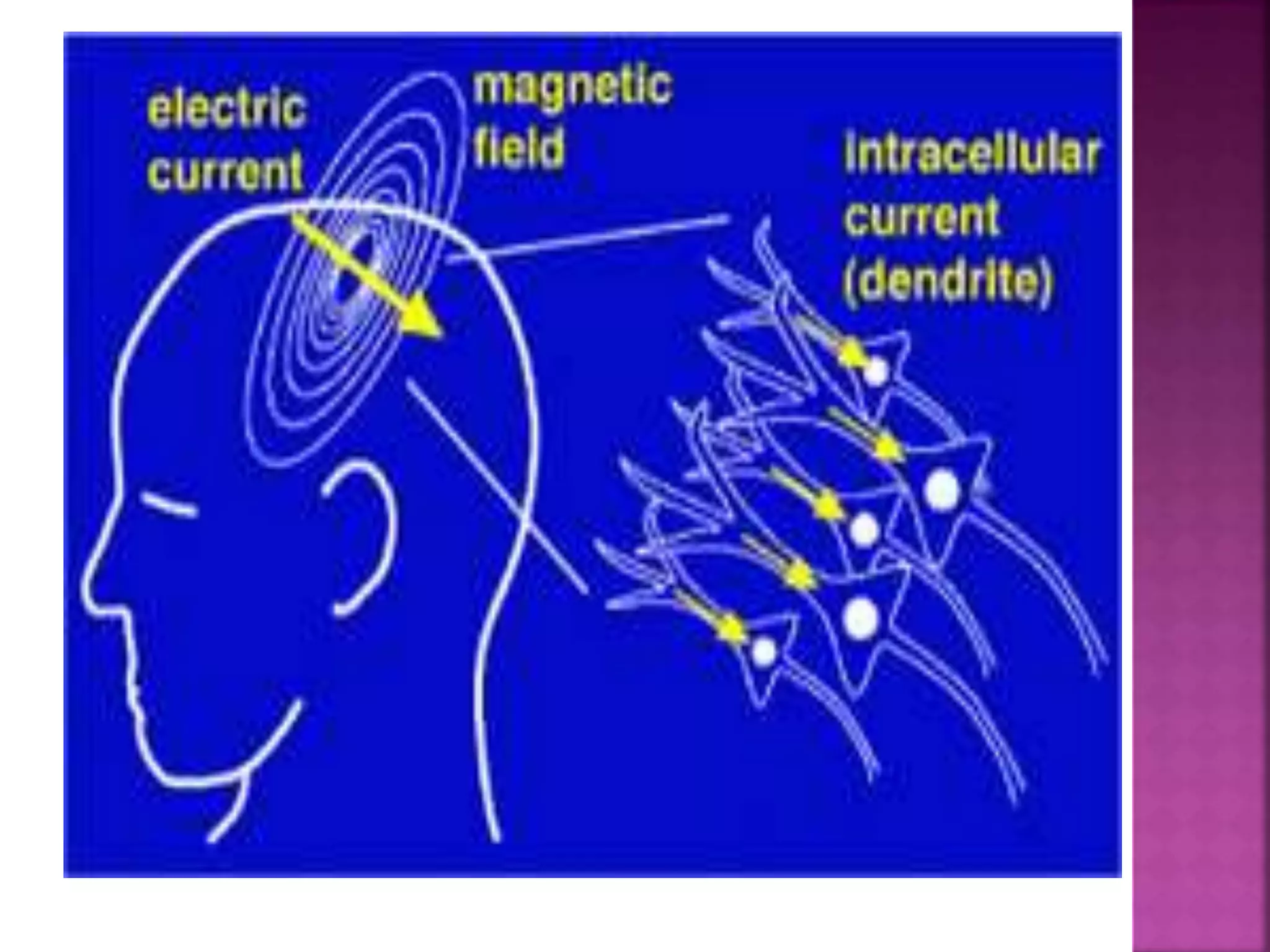
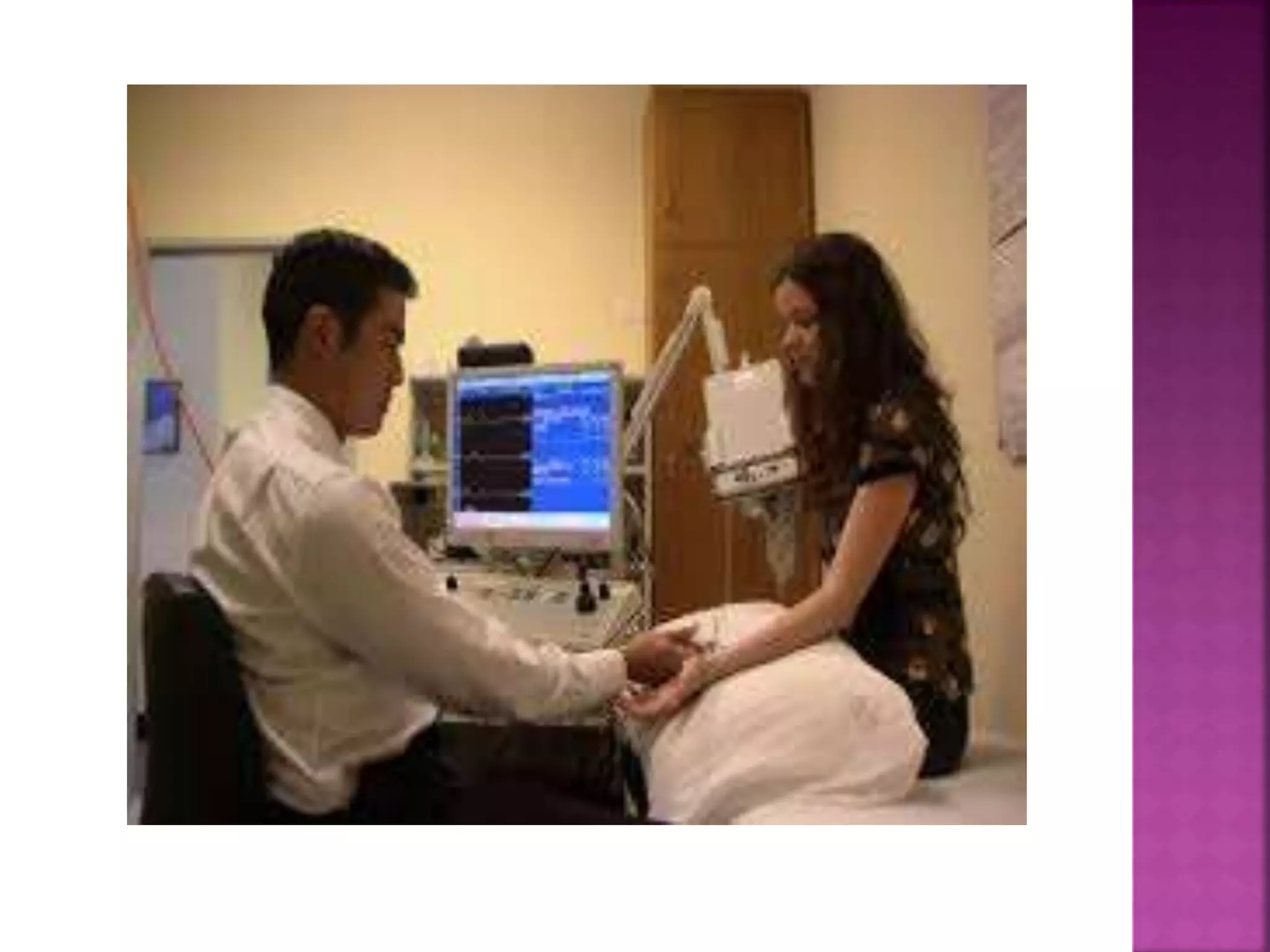
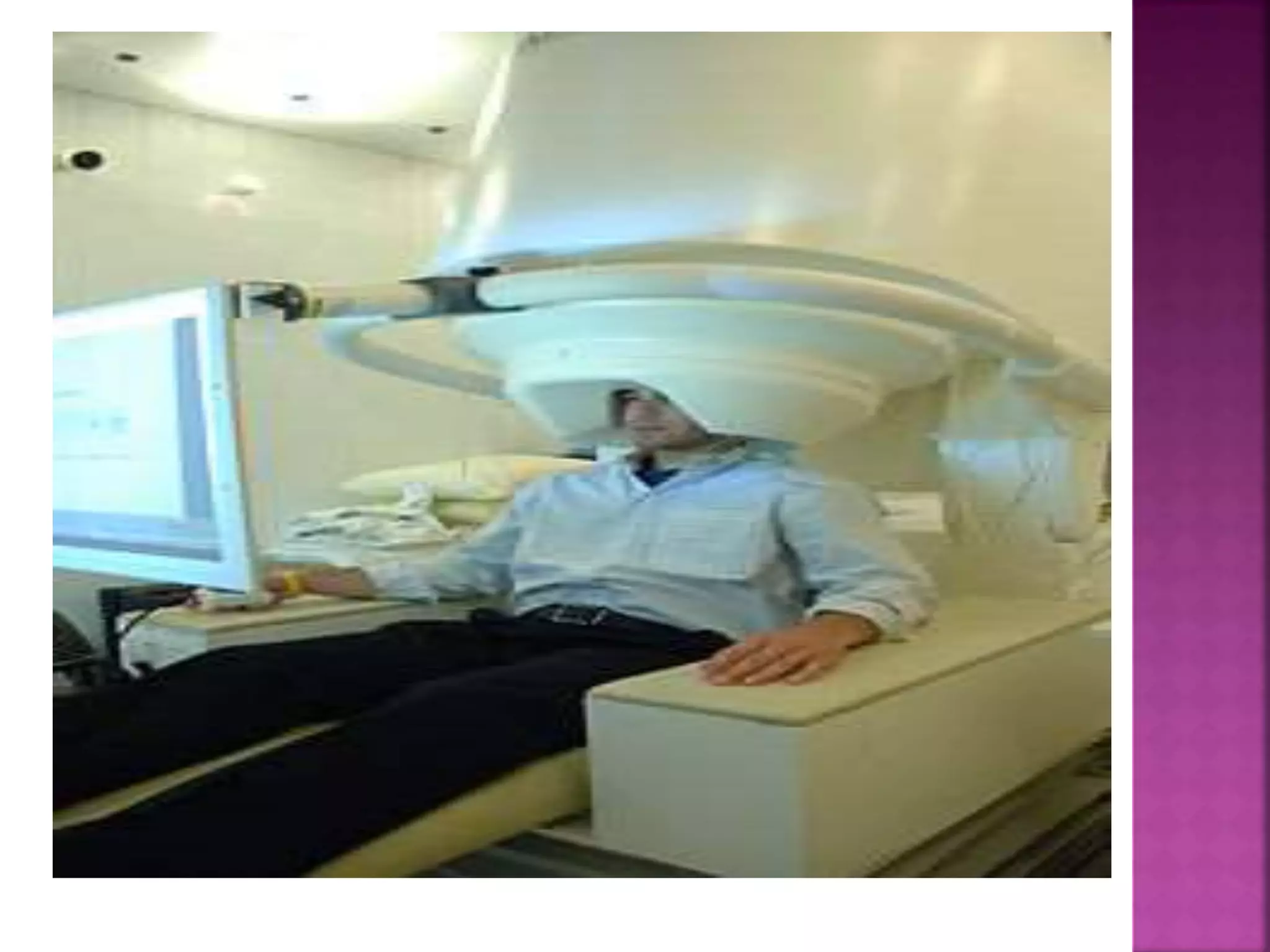
G.VISHWAPRAKASH
M.SC(N) IInd year
NUEROSCIENCE IN NURSING
 An electroencephalogram (EEG) is a test to
measure the electrical activity of the brain.
 An electroencephalogram (EEG) is a painless
procedure that uses small, flat metal discs
(electrodes) attached to your scalp to detect
electrical activity in your brain. Your brain
cells communicate via electrical impulses
and are active all the time, even when you're
asleep. This activity shows up as wavy lines
on an EEG recording.
 Epilepsy or other seizure disorder
 Brain tumor
 Head injury
 Brain dysfunction that may have a variety of
causes (encephalopathy)
 Inflammation of the brain (encephalitis)
 Stroke
 Sleep disorders
 Memory impairment
 EEG is also used to:
 Evaluate problems with sleep ( sleep
disorders)
 Investigate periods of unconsciousness
 Monitor the brain during brain surgery
 Brain cells communicate with each other by
producing tiny electrical signals, called impulses.
 An EEG measures this activity. The test is done by a
EEG specialist.
 Individual will be asked to lie on your back on a bed
or in a reclining chair.
 Flat metal disks called electrodes are placed all
over your scalp. The disks are held in place with a
sticky paste.
 The electrodes are connected by wires to a speaker
and recording machine.
 The recording machine changes the electrical signals
into patterns that can be seen on a computer. It looks
like wavy lines.
 Individual will need to lie still during the test with
your eyes closed because movement can change the
results.
 But one may be asked to do certain things during the
test, such as breathe fast and deeply for several
minutes or look at a bright flashing light.
 EEG ELECTRODES:
 Individual feel little or no discomfort during an
EEG. The electrodes don't transmit any
sensations. They just record brain waves. If you
need to sleep during the EEG, you might be
given a sedative beforehand to help you relax.
During the test:
 A technician measures head and marks scalp
with a type of pencil, to indicate where to
attach the electrodes. Those spots on scalp may
be scrubbed with a gritty cream to improve the
quality of the recording.
 A technician attaches flat metal discs
(electrodes) to scalp using a special adhesive.
The electrodes are connected with wires to an
instrument that amplifies — makes bigger — the
brain waves and records them on computer
equipment. Some people wear an elastic cap
fitted with
 electrodes, instead of having the adhesive
applied to their scalps. Once the electrodes
are in place, an EEG typically takes 30 to 60
minutes.
 Normal Results
 Brain electrical activity has a certain number of
waves per second (frequencies) that are normal
for different levels of alertness. For example,
brain waves are faster when you are awake, and
slower when you are sleeping.
 There are also normal patterns to these waves.
 What Abnormal Results Mean
 Abnormal results on an EEG test may be due to:
 Abnormal bleeding (hemorrhage)
 An abnormal structure in the brain (such as
a brain tumor)
 Attention problems
 Tissue death due to a blockage in blood flow
(cerebral infarction)
 Drug or alcohol abuse
 Head injury
 Migraines (in some cases)
 Seizure disorder (such as epilepsy
or convulsions)
 Sleep disorder (such as narcolepsy)
 Swelling of the brain (encephalitis)
 Note: A normal EEG does not mean that a
seizure did not occur.
 Risks
 The procedure is very safe. The flashing lights or fast
breathing (hyperventilation) required during the test
may trigger seizures in those with seizure disorders.
The health care provider performing the EEG is
trained to take care of you if this happens.
 Alternative Names
 Electroencephalogram; Brain wave test
 What can the test show?
 A normal ('negative') result
 This shows a typical pattern of electrical activity
from the brain. Most people without epilepsy, and
many people with epilepsy, have a normal result. This
is because an EEG only shows the electrical activity
of the brain when the test is
An abnormal ('positive') result
 This shows abnormal patterns of electrical
activity. Some people with certain types of
epilepsy have abnormal patterns all the
time, not just when they have seizures.
(Although, during a seizure the activity is
even more abnormal.) For example, children
with typical 'absence seizures' often have a
characteristic EEG pattern which helps to
confirm this type of epilepsy.
 Strobe lighting
In some cases, a strobe light may be used during
an EEG test. This aims to detect if this alters the
electrical pattern in the brain. (Usually it does
not. However, a small number of people have
seizures triggered by flickering or strobe lights
and so this may help to identify these people.)
 Sleep EEG
This is when an EEG is performed while you are
sleeping. This is usually carried out when you are
in hospital. You may need to have one of these if
your seizures happen when you are asleep or
when you are tired.
 Sleep deprived EEG
There may be a better chance of detecting
abnormal brain activity after a period of
time when you are deprived of sleep.
Therefore, sometimes the EEG test is done
after you have stayed awake for all or most
of the night. It is done in the same way as
the normal test, but with you asleep - after
the period of 'sleep deprivation'.
 Ambulatory EEG
This may be advised in cases where the
diagnosis is not clear. It uses a portable EEG
machine which records the brain's electrical
activity when you are going about your
normal activities. The electrodes can usually
be hidden under your hair, and the wires are
connected to a small machine which you
wear on a belt (a bit like wearing an mp3
player).

 Magnetoencephalography (MEG) is a functional
neuroimaging technique for mapping brain
activity by recording magnetic fields produced
by electrical currents occurring naturally in
the brain, using very sensitive magnetometers.
 A completely noninvasive procedure that uses an
array of highly sensitive sensors to detect and
record the magnetic fields associated with
electrical activity in the brain. Usually
abbreviated as MEG. There are many uses for
MEG, including determining the function of
various parts of the brain and localizing epileptic
activity.
 Arrays of SQUIDs(superconducting quantum
interference devices) are currently the most
common magnetometer, and SERF being
investigated for future machines.
 Applications of MEG include basic research
into perceptual and cognitive brain
processes, localizing regions affected by
pathology before surgical removal,
determining the function of various parts of
the brain, and neurofeedback.
 EMG is performed using an instrumentcalled
an electromyograph, to produce a record
called an electromyogram
 An electromyograph detects the electrical
potentialgenerated by muscle cells.
 when these cells are electrically or
neurologically activated. The signals can be
analyzed to detect medical abnormalities,
activation level, or recruitment order or to
analyze the biomechanicsof human or animal
movement.
Epilepsy is a common chronic neurological
disorder that is characterized by recurrent
unprovoked seizures.
 These seizures occur due to abnormal
neuronal activity in the brain. About 3
million Americans have epilepsy.
 Epilepsy is usually controlled, but not cured,
with medication. Surgery is often the best
option in difficult cases.
 A brain tumor is any intracranial tumor
created by abnormal and uncontrolled cell
division. It can affect almost any part of the
brain.
 Many tumors, depending on their location,
can be successfully removed surgically.
 In more difficult cases, stereotactic
radiosurgery, remains a viable option.
 MEG is increasingly being used in the
preoperative evaluation of patients with
epilepsy and those who will undergo tumor
resection surgery
 In either case, the MEG can localize the
precise areas that are, despite the
pathology, still healthy and functioning.
 This helps the surgeon to determine a
successful surgical approach and also how
aggressively to resect a given area. With
a “roadmap” of which areas to avoid, the
surgeon has a better chance of
performing the procedure without
affecting critical functions such as the
senses, language and motor control.

 These functions are controlled from so
called “eloquent cortex”. For epilepsy
surgery, MEG has the added benefit of
being able to localize, with precise
accuracy, the location(s) where the
epileptic activity originates.
 This information is invaluable in
determining if the patient is a good
candidate for surgery and also to plan the
operation itself.
 The ability to localize pathological areas and
their relationship to eloquent cortex allows the
medical team to more accurately assess the
likelihood of a successful surgery. This is defined
as one where the patient is left free from the
disturbance (for example, the tumor or the
uncontrolled seizures from epilepsy), while
suffering minimal functional deficits (for
example, loss of senses or control).
 For anatomical information, CT and MRI provide
detailed images. For metabolic activity PET,
SPECT and fMRI give useful information on blood
flow, oxygenation, etc.
 Only MEG can measure fast, millisecond
phenomena and also perform localization
accurate to the millimeter level.
 It does this noninvasively (without injections or
radiation of any kind) by measuring the magnetic
fields that naturally emanate whenever electric
current flows within the neurons of the brain.

 The fields being measured are extremely
weak, about a billion times smaller than the
Earth's magnetic field.
 The MEG technique uses very sophisticated
instrumentation, sensitive enough to detect
these weak signals, while simultaneously
discriminating against interference from the
much stronger magnetic background noise.

 After collection, the data will be combined and analyzed
by a trained professional, usually a neurologist.
 From the recorded signals, it will be determined where in
the brain the activity originated from. This applies to both
pathological signals (epileptic spikes) and also healthy
signals (for example, those arising from the sensory
stimuli).
 These locations will then be combined with an MRI, which
shows an image of the brain’s structure. The combined
images are then included in a comprehensive report which
is prepared. When the report is completed, it is forwarded
to the referring physician. This, when pooled with other
information from the patient, forms the basis for
determining whether surgery is the best option for
treatment and, if so, how to plan it.
 .
 Although EEG and MEG signals originate from the
same neurophysiological processes, there are
important differences.
 Magnetic fields are less distorted than electric
fields by the skull and scalp, which results in a
better spatial resolution of the MEG.
 Whereas scalp EEG is sensitive to both tangential
and radial components of a current source in a
spherical volume conductor, MEG detects only its
tangential components.
 Scalp EEG can, therefore, detect activity both in
the sulci and at the top of the cortical gyri,
whereas MEG is most sensitive to activity
originating in sulci.
 EEG is, therefore, sensitive to activity in
more brain areas, but activity that is visible
in MEG can also be localized with more
accuracy.
 Scalp EEG is sensitive to extracellular volume
currents produced by postsynaptic
potentials. MEG detects intracellular
currents associated primarily with these
synaptic potentials because the field
components generated by volume currents
tend to cancel out in a spherical volume
conductor.
 The decay of magnetic fields as a function of
distance is more pronounced than for
electric fields.
 Therefore, MEG is more sensitive to
superficial cortical activity, which makes it
useful for the study of neocortical epilepsy.
Finally, MEG is reference-free, while scalp
EEG relies on a reference that, when active,
makes interpretation of the data difficult.
 Electromyography (EMG) is a technique for
evaluating and recording the electrical activity
produced by skeletal muscles.EMG is performed
using an instrument called an electromyograph,
to produce a record called an electromyogram.
 An electromyograph detects the electrical
potential generated by muscle cells when these
cells are electrically or neurologically activated.
The signals can be analyzed to detect medical
abnormalities, activation level, or recruitment
order or to analyze the biomechanics of human
or animal movement
 There are two kinds of EMG in widespread use: surface
EMG and intramuscular (needle and fine-wire) EMG.
 To perform intramuscular EMG, a needle electrode or a
needle containing two fine-wire electrodes is inserted
through the skin into the muscle tissue.
 A trained professional (such as
a neurologist, physiatrist,chiropractor, or physical
therapist) observes the electrical activity while inserting
the electrode.
 Certain places limit the performance of needle EMG by
non-physicians. A recent case ruling in the state of New
Jersey declared that it cannot be delegated to a
physician's assistant.
 The insertional activity provides valuable information
about the state of the muscle and its innervating nerve.
 Normal muscles at rest make certain, normal
electrical signals when the needle is inserted
into them.
 Then the electrical activity when the muscle is
at rest is studied. Abnormal spontaneous activity
might indicate some nerve and/or muscle
damage.
 Then the patient is asked to contract the muscle
smoothly.
 The shape, size, and frequency of the resulting
electrical signals are judged. Then the electrode
is retracted a few millimetres, and again the
activity is analyzed until at least 10–20 motor
units have been collected.
 Each electrode track gives only a very local
picture of the activity of the whole muscle.
 Because skeletal muscles differ in the inner
structure, the electrode has to be placed at
various locations to obtain an accurate study.
 Intramuscular EMG may be considered too
invasive or unnecessary in some cases.
Instead, a surface electrode may be used to
monitor the general picture of muscle
activation, as opposed to the activity of only
a few fibres as observed using an
intramuscular EMG.
 This technique is used in a number of
settings; for example, in the physiotherapy
clinic, muscle activation is monitored using
surface EMG and patients have an auditory or
visual stimulus to help them know when they
are activating the muscle (biofeedback).
 A motor unitis defined as one motor neuron
and all of the muscle fibers it innervates.
When a motor unit fires, the impulse (called
an action potential) is carried down the
motor neuron to the muscle.
 The area where the nerve contacts the
muscle is called the neuromuscular junction,
or the motor end plate. After the action
potential is transmitted across the
neuromuscular junction, an action potential
is elicited in all of the innervated muscle
fibers of that particular motor unit.
 The sum of all this electrical activity is
known as a motor unit action potential
(MUAP). This electrophysiologic activity from
multiple motor units is the signal typically
evaluated during an EMG.
 The composition of the motor unit, the
number of muscle fibres per motor unit, the
metabolic type of muscle fibres and many
other factors affect the shape of the motor
unit potentials in the myogram.
 Muscle tissue at rest is normally electrically inactive. After
the electrical activity caused by the irritation of needle
insertion subsides, the electromyograph should detect no
abnormal spontaneous activity (i.e., a muscle at rest
should be electrically silent, with the exception of the
area of the neuromuscular junction, which is, under
normal circumstances, very spontaneously active).
 When the muscle is voluntarily contracted, action
potentials begin to appear. As the strength of the muscle
contraction is increased, more and more muscle fibers
produce action potentials.
 When the muscle is fully contracted, there should appear a
disorderly group of action potentials of varying rates and
amplitudes (a complete recruitment and interference
pattern).
 EMG is used to diagnose diseases that generally
may be classified into one of the following
categories: neuropathies,neuromuscular junction
diseases and myopathies.
 Neuropathic disease has the following defining
EMG characteristics:
 An action potentialamplitude that is twice
normal due to the increased number of fibresper
motor unit because of reinnervationof
denervated fibres
 An increase in duration of the action potential
 A decrease in the number of motor unitsin the
muscle (as found using motor unit number
estimation techniques)
 Myopathic disease has these defining EMG
characteristics:
 A decrease in duration of the action potential
 A reduction in the areato amplituderatio of the
action potential
 A decrease in the number of motor units in the
muscle (in extremely severe cases only)
 Because of the individuality of each patient and
disease, some of these characteristics may not
appear in every case.
 APPLICATIONS OF EMG
 EMG signals are used in many clinical and
biomedical applications. EMG is used as a
diagnostics tool for identifying neuromuscular
diseases, assessing low-back pain, kinesiology,
and disorders of motor control.
 EMG signals are also used as a control signal
for prosthetic devices such as prosthetic hands,
arms, and lower limbs.
 EMG can be used to sense isometric muscular
activity where no movement is produced.

 This enables definition of a class of subtle
motionless gestures to control interfaces
without being noticed and without disrupting
the surrounding environment. These signals
can be used to control a prosthesis or as a
control signal for an electronic device such
as a mobile phone or PDA.
 EMG then acceleromyograph may be used
for neuromuscular monitoring in general
anesthesia with neuromuscular-blocking
drugs, in order to avoid postoperative
residual curarization PORC).
 EMG signals have been targeted as control
for flight systems. The Human Senses Group
at the NASA Ames Research Centerat Moffett
Field, CA seeks to advance man-machine
interfaces by directly connecting a person to
a computer. In this project, an EMG signal is
used to substitute for mechanical joysticks
and keyboards. EMG has also been used in
research towards a "wearable cockpit," which
employs EMG-based gestures to manipulate
switches and control sticks necessary for
flight in conjunction with a goggle-based
display.
Electroencephal ogram (eeg)

Electroencephal ogram (eeg)

  • 1.
  • 2.
     An electroencephalogram(EEG) is a test to measure the electrical activity of the brain.  An electroencephalogram (EEG) is a painless procedure that uses small, flat metal discs (electrodes) attached to your scalp to detect electrical activity in your brain. Your brain cells communicate via electrical impulses and are active all the time, even when you're asleep. This activity shows up as wavy lines on an EEG recording.
  • 3.
     Epilepsy orother seizure disorder  Brain tumor  Head injury  Brain dysfunction that may have a variety of causes (encephalopathy)  Inflammation of the brain (encephalitis)  Stroke
  • 4.
     Sleep disorders Memory impairment  EEG is also used to:  Evaluate problems with sleep ( sleep disorders)  Investigate periods of unconsciousness  Monitor the brain during brain surgery
  • 7.
     Brain cellscommunicate with each other by producing tiny electrical signals, called impulses.  An EEG measures this activity. The test is done by a EEG specialist.  Individual will be asked to lie on your back on a bed or in a reclining chair.  Flat metal disks called electrodes are placed all over your scalp. The disks are held in place with a sticky paste.  The electrodes are connected by wires to a speaker and recording machine.  The recording machine changes the electrical signals into patterns that can be seen on a computer. It looks like wavy lines.  Individual will need to lie still during the test with your eyes closed because movement can change the results.  But one may be asked to do certain things during the test, such as breathe fast and deeply for several minutes or look at a bright flashing light.
  • 8.
     EEG ELECTRODES: Individual feel little or no discomfort during an EEG. The electrodes don't transmit any sensations. They just record brain waves. If you need to sleep during the EEG, you might be given a sedative beforehand to help you relax. During the test:  A technician measures head and marks scalp with a type of pencil, to indicate where to attach the electrodes. Those spots on scalp may be scrubbed with a gritty cream to improve the quality of the recording.  A technician attaches flat metal discs (electrodes) to scalp using a special adhesive. The electrodes are connected with wires to an instrument that amplifies — makes bigger — the brain waves and records them on computer equipment. Some people wear an elastic cap fitted with
  • 9.
     electrodes, insteadof having the adhesive applied to their scalps. Once the electrodes are in place, an EEG typically takes 30 to 60 minutes.
  • 10.
     Normal Results Brain electrical activity has a certain number of waves per second (frequencies) that are normal for different levels of alertness. For example, brain waves are faster when you are awake, and slower when you are sleeping.  There are also normal patterns to these waves.  What Abnormal Results Mean  Abnormal results on an EEG test may be due to:  Abnormal bleeding (hemorrhage)  An abnormal structure in the brain (such as a brain tumor)  Attention problems  Tissue death due to a blockage in blood flow (cerebral infarction)
  • 11.
     Drug oralcohol abuse  Head injury  Migraines (in some cases)  Seizure disorder (such as epilepsy or convulsions)  Sleep disorder (such as narcolepsy)  Swelling of the brain (encephalitis)  Note: A normal EEG does not mean that a seizure did not occur.
  • 12.
     Risks  Theprocedure is very safe. The flashing lights or fast breathing (hyperventilation) required during the test may trigger seizures in those with seizure disorders. The health care provider performing the EEG is trained to take care of you if this happens.  Alternative Names  Electroencephalogram; Brain wave test  What can the test show?  A normal ('negative') result  This shows a typical pattern of electrical activity from the brain. Most people without epilepsy, and many people with epilepsy, have a normal result. This is because an EEG only shows the electrical activity of the brain when the test is
  • 13.
    An abnormal ('positive')result  This shows abnormal patterns of electrical activity. Some people with certain types of epilepsy have abnormal patterns all the time, not just when they have seizures. (Although, during a seizure the activity is even more abnormal.) For example, children with typical 'absence seizures' often have a characteristic EEG pattern which helps to confirm this type of epilepsy.
  • 14.
     Strobe lighting Insome cases, a strobe light may be used during an EEG test. This aims to detect if this alters the electrical pattern in the brain. (Usually it does not. However, a small number of people have seizures triggered by flickering or strobe lights and so this may help to identify these people.)  Sleep EEG This is when an EEG is performed while you are sleeping. This is usually carried out when you are in hospital. You may need to have one of these if your seizures happen when you are asleep or when you are tired.
  • 15.
     Sleep deprivedEEG There may be a better chance of detecting abnormal brain activity after a period of time when you are deprived of sleep. Therefore, sometimes the EEG test is done after you have stayed awake for all or most of the night. It is done in the same way as the normal test, but with you asleep - after the period of 'sleep deprivation'.
  • 16.
     Ambulatory EEG Thismay be advised in cases where the diagnosis is not clear. It uses a portable EEG machine which records the brain's electrical activity when you are going about your normal activities. The electrodes can usually be hidden under your hair, and the wires are connected to a small machine which you wear on a belt (a bit like wearing an mp3 player).
  • 17.
      Magnetoencephalography (MEG)is a functional neuroimaging technique for mapping brain activity by recording magnetic fields produced by electrical currents occurring naturally in the brain, using very sensitive magnetometers.  A completely noninvasive procedure that uses an array of highly sensitive sensors to detect and record the magnetic fields associated with electrical activity in the brain. Usually abbreviated as MEG. There are many uses for MEG, including determining the function of various parts of the brain and localizing epileptic activity.
  • 20.
     Arrays ofSQUIDs(superconducting quantum interference devices) are currently the most common magnetometer, and SERF being investigated for future machines.  Applications of MEG include basic research into perceptual and cognitive brain processes, localizing regions affected by pathology before surgical removal, determining the function of various parts of the brain, and neurofeedback.
  • 21.
     EMG isperformed using an instrumentcalled an electromyograph, to produce a record called an electromyogram  An electromyograph detects the electrical potentialgenerated by muscle cells.  when these cells are electrically or neurologically activated. The signals can be analyzed to detect medical abnormalities, activation level, or recruitment order or to analyze the biomechanicsof human or animal movement.
  • 22.
    Epilepsy is acommon chronic neurological disorder that is characterized by recurrent unprovoked seizures.  These seizures occur due to abnormal neuronal activity in the brain. About 3 million Americans have epilepsy.  Epilepsy is usually controlled, but not cured, with medication. Surgery is often the best option in difficult cases.
  • 23.
     A braintumor is any intracranial tumor created by abnormal and uncontrolled cell division. It can affect almost any part of the brain.  Many tumors, depending on their location, can be successfully removed surgically.  In more difficult cases, stereotactic radiosurgery, remains a viable option.  MEG is increasingly being used in the preoperative evaluation of patients with epilepsy and those who will undergo tumor resection surgery
  • 24.
     In eithercase, the MEG can localize the precise areas that are, despite the pathology, still healthy and functioning.  This helps the surgeon to determine a successful surgical approach and also how aggressively to resect a given area. With a “roadmap” of which areas to avoid, the surgeon has a better chance of performing the procedure without affecting critical functions such as the senses, language and motor control. 
  • 25.
     These functionsare controlled from so called “eloquent cortex”. For epilepsy surgery, MEG has the added benefit of being able to localize, with precise accuracy, the location(s) where the epileptic activity originates.  This information is invaluable in determining if the patient is a good candidate for surgery and also to plan the operation itself.
  • 26.
     The abilityto localize pathological areas and their relationship to eloquent cortex allows the medical team to more accurately assess the likelihood of a successful surgery. This is defined as one where the patient is left free from the disturbance (for example, the tumor or the uncontrolled seizures from epilepsy), while suffering minimal functional deficits (for example, loss of senses or control).
  • 27.
     For anatomicalinformation, CT and MRI provide detailed images. For metabolic activity PET, SPECT and fMRI give useful information on blood flow, oxygenation, etc.  Only MEG can measure fast, millisecond phenomena and also perform localization accurate to the millimeter level.  It does this noninvasively (without injections or radiation of any kind) by measuring the magnetic fields that naturally emanate whenever electric current flows within the neurons of the brain. 
  • 28.
     The fieldsbeing measured are extremely weak, about a billion times smaller than the Earth's magnetic field.  The MEG technique uses very sophisticated instrumentation, sensitive enough to detect these weak signals, while simultaneously discriminating against interference from the much stronger magnetic background noise. 
  • 30.
     After collection,the data will be combined and analyzed by a trained professional, usually a neurologist.  From the recorded signals, it will be determined where in the brain the activity originated from. This applies to both pathological signals (epileptic spikes) and also healthy signals (for example, those arising from the sensory stimuli).  These locations will then be combined with an MRI, which shows an image of the brain’s structure. The combined images are then included in a comprehensive report which is prepared. When the report is completed, it is forwarded to the referring physician. This, when pooled with other information from the patient, forms the basis for determining whether surgery is the best option for treatment and, if so, how to plan it.  .
  • 31.
     Although EEGand MEG signals originate from the same neurophysiological processes, there are important differences.  Magnetic fields are less distorted than electric fields by the skull and scalp, which results in a better spatial resolution of the MEG.  Whereas scalp EEG is sensitive to both tangential and radial components of a current source in a spherical volume conductor, MEG detects only its tangential components.  Scalp EEG can, therefore, detect activity both in the sulci and at the top of the cortical gyri, whereas MEG is most sensitive to activity originating in sulci.
  • 32.
     EEG is,therefore, sensitive to activity in more brain areas, but activity that is visible in MEG can also be localized with more accuracy.  Scalp EEG is sensitive to extracellular volume currents produced by postsynaptic potentials. MEG detects intracellular currents associated primarily with these synaptic potentials because the field components generated by volume currents tend to cancel out in a spherical volume conductor.
  • 33.
     The decayof magnetic fields as a function of distance is more pronounced than for electric fields.  Therefore, MEG is more sensitive to superficial cortical activity, which makes it useful for the study of neocortical epilepsy. Finally, MEG is reference-free, while scalp EEG relies on a reference that, when active, makes interpretation of the data difficult.
  • 35.
     Electromyography (EMG)is a technique for evaluating and recording the electrical activity produced by skeletal muscles.EMG is performed using an instrument called an electromyograph, to produce a record called an electromyogram.  An electromyograph detects the electrical potential generated by muscle cells when these cells are electrically or neurologically activated. The signals can be analyzed to detect medical abnormalities, activation level, or recruitment order or to analyze the biomechanics of human or animal movement
  • 36.
     There aretwo kinds of EMG in widespread use: surface EMG and intramuscular (needle and fine-wire) EMG.  To perform intramuscular EMG, a needle electrode or a needle containing two fine-wire electrodes is inserted through the skin into the muscle tissue.  A trained professional (such as a neurologist, physiatrist,chiropractor, or physical therapist) observes the electrical activity while inserting the electrode.  Certain places limit the performance of needle EMG by non-physicians. A recent case ruling in the state of New Jersey declared that it cannot be delegated to a physician's assistant.  The insertional activity provides valuable information about the state of the muscle and its innervating nerve.
  • 37.
     Normal musclesat rest make certain, normal electrical signals when the needle is inserted into them.  Then the electrical activity when the muscle is at rest is studied. Abnormal spontaneous activity might indicate some nerve and/or muscle damage.  Then the patient is asked to contract the muscle smoothly.  The shape, size, and frequency of the resulting electrical signals are judged. Then the electrode is retracted a few millimetres, and again the activity is analyzed until at least 10–20 motor units have been collected.
  • 38.
     Each electrodetrack gives only a very local picture of the activity of the whole muscle.  Because skeletal muscles differ in the inner structure, the electrode has to be placed at various locations to obtain an accurate study.  Intramuscular EMG may be considered too invasive or unnecessary in some cases. Instead, a surface electrode may be used to monitor the general picture of muscle activation, as opposed to the activity of only a few fibres as observed using an intramuscular EMG.
  • 39.
     This techniqueis used in a number of settings; for example, in the physiotherapy clinic, muscle activation is monitored using surface EMG and patients have an auditory or visual stimulus to help them know when they are activating the muscle (biofeedback).  A motor unitis defined as one motor neuron and all of the muscle fibers it innervates. When a motor unit fires, the impulse (called an action potential) is carried down the motor neuron to the muscle.
  • 40.
     The areawhere the nerve contacts the muscle is called the neuromuscular junction, or the motor end plate. After the action potential is transmitted across the neuromuscular junction, an action potential is elicited in all of the innervated muscle fibers of that particular motor unit.  The sum of all this electrical activity is known as a motor unit action potential (MUAP). This electrophysiologic activity from multiple motor units is the signal typically evaluated during an EMG.
  • 41.
     The compositionof the motor unit, the number of muscle fibres per motor unit, the metabolic type of muscle fibres and many other factors affect the shape of the motor unit potentials in the myogram.
  • 42.
     Muscle tissueat rest is normally electrically inactive. After the electrical activity caused by the irritation of needle insertion subsides, the electromyograph should detect no abnormal spontaneous activity (i.e., a muscle at rest should be electrically silent, with the exception of the area of the neuromuscular junction, which is, under normal circumstances, very spontaneously active).  When the muscle is voluntarily contracted, action potentials begin to appear. As the strength of the muscle contraction is increased, more and more muscle fibers produce action potentials.  When the muscle is fully contracted, there should appear a disorderly group of action potentials of varying rates and amplitudes (a complete recruitment and interference pattern).
  • 43.
     EMG isused to diagnose diseases that generally may be classified into one of the following categories: neuropathies,neuromuscular junction diseases and myopathies.  Neuropathic disease has the following defining EMG characteristics:  An action potentialamplitude that is twice normal due to the increased number of fibresper motor unit because of reinnervationof denervated fibres  An increase in duration of the action potential
  • 44.
     A decreasein the number of motor unitsin the muscle (as found using motor unit number estimation techniques)  Myopathic disease has these defining EMG characteristics:  A decrease in duration of the action potential  A reduction in the areato amplituderatio of the action potential  A decrease in the number of motor units in the muscle (in extremely severe cases only)  Because of the individuality of each patient and disease, some of these characteristics may not appear in every case.
  • 45.
     APPLICATIONS OFEMG  EMG signals are used in many clinical and biomedical applications. EMG is used as a diagnostics tool for identifying neuromuscular diseases, assessing low-back pain, kinesiology, and disorders of motor control.  EMG signals are also used as a control signal for prosthetic devices such as prosthetic hands, arms, and lower limbs.  EMG can be used to sense isometric muscular activity where no movement is produced. 
  • 46.
     This enablesdefinition of a class of subtle motionless gestures to control interfaces without being noticed and without disrupting the surrounding environment. These signals can be used to control a prosthesis or as a control signal for an electronic device such as a mobile phone or PDA.  EMG then acceleromyograph may be used for neuromuscular monitoring in general anesthesia with neuromuscular-blocking drugs, in order to avoid postoperative residual curarization PORC).
  • 47.
     EMG signalshave been targeted as control for flight systems. The Human Senses Group at the NASA Ames Research Centerat Moffett Field, CA seeks to advance man-machine interfaces by directly connecting a person to a computer. In this project, an EMG signal is used to substitute for mechanical joysticks and keyboards. EMG has also been used in research towards a "wearable cockpit," which employs EMG-based gestures to manipulate switches and control sticks necessary for flight in conjunction with a goggle-based display.