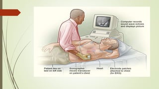
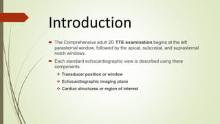
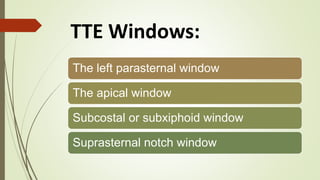
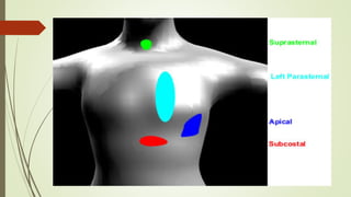
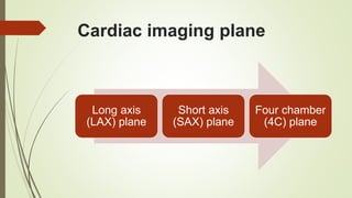
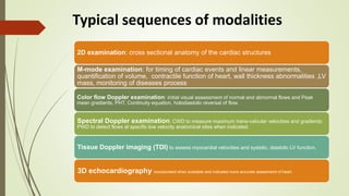
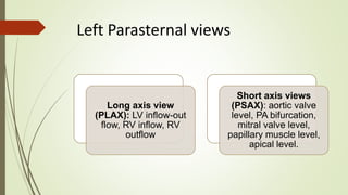
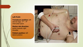
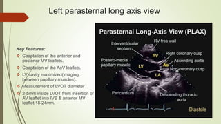
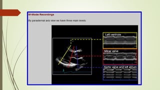
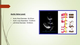
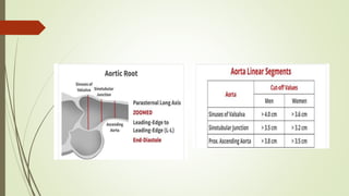
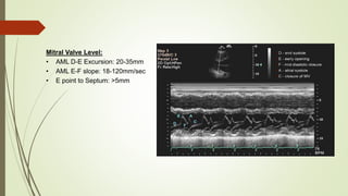
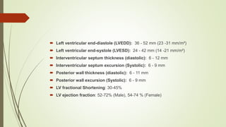
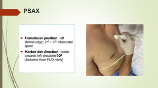
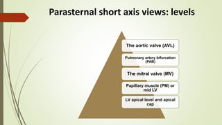
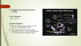
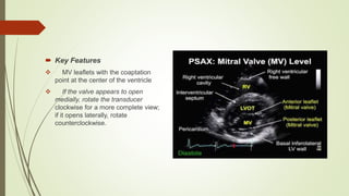
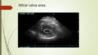
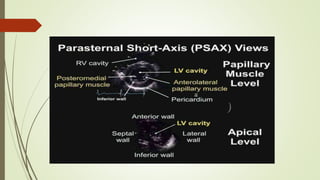
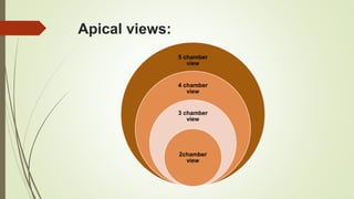
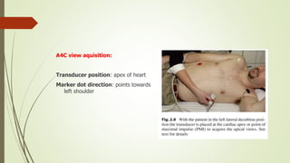
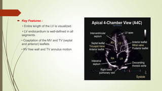
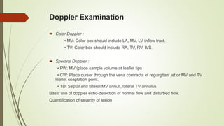
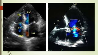
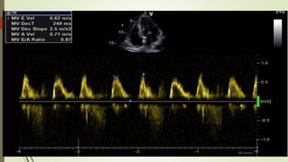
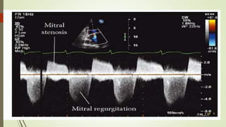
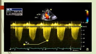
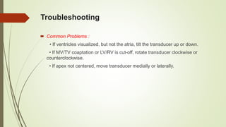
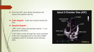
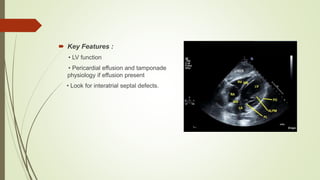
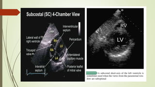
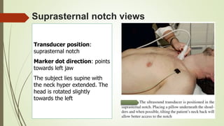
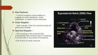
SlideShare a Scribd company logo
ECHO views and measurements
Chairperson :
Assoc. Prof. Dr. Naveen Sheikh
Dept of Cardiology,UCC, BSMMU
Dr. Md. Reza-ud-doulla Razu
D-card
Introduction
 The Comprehensive adult 2D TTE examination begins at the left
parasternal window, followed by the apical, subcostal, and suprasternal
notch windows.
 Each standard echocardiographic view is described using there
components
 Transducer position or window
 Echocardiographic imaging plane
 Cardiac structures or region of interest
TTE Windows:
The left parasternal window
The apical window
Subcostal or subxiphoid window
Suprasternal notch window
Cardiac imaging plane
Long axis
(LAX) plane
Short axis
(SAX) plane
Four chamber
(4C) plane
Typical sequences of modalities
2D examination: cross sectional anatomy of the cardiac structures
M-mode examination: for timing of cardiac events and linear measurements,
quantification of volume, contractile function of heart, wall thickness abnormalities ,LV
mass, monitoring of diseases process
Color flow Doppler examination: initial visual assessment of normal and abnormal flows and Peak
mean gradients, PHT, Continuity equation, holodiastolic reversal of flow.
Spectral Doppler examination: CWD to measure maximum trans-valvular velocities and gradients;
PWD to detect flows at specific low velocity anatomical sites when indicated.
Tissue Doppler imaging (TDI) to assess myocardial velocities and systolic, diastolic LV function.
3D echocardiography incorporated when available and indicated more accurate assesmemt of heart.
Left Parasternal views
Long axis view
(PLAX): LV inflow-out
flow, RV inflow, RV
outflow
Short axis views
(PSAX): aortic valve
level, PA bifurcation,
mitral valve level,
papillary muscle level,
apical level.
Left PLAX:
Transducer position: left
sternal edge; 3rd – 4th
intercostal space
Marker dot direction:
points towards right
shoulder
Patient position: left
lateral position
Left parasternal long axis view
Key Features:
 Coaptation of the anterior and
posterior MV leaflets.
 Coaptation of the AoV leaflets.
 LV cavity maximized(imaging
between papillary muscles).
 Measurement of LVOT diameter
 2-5mm inside LVOT from insertion of
AV leaflet into IVS & anterior MV
leaflet.18-24mm.
Aortic Valve Level:
• Aortic Root Diameter: 30-37mm
• Aortic cusp Separation: 15-26mm
• Left Atrial Diameter: 19-40mm
Mitral Valve Level:
• AML D-E Excursion: 20-35mm
• AML E-F slope: 18-120mm/sec
• E point to Septum: >5mm
 Left ventricular end-diastole (LVEDD): 36 - 52 mm (23 -31 mm/m²)
 Left ventricular end-systole (LVESD): 24 - 42 mm (14 -21 mm/m²)
 Interventricular septum thickness (diastolic): 6 - 12 mm
 Interventricular septum excursion (Systolic): 6 - 9 mm
 Posterior wall thickness (diastolic): 6 - 11 mm
 Posterior wall excursion (Systolic): 6 - 9 mm
 LV fractional Shortening: 30-45%
 LV ejection fraction: 52-72% (Male), 54-74 % (Female)
PSAX
 Transducer position: left
sternal edge; 2nd – 4th intercostal
space
 Marker dot direction: points
towards left shoulder(900
clockwise from PLAX view)
Parasternal short axis views: levels
The aortic valve (AVL)
Pulmonary artery bifurcation
(PAB)
The mitral valve (MV)
Papillary muscle (PM) or
mid LV
LV apical level and apical
cap
 Doppler Examination (AoV
Level):
Color Doppler :
AoV, PV, TV
Spectral Doppler :
 PW: PV (place sample volume in the
RVOT, 1 cm proximal to PV).
 CW: Place cursor through the vena
contracta of the TV or PV regurgitant jet,
or the valve leaflet coaptation point.
 Key Features
 MV leaflets with the coaptation
point at the center of the ventricle
 If the valve appears to open
medially, rotate the transducer
clockwise for a more complete view;
if it opens laterally, rotate
counterclockwise.
Mitral valve area
Apical views:
5 chamber
view
4 chamber
view
3 chamber
view
2chamber
view
A4C view aquisition:
Transducer position: apex of heart
Marker dot direction: points towards
left shoulder
 Key Features :
• Entire length of the LV is visualized.
• LV endocardium is well-defined in all
segments.
• Coaptation of the MV and TV (septal
and anterior) leaflets
• RV free wall and TV annulus motion
Doppler Examination
 Color Doppler :
• MV: Color box should include LA, MV, LV inflow tract.
• TV: Color box should include RA, TV, RV, IVS.
 Spectral Doppler :
• PW: MV (place sample volume at leaflet tips
• CW: Place cursor through the vena contracta of regurgitant jet or MV and TV
leaflet coaptation point.
• TD: Septal and lateral MV annuli, lateral TV annulus
Basic use of doppler echo-detection of normal flow and disturbed flow.
Quentification of severity of lesion
Troubleshooting
 Common Problems :
• If ventricles visualized, but not the atria, tilt the transducer up or down.
• If MV/TV coaptation or LV/RV is cut-off, rotate transducer clockwise or
counterclockwise.
• If apex not centered, move transducer medially or laterally.
 From the A4C view, tilt the transducer tail
toward the patient’s left hip.
 Color Doppler : Color box should include the
AoV.
 Spectral Doppler :
• PW: LVOT (place the sample volume ∼1 cm
proximal to the AoV).
• CW: Place cursor through the vena contracta
of the AoV regurgitant jet or the valve leaflet
coaptation point to evaluate aortic stenosis
(AS).
 From the A4C view, rotate the
transducer roughly 30°
counterclockwise.
 Note: Be careful not to foreshorten
the LV by moving the transducer
medially.
 Key Features:
• Coaptation of the MV and AoV leaflets.
 Doppler Examination :
Color Doppler
• MV and AoV: Color box should include the
IVS, AoV, and MV.
Spectral Doppler
• PW: LVOT, MV inflow
• CW: MV for regurgitation and AoV for AS
Subcostal view
Transducer position: under the
xiphisternum
Marker dot position: points towards
left shoulder
The subject lies supine with head
slightly low (no pillow). With feet on
the bed, the knees are slightly
elevated
Better images are obtained with the
abdomen relaxed and during
inspiration
Subcostal views
Subcostal 4
chamber view
Subcostal short
axis view
Subcostal great
vessel imaging:
IVC, abdominal
aorta, hepatic vein
 Key Features :
• LV function
• Pericardial effusion and tamponade
physiology if effusion present
• Look for interatrial septal defects.
 Troubleshooting :
• Utilize the liver’s low acoustic impedance by imaging slightly to the right of the
xiphoid process.
• Image at end-inspiration.
• Relax the abdominal muscles by bending the patient’s knees.
• Decrease the transducer frequency to increase the depth of ultrasound
penetration.
Suprasternal notch views
Transducer position:
suprasternal notch
Marker dot direction: points
towards left jaw
The subject lies supine with
the neck hyper extended. The
head is rotated slightly
towards the left
 Key Features
• Look for anatomic abnormalities to
suggest an aortic dissection, aortic
coarctation, or patent ductus arteriosis
 Color Doppler :
• Arch vessels: Color box should include all
of the arch vessels.
 Spectral Doppler :
• PW sampling of the proximal DAo
(holodiastolic flow reversal seen in moderate
to severe AR) or any areas of turbulence.
• CW of AoV for aortic valve jet.
Tips For Technically Difficult Studies
 Patients with Hyperinflated Lungs :
• Lower-than-normal parasternal windows should be attempted. It is not uncommon
to obtain the best parasternal orientation from the subcostal view.
• Have the patient lie flat.
• Transducer orientation is the same, and all axis views are often attainable.
 Obesity :
• Reduce transducer frequency for deeper penetration.
• Increase transducer pressure slightly for better tissue compression.
• Optimize width and depth.
• Decrease frame rates.
ECHO views and measurements-Dr. Razu.pptx

More Related Content

Similar to ECHO views and measurements-Dr. Razu.pptx

Upper Extrem CT Venography_Hallett_2017_sm.pdf
Upper Extrem CT Venography_Hallett_2017_sm.pdfUpper Extrem CT Venography_Hallett_2017_sm.pdf
Upper Extrem CT Venography_Hallett_2017_sm.pdf
MonicaKamal5
 
LEFT HEART CATHETERIZATION
LEFT HEART CATHETERIZATIONLEFT HEART CATHETERIZATION
LEFT HEART CATHETERIZATIONPraveen Nagula
 
CARDIAC INDICES(1)-1.pptx
CARDIAC INDICES(1)-1.pptxCARDIAC INDICES(1)-1.pptx
CARDIAC INDICES(1)-1.pptx
Kemi Adaramola
 
Synopses in vsd
Synopses in vsdSynopses in vsd
Synopses in vsd
Aliaa Shaban
 
Renal artery Doppler and renal transplant
Renal artery Doppler and renal transplantRenal artery Doppler and renal transplant
Renal artery Doppler and renal transplant
Milan Silwal
 
emergency echo in critically ill patients.ppt
emergency echo in critically ill patients.pptemergency echo in critically ill patients.ppt
emergency echo in critically ill patients.ppt
Shivani Rao
 
Renal artery doppler study ppt
Renal artery doppler study pptRenal artery doppler study ppt
Renal artery doppler study ppt
Subash Pathak
 
Ventricular septal defect (vsd)
Ventricular septal defect (vsd)Ventricular septal defect (vsd)
Ventricular septal defect (vsd)
Priyanka Thakur
 
TRANSESOPHAGEAL ECHOCARDIOGRAPHY
TRANSESOPHAGEAL ECHOCARDIOGRAPHYTRANSESOPHAGEAL ECHOCARDIOGRAPHY
TRANSESOPHAGEAL ECHOCARDIOGRAPHY
SYED ALI AFRIN
 
Canine Echocardiography -Dr Venkatesh S M.pptx
Canine Echocardiography -Dr Venkatesh S M.pptxCanine Echocardiography -Dr Venkatesh S M.pptx
Canine Echocardiography -Dr Venkatesh S M.pptx
VenkateshSM2
 
Ebstein anomaly
Ebstein anomalyEbstein anomaly
Ebstein anomaly
degnarog
 
39.7.Rajiah.pptx
39.7.Rajiah.pptx39.7.Rajiah.pptx
39.7.Rajiah.pptx
GAUTAMKUMAR763954
 
Mitral stenosis Echocardiography
Mitral stenosis EchocardiographyMitral stenosis Echocardiography
Mitral stenosis Echocardiography
Sruthi Meenaxshi
 
Echocardiography
EchocardiographyEchocardiography
Echocardiography
Gobardhan Thapa
 
Echocardiography
EchocardiographyEchocardiography
Echocardiography
Dr.Debmalya Saha
 
Coronary angiography
Coronary angiography Coronary angiography
Coronary angiography
Madhu Reddy
 
coronaryangiographymadhu-151130170912-lva1-app6892 (1).pdf
coronaryangiographymadhu-151130170912-lva1-app6892 (1).pdfcoronaryangiographymadhu-151130170912-lva1-app6892 (1).pdf
coronaryangiographymadhu-151130170912-lva1-app6892 (1).pdf
jiregnaetichadako
 
1 Monitoring of Central Venous Pressure & Its Techniques
1 Monitoring of Central Venous Pressure & Its Techniques1 Monitoring of Central Venous Pressure & Its Techniques
1 Monitoring of Central Venous Pressure & Its Techniques
srinivas8990
 
Qi carotid study 5 10-12 -cme
Qi carotid study 5 10-12 -cmeQi carotid study 5 10-12 -cme
Qi carotid study 5 10-12 -cmeinertia182
 
ECHOcardiography WORKSHOP dr Jane Nader.pdf
ECHOcardiography WORKSHOP dr Jane Nader.pdfECHOcardiography WORKSHOP dr Jane Nader.pdf
ECHOcardiography WORKSHOP dr Jane Nader.pdf
Jane390174
 

Similar to ECHO views and measurements-Dr. Razu.pptx (20)

Upper Extrem CT Venography_Hallett_2017_sm.pdf
Upper Extrem CT Venography_Hallett_2017_sm.pdfUpper Extrem CT Venography_Hallett_2017_sm.pdf
Upper Extrem CT Venography_Hallett_2017_sm.pdf
 
LEFT HEART CATHETERIZATION
LEFT HEART CATHETERIZATIONLEFT HEART CATHETERIZATION
LEFT HEART CATHETERIZATION
 
CARDIAC INDICES(1)-1.pptx
CARDIAC INDICES(1)-1.pptxCARDIAC INDICES(1)-1.pptx
CARDIAC INDICES(1)-1.pptx
 
Synopses in vsd
Synopses in vsdSynopses in vsd
Synopses in vsd
 
Renal artery Doppler and renal transplant
Renal artery Doppler and renal transplantRenal artery Doppler and renal transplant
Renal artery Doppler and renal transplant
 
emergency echo in critically ill patients.ppt
emergency echo in critically ill patients.pptemergency echo in critically ill patients.ppt
emergency echo in critically ill patients.ppt
 
Renal artery doppler study ppt
Renal artery doppler study pptRenal artery doppler study ppt
Renal artery doppler study ppt
 
Ventricular septal defect (vsd)
Ventricular septal defect (vsd)Ventricular septal defect (vsd)
Ventricular septal defect (vsd)
 
TRANSESOPHAGEAL ECHOCARDIOGRAPHY
TRANSESOPHAGEAL ECHOCARDIOGRAPHYTRANSESOPHAGEAL ECHOCARDIOGRAPHY
TRANSESOPHAGEAL ECHOCARDIOGRAPHY
 
Canine Echocardiography -Dr Venkatesh S M.pptx
Canine Echocardiography -Dr Venkatesh S M.pptxCanine Echocardiography -Dr Venkatesh S M.pptx
Canine Echocardiography -Dr Venkatesh S M.pptx
 
Ebstein anomaly
Ebstein anomalyEbstein anomaly
Ebstein anomaly
 
39.7.Rajiah.pptx
39.7.Rajiah.pptx39.7.Rajiah.pptx
39.7.Rajiah.pptx
 
Mitral stenosis Echocardiography
Mitral stenosis EchocardiographyMitral stenosis Echocardiography
Mitral stenosis Echocardiography
 
Echocardiography
EchocardiographyEchocardiography
Echocardiography
 
Echocardiography
EchocardiographyEchocardiography
Echocardiography
 
Coronary angiography
Coronary angiography Coronary angiography
Coronary angiography
 
coronaryangiographymadhu-151130170912-lva1-app6892 (1).pdf
coronaryangiographymadhu-151130170912-lva1-app6892 (1).pdfcoronaryangiographymadhu-151130170912-lva1-app6892 (1).pdf
coronaryangiographymadhu-151130170912-lva1-app6892 (1).pdf
 
1 Monitoring of Central Venous Pressure & Its Techniques
1 Monitoring of Central Venous Pressure & Its Techniques1 Monitoring of Central Venous Pressure & Its Techniques
1 Monitoring of Central Venous Pressure & Its Techniques
 
Qi carotid study 5 10-12 -cme
Qi carotid study 5 10-12 -cmeQi carotid study 5 10-12 -cme
Qi carotid study 5 10-12 -cme
 
ECHOcardiography WORKSHOP dr Jane Nader.pdf
ECHOcardiography WORKSHOP dr Jane Nader.pdfECHOcardiography WORKSHOP dr Jane Nader.pdf
ECHOcardiography WORKSHOP dr Jane Nader.pdf
 

Recently uploaded

Ozempic: Preoperative Management of Patients on GLP-1 Receptor Agonists
Ozempic: Preoperative Management of Patients on GLP-1 Receptor Agonists  Ozempic: Preoperative Management of Patients on GLP-1 Receptor Agonists
Ozempic: Preoperative Management of Patients on GLP-1 Receptor Agonists
Saeid Safari
 
Prix Galien International 2024 Forum Program
Prix Galien International 2024 Forum ProgramPrix Galien International 2024 Forum Program
Prix Galien International 2024 Forum Program
Levi Shapiro
 
ANATOMY AND PHYSIOLOGY OF URINARY SYSTEM.pptx
ANATOMY AND PHYSIOLOGY OF URINARY SYSTEM.pptxANATOMY AND PHYSIOLOGY OF URINARY SYSTEM.pptx
ANATOMY AND PHYSIOLOGY OF URINARY SYSTEM.pptx
Swetaba Besh
 
Hemodialysis: Chapter 3, Dialysis Water Unit - Dr.Gawad
Hemodialysis: Chapter 3, Dialysis Water Unit - Dr.GawadHemodialysis: Chapter 3, Dialysis Water Unit - Dr.Gawad
Hemodialysis: Chapter 3, Dialysis Water Unit - Dr.Gawad
NephroTube - Dr.Gawad
 
BENIGN PROSTATIC HYPERPLASIA.BPH. BPHpdf
BENIGN PROSTATIC HYPERPLASIA.BPH. BPHpdfBENIGN PROSTATIC HYPERPLASIA.BPH. BPHpdf
BENIGN PROSTATIC HYPERPLASIA.BPH. BPHpdf
DR SETH JOTHAM
 
HOT NEW PRODUCT! BIG SALES FAST SHIPPING NOW FROM CHINA!! EU KU DB BK substit...
HOT NEW PRODUCT! BIG SALES FAST SHIPPING NOW FROM CHINA!! EU KU DB BK substit...HOT NEW PRODUCT! BIG SALES FAST SHIPPING NOW FROM CHINA!! EU KU DB BK substit...
HOT NEW PRODUCT! BIG SALES FAST SHIPPING NOW FROM CHINA!! EU KU DB BK substit...
GL Anaacs
 
24 Upakrama.pptx class ppt useful in all
24 Upakrama.pptx class ppt useful in all24 Upakrama.pptx class ppt useful in all
24 Upakrama.pptx class ppt useful in all
DrSathishMS1
 
Flu Vaccine Alert in Bangalore Karnataka
Flu Vaccine Alert in Bangalore KarnatakaFlu Vaccine Alert in Bangalore Karnataka
Flu Vaccine Alert in Bangalore Karnataka
addon Scans
 
Superficial & Deep Fascia of the NECK.pptx
Superficial & Deep Fascia of the NECK.pptxSuperficial & Deep Fascia of the NECK.pptx
Superficial & Deep Fascia of the NECK.pptx
Dr. Rabia Inam Gandapore
 
New Drug Discovery and Development .....
New Drug Discovery and Development .....New Drug Discovery and Development .....
New Drug Discovery and Development .....
NEHA GUPTA
 
ACUTE SCROTUM.....pdf. ACUTE SCROTAL CONDITIOND
ACUTE SCROTUM.....pdf. ACUTE SCROTAL CONDITIONDACUTE SCROTUM.....pdf. ACUTE SCROTAL CONDITIOND
ACUTE SCROTUM.....pdf. ACUTE SCROTAL CONDITIOND
DR SETH JOTHAM
 
Couples presenting to the infertility clinic- Do they really have infertility...
Couples presenting to the infertility clinic- Do they really have infertility...Couples presenting to the infertility clinic- Do they really have infertility...
Couples presenting to the infertility clinic- Do they really have infertility...
Sujoy Dasgupta
 
Alcohol_Dr. Jeenal Mistry MD Pharmacology.pdf
Alcohol_Dr. Jeenal Mistry MD Pharmacology.pdfAlcohol_Dr. Jeenal Mistry MD Pharmacology.pdf
Alcohol_Dr. Jeenal Mistry MD Pharmacology.pdf
Dr Jeenal Mistry
 
Maxilla, Mandible & Hyoid Bone & Clinical Correlations by Dr. RIG.pptx
Maxilla, Mandible & Hyoid Bone & Clinical Correlations by Dr. RIG.pptxMaxilla, Mandible & Hyoid Bone & Clinical Correlations by Dr. RIG.pptx
Maxilla, Mandible & Hyoid Bone & Clinical Correlations by Dr. RIG.pptx
Dr. Rabia Inam Gandapore
 
The Normal Electrocardiogram - Part I of II
The Normal Electrocardiogram - Part I of IIThe Normal Electrocardiogram - Part I of II
The Normal Electrocardiogram - Part I of II
MedicoseAcademics
 
Evaluation of antidepressant activity of clitoris ternatea in animals
Evaluation of antidepressant activity of clitoris ternatea in animalsEvaluation of antidepressant activity of clitoris ternatea in animals
Evaluation of antidepressant activity of clitoris ternatea in animals
Shweta
 
Tom Selleck Health: A Comprehensive Look at the Iconic Actor’s Wellness Journey
Tom Selleck Health: A Comprehensive Look at the Iconic Actor’s Wellness JourneyTom Selleck Health: A Comprehensive Look at the Iconic Actor’s Wellness Journey
Tom Selleck Health: A Comprehensive Look at the Iconic Actor’s Wellness Journey
greendigital
 
Cervical & Brachial Plexus By Dr. RIG.pptx
Cervical & Brachial Plexus By Dr. RIG.pptxCervical & Brachial Plexus By Dr. RIG.pptx
Cervical & Brachial Plexus By Dr. RIG.pptx
Dr. Rabia Inam Gandapore
 
How to Give Better Lectures: Some Tips for Doctors
How to Give Better Lectures: Some Tips for DoctorsHow to Give Better Lectures: Some Tips for Doctors
How to Give Better Lectures: Some Tips for Doctors
LanceCatedral
 
For Better Surat #ℂall #Girl Service ❤85270-49040❤ Surat #ℂall #Girls
For Better Surat #ℂall #Girl Service ❤85270-49040❤ Surat #ℂall #GirlsFor Better Surat #ℂall #Girl Service ❤85270-49040❤ Surat #ℂall #Girls
For Better Surat #ℂall #Girl Service ❤85270-49040❤ Surat #ℂall #Girls
Savita Shen $i11
 

Recently uploaded (20)

Ozempic: Preoperative Management of Patients on GLP-1 Receptor Agonists
Ozempic: Preoperative Management of Patients on GLP-1 Receptor Agonists  Ozempic: Preoperative Management of Patients on GLP-1 Receptor Agonists
Ozempic: Preoperative Management of Patients on GLP-1 Receptor Agonists
 
Prix Galien International 2024 Forum Program
Prix Galien International 2024 Forum ProgramPrix Galien International 2024 Forum Program
Prix Galien International 2024 Forum Program
 
ANATOMY AND PHYSIOLOGY OF URINARY SYSTEM.pptx
ANATOMY AND PHYSIOLOGY OF URINARY SYSTEM.pptxANATOMY AND PHYSIOLOGY OF URINARY SYSTEM.pptx
ANATOMY AND PHYSIOLOGY OF URINARY SYSTEM.pptx
 
Hemodialysis: Chapter 3, Dialysis Water Unit - Dr.Gawad
Hemodialysis: Chapter 3, Dialysis Water Unit - Dr.GawadHemodialysis: Chapter 3, Dialysis Water Unit - Dr.Gawad
Hemodialysis: Chapter 3, Dialysis Water Unit - Dr.Gawad
 
BENIGN PROSTATIC HYPERPLASIA.BPH. BPHpdf
BENIGN PROSTATIC HYPERPLASIA.BPH. BPHpdfBENIGN PROSTATIC HYPERPLASIA.BPH. BPHpdf
BENIGN PROSTATIC HYPERPLASIA.BPH. BPHpdf
 
HOT NEW PRODUCT! BIG SALES FAST SHIPPING NOW FROM CHINA!! EU KU DB BK substit...
HOT NEW PRODUCT! BIG SALES FAST SHIPPING NOW FROM CHINA!! EU KU DB BK substit...HOT NEW PRODUCT! BIG SALES FAST SHIPPING NOW FROM CHINA!! EU KU DB BK substit...
HOT NEW PRODUCT! BIG SALES FAST SHIPPING NOW FROM CHINA!! EU KU DB BK substit...
 
24 Upakrama.pptx class ppt useful in all
24 Upakrama.pptx class ppt useful in all24 Upakrama.pptx class ppt useful in all
24 Upakrama.pptx class ppt useful in all
 
Flu Vaccine Alert in Bangalore Karnataka
Flu Vaccine Alert in Bangalore KarnatakaFlu Vaccine Alert in Bangalore Karnataka
Flu Vaccine Alert in Bangalore Karnataka
 
Superficial & Deep Fascia of the NECK.pptx
Superficial & Deep Fascia of the NECK.pptxSuperficial & Deep Fascia of the NECK.pptx
Superficial & Deep Fascia of the NECK.pptx
 
New Drug Discovery and Development .....
New Drug Discovery and Development .....New Drug Discovery and Development .....
New Drug Discovery and Development .....
 
ACUTE SCROTUM.....pdf. ACUTE SCROTAL CONDITIOND
ACUTE SCROTUM.....pdf. ACUTE SCROTAL CONDITIONDACUTE SCROTUM.....pdf. ACUTE SCROTAL CONDITIOND
ACUTE SCROTUM.....pdf. ACUTE SCROTAL CONDITIOND
 
Couples presenting to the infertility clinic- Do they really have infertility...
Couples presenting to the infertility clinic- Do they really have infertility...Couples presenting to the infertility clinic- Do they really have infertility...
Couples presenting to the infertility clinic- Do they really have infertility...
 
Alcohol_Dr. Jeenal Mistry MD Pharmacology.pdf
Alcohol_Dr. Jeenal Mistry MD Pharmacology.pdfAlcohol_Dr. Jeenal Mistry MD Pharmacology.pdf
Alcohol_Dr. Jeenal Mistry MD Pharmacology.pdf
 
Maxilla, Mandible & Hyoid Bone & Clinical Correlations by Dr. RIG.pptx
Maxilla, Mandible & Hyoid Bone & Clinical Correlations by Dr. RIG.pptxMaxilla, Mandible & Hyoid Bone & Clinical Correlations by Dr. RIG.pptx
Maxilla, Mandible & Hyoid Bone & Clinical Correlations by Dr. RIG.pptx
 
The Normal Electrocardiogram - Part I of II
The Normal Electrocardiogram - Part I of IIThe Normal Electrocardiogram - Part I of II
The Normal Electrocardiogram - Part I of II
 
Evaluation of antidepressant activity of clitoris ternatea in animals
Evaluation of antidepressant activity of clitoris ternatea in animalsEvaluation of antidepressant activity of clitoris ternatea in animals
Evaluation of antidepressant activity of clitoris ternatea in animals
 
Tom Selleck Health: A Comprehensive Look at the Iconic Actor’s Wellness Journey
Tom Selleck Health: A Comprehensive Look at the Iconic Actor’s Wellness JourneyTom Selleck Health: A Comprehensive Look at the Iconic Actor’s Wellness Journey
Tom Selleck Health: A Comprehensive Look at the Iconic Actor’s Wellness Journey
 
Cervical & Brachial Plexus By Dr. RIG.pptx
Cervical & Brachial Plexus By Dr. RIG.pptxCervical & Brachial Plexus By Dr. RIG.pptx
Cervical & Brachial Plexus By Dr. RIG.pptx
 
How to Give Better Lectures: Some Tips for Doctors
How to Give Better Lectures: Some Tips for DoctorsHow to Give Better Lectures: Some Tips for Doctors
How to Give Better Lectures: Some Tips for Doctors
 
For Better Surat #ℂall #Girl Service ❤85270-49040❤ Surat #ℂall #Girls
For Better Surat #ℂall #Girl Service ❤85270-49040❤ Surat #ℂall #GirlsFor Better Surat #ℂall #Girl Service ❤85270-49040❤ Surat #ℂall #Girls
For Better Surat #ℂall #Girl Service ❤85270-49040❤ Surat #ℂall #Girls
 

ECHO views and measurements-Dr. Razu.pptx

  • 1. ECHO views and measurements Chairperson : Assoc. Prof. Dr. Naveen Sheikh Dept of Cardiology,UCC, BSMMU Dr. Md. Reza-ud-doulla Razu D-card
  • 2.
  • 3. Introduction  The Comprehensive adult 2D TTE examination begins at the left parasternal window, followed by the apical, subcostal, and suprasternal notch windows.  Each standard echocardiographic view is described using there components  Transducer position or window  Echocardiographic imaging plane  Cardiac structures or region of interest
  • 4. TTE Windows: The left parasternal window The apical window Subcostal or subxiphoid window Suprasternal notch window
  • 5.
  • 6. Cardiac imaging plane Long axis (LAX) plane Short axis (SAX) plane Four chamber (4C) plane
  • 7. Typical sequences of modalities 2D examination: cross sectional anatomy of the cardiac structures M-mode examination: for timing of cardiac events and linear measurements, quantification of volume, contractile function of heart, wall thickness abnormalities ,LV mass, monitoring of diseases process Color flow Doppler examination: initial visual assessment of normal and abnormal flows and Peak mean gradients, PHT, Continuity equation, holodiastolic reversal of flow. Spectral Doppler examination: CWD to measure maximum trans-valvular velocities and gradients; PWD to detect flows at specific low velocity anatomical sites when indicated. Tissue Doppler imaging (TDI) to assess myocardial velocities and systolic, diastolic LV function. 3D echocardiography incorporated when available and indicated more accurate assesmemt of heart.
  • 8. Left Parasternal views Long axis view (PLAX): LV inflow-out flow, RV inflow, RV outflow Short axis views (PSAX): aortic valve level, PA bifurcation, mitral valve level, papillary muscle level, apical level.
  • 9. Left PLAX: Transducer position: left sternal edge; 3rd – 4th intercostal space Marker dot direction: points towards right shoulder Patient position: left lateral position
  • 10. Left parasternal long axis view Key Features:  Coaptation of the anterior and posterior MV leaflets.  Coaptation of the AoV leaflets.  LV cavity maximized(imaging between papillary muscles).  Measurement of LVOT diameter  2-5mm inside LVOT from insertion of AV leaflet into IVS & anterior MV leaflet.18-24mm.
  • 11.
  • 12. Aortic Valve Level: • Aortic Root Diameter: 30-37mm • Aortic cusp Separation: 15-26mm • Left Atrial Diameter: 19-40mm
  • 13.
  • 14. Mitral Valve Level: • AML D-E Excursion: 20-35mm • AML E-F slope: 18-120mm/sec • E point to Septum: >5mm
  • 15.
  • 16.  Left ventricular end-diastole (LVEDD): 36 - 52 mm (23 -31 mm/m²)  Left ventricular end-systole (LVESD): 24 - 42 mm (14 -21 mm/m²)  Interventricular septum thickness (diastolic): 6 - 12 mm  Interventricular septum excursion (Systolic): 6 - 9 mm  Posterior wall thickness (diastolic): 6 - 11 mm  Posterior wall excursion (Systolic): 6 - 9 mm  LV fractional Shortening: 30-45%  LV ejection fraction: 52-72% (Male), 54-74 % (Female)
  • 17. PSAX  Transducer position: left sternal edge; 2nd – 4th intercostal space  Marker dot direction: points towards left shoulder(900 clockwise from PLAX view)
  • 18. Parasternal short axis views: levels The aortic valve (AVL) Pulmonary artery bifurcation (PAB) The mitral valve (MV) Papillary muscle (PM) or mid LV LV apical level and apical cap
  • 19.  Doppler Examination (AoV Level): Color Doppler : AoV, PV, TV Spectral Doppler :  PW: PV (place sample volume in the RVOT, 1 cm proximal to PV).  CW: Place cursor through the vena contracta of the TV or PV regurgitant jet, or the valve leaflet coaptation point.
  • 20.  Key Features  MV leaflets with the coaptation point at the center of the ventricle  If the valve appears to open medially, rotate the transducer clockwise for a more complete view; if it opens laterally, rotate counterclockwise.
  • 22.
  • 23. Apical views: 5 chamber view 4 chamber view 3 chamber view 2chamber view
  • 24. A4C view aquisition: Transducer position: apex of heart Marker dot direction: points towards left shoulder
  • 25.  Key Features : • Entire length of the LV is visualized. • LV endocardium is well-defined in all segments. • Coaptation of the MV and TV (septal and anterior) leaflets • RV free wall and TV annulus motion
  • 26. Doppler Examination  Color Doppler : • MV: Color box should include LA, MV, LV inflow tract. • TV: Color box should include RA, TV, RV, IVS.  Spectral Doppler : • PW: MV (place sample volume at leaflet tips • CW: Place cursor through the vena contracta of regurgitant jet or MV and TV leaflet coaptation point. • TD: Septal and lateral MV annuli, lateral TV annulus Basic use of doppler echo-detection of normal flow and disturbed flow. Quentification of severity of lesion
  • 27.
  • 28.
  • 29.
  • 30.
  • 31. Troubleshooting  Common Problems : • If ventricles visualized, but not the atria, tilt the transducer up or down. • If MV/TV coaptation or LV/RV is cut-off, rotate transducer clockwise or counterclockwise. • If apex not centered, move transducer medially or laterally.
  • 32.  From the A4C view, tilt the transducer tail toward the patient’s left hip.  Color Doppler : Color box should include the AoV.  Spectral Doppler : • PW: LVOT (place the sample volume ∼1 cm proximal to the AoV). • CW: Place cursor through the vena contracta of the AoV regurgitant jet or the valve leaflet coaptation point to evaluate aortic stenosis (AS).
  • 33.  From the A4C view, rotate the transducer roughly 30° counterclockwise.  Note: Be careful not to foreshorten the LV by moving the transducer medially.
  • 34.  Key Features: • Coaptation of the MV and AoV leaflets.  Doppler Examination : Color Doppler • MV and AoV: Color box should include the IVS, AoV, and MV. Spectral Doppler • PW: LVOT, MV inflow • CW: MV for regurgitation and AoV for AS
  • 35. Subcostal view Transducer position: under the xiphisternum Marker dot position: points towards left shoulder The subject lies supine with head slightly low (no pillow). With feet on the bed, the knees are slightly elevated Better images are obtained with the abdomen relaxed and during inspiration
  • 36. Subcostal views Subcostal 4 chamber view Subcostal short axis view Subcostal great vessel imaging: IVC, abdominal aorta, hepatic vein
  • 37.  Key Features : • LV function • Pericardial effusion and tamponade physiology if effusion present • Look for interatrial septal defects.
  • 38.
  • 39.  Troubleshooting : • Utilize the liver’s low acoustic impedance by imaging slightly to the right of the xiphoid process. • Image at end-inspiration. • Relax the abdominal muscles by bending the patient’s knees. • Decrease the transducer frequency to increase the depth of ultrasound penetration.
  • 40. Suprasternal notch views Transducer position: suprasternal notch Marker dot direction: points towards left jaw The subject lies supine with the neck hyper extended. The head is rotated slightly towards the left
  • 41.  Key Features • Look for anatomic abnormalities to suggest an aortic dissection, aortic coarctation, or patent ductus arteriosis  Color Doppler : • Arch vessels: Color box should include all of the arch vessels.  Spectral Doppler : • PW sampling of the proximal DAo (holodiastolic flow reversal seen in moderate to severe AR) or any areas of turbulence. • CW of AoV for aortic valve jet.
  • 42. Tips For Technically Difficult Studies  Patients with Hyperinflated Lungs : • Lower-than-normal parasternal windows should be attempted. It is not uncommon to obtain the best parasternal orientation from the subcostal view. • Have the patient lie flat. • Transducer orientation is the same, and all axis views are often attainable.
  • 43.  Obesity : • Reduce transducer frequency for deeper penetration. • Increase transducer pressure slightly for better tissue compression. • Optimize width and depth. • Decrease frame rates.