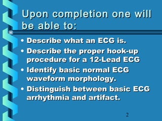
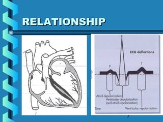
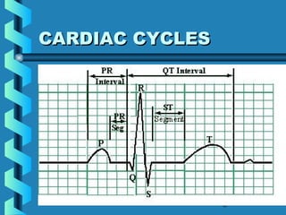
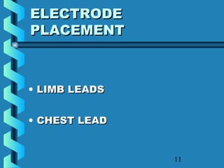
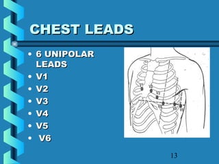
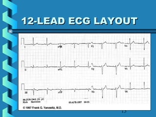
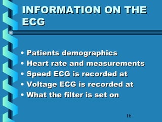
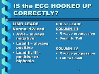
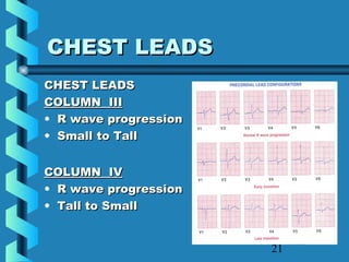
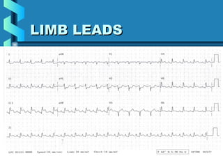
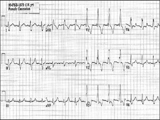
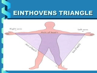
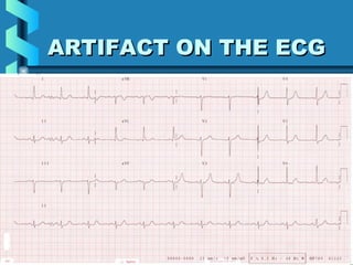
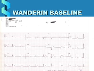
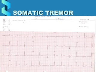
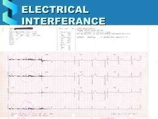
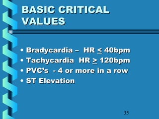
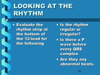
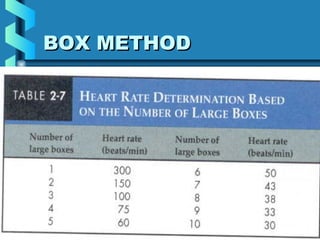
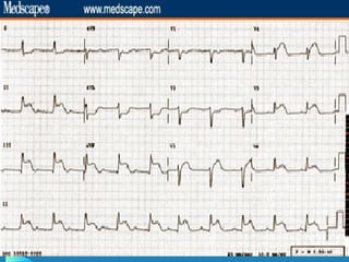
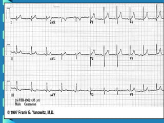
This document provides instructions for performing and interpreting a 12-lead electrocardiogram (ECG). It describes the proper procedure for electrode placement on the limbs and chest to produce the 12 leads. It also covers evaluating the ECG for correct lead placement, artifacts, and basic abnormalities like arrhythmias, ectopic beats, and critical values such as ST elevation. The overall goal is to produce high quality ECGs and correctly interpret basic findings.
This presentation covers few basic things about ECG, especially for UG Medical students like ECG leads, normal ECG waves, axis of ECG and how to look for common ECG misplacements.
The ECG is an essential tool for health professionals in making a diagnosis of abnormal heart rhythms when one is suspected. It is a routine investigation in people who complain of symptoms such as chest pain as well as breathlessness. The findings of the ECG can also have an effect on treatment of certain conditions, for example it is helpful in deciding some aspects of treatment in people who have suffered from a heart attack.
Salient features of the book are -
- The book provides a shortcut to understand and remember certain specific formulae and points you require to interpret the 12-lead ECG.
- Treatment protocols (in green boxes) for most of the important conditions are also included.
- View sample ECGs as you read along the topics.
- The content is explained in a very simple language to provide good conceptions, written from a student’s point of view.
- People can gain their belief in the book after going through sample ECGs which would be available at www.themedicalpost.net/ecg
- The book competes with the other books available in the market in simplicity, summaries, treatment protocols, live diagrams and regularly updated sample ECGs on the website.
Presentation for Medical undergraduates for teaching pharmacology. It deals with Physiology of steroid hormones and their action along with agents which are used therapeutically with their action, adverse effects and therapeutic uses.
ECG In Ischemic Heart Disease - Dr Vivek Baliga ReviewDr Vivek Baliga
Dr Vivek Baliga Presentation on the role of ECG in the diagnosis of ischemic heart disease. Here, he covers the very basics in ECG diagnosis of heart disease. Suitable for medical students and physicians alike. For more health articles for patients, visit http://baligadiagnostics.com/category/dr-vivek-baliga/
Topic; "ECG"
An Electrocardiogram (ECG) is a simple test that can be used to check your heart's rhythm and electrical activity.
Sensors attached to the skin are used to detect the electrical signals produced by your heart each time it beats. An ECG is often used alongside other tests to help diagnose and monitor conditions affecting the heart.
It can be used to investigate symptoms of a possible heart problem, such as chest pain, palpitations (suddenly noticeable heartbeats), dizziness and shortness of breath.
An ECG can help detect:
arrhythmias – where the heart beats too slowly, too quickly, or irregularly
coronary heart disease – where the heart's blood supply is blocked or interrupted by a build-up of fatty substances
heart attacks – where the supply of blood to the heart is suddenly blocked
cardiomyopathy – where the heart walls become thickened or enlarged.
Submitted by
Rida Batool
Pulmonary Thromboembolism - etilogy, types, medical- Surgical and nursing man...VarunMahajani
Disruption of blood supply to lung alveoli due to blockage of one or more pulmonary blood vessels is called as Pulmonary thromboembolism. In this presentation we will discuss its causes, types and its management in depth.
New Directions in Targeted Therapeutic Approaches for Older Adults With Mantl...i3 Health
i3 Health is pleased to make the speaker slides from this activity available for use as a non-accredited self-study or teaching resource.
This slide deck presented by Dr. Kami Maddocks, Professor-Clinical in the Division of Hematology and
Associate Division Director for Ambulatory Operations
The Ohio State University Comprehensive Cancer Center, will provide insight into new directions in targeted therapeutic approaches for older adults with mantle cell lymphoma.
STATEMENT OF NEED
Mantle cell lymphoma (MCL) is a rare, aggressive B-cell non-Hodgkin lymphoma (NHL) accounting for 5% to 7% of all lymphomas. Its prognosis ranges from indolent disease that does not require treatment for years to very aggressive disease, which is associated with poor survival (Silkenstedt et al, 2021). Typically, MCL is diagnosed at advanced stage and in older patients who cannot tolerate intensive therapy (NCCN, 2022). Although recent advances have slightly increased remission rates, recurrence and relapse remain very common, leading to a median overall survival between 3 and 6 years (LLS, 2021). Though there are several effective options, progress is still needed towards establishing an accepted frontline approach for MCL (Castellino et al, 2022). Treatment selection and management of MCL are complicated by the heterogeneity of prognosis, advanced age and comorbidities of patients, and lack of an established standard approach for treatment, making it vital that clinicians be familiar with the latest research and advances in this area. In this activity chaired by Michael Wang, MD, Professor in the Department of Lymphoma & Myeloma at MD Anderson Cancer Center, expert faculty will discuss prognostic factors informing treatment, the promising results of recent trials in new therapeutic approaches, and the implications of treatment resistance in therapeutic selection for MCL.
Target Audience
Hematology/oncology fellows, attending faculty, and other health care professionals involved in the treatment of patients with mantle cell lymphoma (MCL).
Learning Objectives
1.) Identify clinical and biological prognostic factors that can guide treatment decision making for older adults with MCL
2.) Evaluate emerging data on targeted therapeutic approaches for treatment-naive and relapsed/refractory MCL and their applicability to older adults
3.) Assess mechanisms of resistance to targeted therapies for MCL and their implications for treatment selection
The prostate is an exocrine gland of the male mammalian reproductive system
It is a walnut-sized gland that forms part of the male reproductive system and is located in front of the rectum and just below the urinary bladder
Function is to store and secrete a clear, slightly alkaline fluid that constitutes 10-30% of the volume of the seminal fluid that along with the spermatozoa, constitutes semen
A healthy human prostate measures (4cm-vertical, by 3cm-horizontal, 2cm ant-post ).
It surrounds the urethra just below the urinary bladder. It has anterior, median, posterior and two lateral lobes
It’s work is regulated by androgens which are responsible for male sex characteristics
Generalised disease of the prostate due to hormonal derangement which leads to non malignant enlargement of the gland (increase in the number of epithelial cells and stromal tissue)to cause compression of the urethra leading to symptoms (LUTS
micro teaching on communication m.sc nursing.pdfAnurag Sharma
Microteaching is a unique model of practice teaching. It is a viable instrument for the. desired change in the teaching behavior or the behavior potential which, in specified types of real. classroom situations, tends to facilitate the achievement of specified types of objectives.
Explore natural remedies for syphilis treatment in Singapore. Discover alternative therapies, herbal remedies, and lifestyle changes that may complement conventional treatments. Learn about holistic approaches to managing syphilis symptoms and supporting overall health.
Factory Supply Best Quality Pmk Oil CAS 28578–16–7 PMK Powder in Stockrebeccabio
Factory Supply Best Quality Pmk Oil CAS 28578–16–7 PMK Powder in Stock
Telegram: bmksupplier
signal: +85264872720
threema: TUD4A6YC
You can contact me on Telegram or Threema
Communicate promptly and reply
Free of customs clearance, Double Clearance 100% pass delivery to USA, Canada, Spain, Germany, Netherland, Poland, Italy, Sweden, UK, Czech Republic, Australia, Mexico, Russia, Ukraine, Kazakhstan.Door to door service
Hot Selling Organic intermediates
Anti ulcer drugs and their Advance pharmacology ||
Anti-ulcer drugs are medications used to prevent and treat ulcers in the stomach and upper part of the small intestine (duodenal ulcers). These ulcers are often caused by an imbalance between stomach acid and the mucosal lining, which protects the stomach lining.
||Scope: Overview of various classes of anti-ulcer drugs, their mechanisms of action, indications, side effects, and clinical considerations.
These lecture slides, by Dr Sidra Arshad, offer a quick overview of physiological basis of a normal electrocardiogram.
Learning objectives:
1. Define an electrocardiogram (ECG) and electrocardiography
2. Describe how dipoles generated by the heart produce the waveforms of the ECG
3. Describe the components of a normal electrocardiogram of a typical bipolar leads (limb II)
4. Differentiate between intervals and segments
5. Enlist some common indications for obtaining an ECG
Study Resources:
1. Chapter 11, Guyton and Hall Textbook of Medical Physiology, 14th edition
2. Chapter 9, Human Physiology - From Cells to Systems, Lauralee Sherwood, 9th edition
3. Chapter 29, Ganong’s Review of Medical Physiology, 26th edition
4. Electrocardiogram, StatPearls - https://www.ncbi.nlm.nih.gov/books/NBK549803/
5. ECG in Medical Practice by ABM Abdullah, 4th edition
6. ECG Basics, http://www.nataliescasebook.com/tag/e-c-g-basics
- Video recording of this lecture in English language: https://youtu.be/lK81BzxMqdo
- Video recording of this lecture in Arabic language: https://youtu.be/Ve4P0COk9OI
- Link to download the book free: https://nephrotube.blogspot.com/p/nephrotube-nephrology-books.html
- Link to NephroTube website: www.NephroTube.com
- Link to NephroTube social media accounts: https://nephrotube.blogspot.com/p/join-nephrotube-on-social-media.html
1. MAKING ECG’S EASYMAKING ECG’S EASY
APPLICATION FOR THEAPPLICATION FOR THE
ECGECG
EVALUATING THE ECGEVALUATING THE ECG
2. 2
Upon completion one willUpon completion one will
be able to:be able to:
• Describe what an ECG is.Describe what an ECG is.
• Describe the proper hook-upDescribe the proper hook-up
procedure for a 12-Lead ECGprocedure for a 12-Lead ECG
• Identify basic normal ECGIdentify basic normal ECG
waveform morphology.waveform morphology.
• Distinguish between basic ECGDistinguish between basic ECG
arrhythmia and artifact.arrhythmia and artifact.
7. 7
12 LEAD ECG12 LEAD ECG
Learning objectives:Learning objectives:
Describe the correctDescribe the correct
placement of all electrodesplacement of all electrodes
Proper Skin preparationProper Skin preparation
Care of the ECG machine, wiresCare of the ECG machine, wires
and electrodesand electrodes
8. 8
Principles ofPrinciples of
ElectrocardiographElectrocardiograph
• Electrocardiograph – is theElectrocardiograph – is the
instrument that records theinstrument that records the
electrical activity of the heartelectrical activity of the heart
• Electrocardiogram (ECG) is theElectrocardiogram (ECG) is the
record of that activityrecord of that activity
9. 9
HOOKING UP THEHOOKING UP THE
12-LEAD ECG12-LEAD ECG
• Proper skin prepProper skin prep
• Placement of the limbPlacement of the limb
electrodeselectrodes
• Placement of the chestPlacement of the chest
electrodeselectrodes
14. 14
LETS REVIEWLETS REVIEW
• What is an ECGWhat is an ECG
• What are the limb leads?What are the limb leads?
• What are the chest leads?What are the chest leads?
• Why do skin prep?Why do skin prep?
16. 16
INFORMATION ON THEINFORMATION ON THE
ECGECG
• Patients demographicsPatients demographics
• Heart rate and measurementsHeart rate and measurements
• Speed ECG is recorded atSpeed ECG is recorded at
• Voltage ECG is recorded atVoltage ECG is recorded at
• What the filter is set onWhat the filter is set on
18. 18
WHAT YOU NEED TOWHAT YOU NEED TO
LOOK FORLOOK FOR
• Are the limb leads hooked upAre the limb leads hooked up
correctly?correctly?
• Are the chest leads hooked upAre the chest leads hooked up
correctly?correctly?
• Is the ECG free of artifact.Is the ECG free of artifact.
• Is this ECG a Critical ValueIs this ECG a Critical Value
19. 19
IS the ECG HOOKED UPIS the ECG HOOKED UP
CORRECTLY?CORRECTLY?
LIMB LEADSLIMB LEADS
Normal 12-leadNormal 12-lead
• AVR – alwaysAVR – always
negativenegative
• Lead I – alwaysLead I – always
positivepositive
• Lead II, III –Lead II, III –
positive orpositive or
biphasicbiphasic
CHEST LEADSCHEST LEADS
COLUMN IIICOLUMN III
• R wave progressionR wave progression
• Small to TallSmall to Tall
COLUMN IVCOLUMN IV
• R wave progressionR wave progression
• Tall to SmallTall to Small
20. 20
RULING OUT LIMB LEADRULING OUT LIMB LEAD
REVERSALREVERSAL
• Avr is always negativeAvr is always negative
• Lead I is always positiveLead I is always positive
• Lead II and III positive for the PLead II and III positive for the P
wave and usually the QRSwave and usually the QRS
complexcomplex
21. 21
CHEST LEADSCHEST LEADS
COLUMN IIICOLUMN III
• R wave progressionR wave progression
• Small to TallSmall to Tall
COLUMN IVCOLUMN IV
• R wave progressionR wave progression
• Tall to SmallTall to Small
35. 35
BASIC CRITICALBASIC CRITICAL
VALUESVALUES
• Bradycardia – HRBradycardia – HR << 40bpm40bpm
• Tachycardia HRTachycardia HR >> 120bpm120bpm
• PVC’s - 4 or more in a rowPVC’s - 4 or more in a row
• ST ElevationST Elevation
36. 36
LOOKING AT THELOOKING AT THE
RHYTHMRHYTHM
• Evaluate theEvaluate the
rhythm strip atrhythm strip at
the bottom ofthe bottom of
the 12-lead forthe 12-lead for
the followingthe following
• Is the rhythmIs the rhythm
regular orregular or
irregular?irregular?
• Is there a PIs there a P
wave beforewave before
every QRSevery QRS
complexcomplex
• Are they anyAre they any
abnormal beats.abnormal beats.
45. 45
LETS SUMMARIZELETS SUMMARIZE
How do we produce an excellentHow do we produce an excellent
12-lead ECG?12-lead ECG?
Proper skin prepProper skin prep
Correct electrode placementCorrect electrode placement
Recognize and know how toRecognize and know how to
correct problemscorrect problems
Recognize basic critical valuesRecognize basic critical values
Editor's Notes
In an adult with a healthy heart, the heart rate is usually about 72 beats per minute.
The excitatory and electrical conduction system of the heart is responsible for the contraction and relaxation of the heart muscle.
The heart is divided into four chambers, but it functions as a two sided pump.
Top is the right and left atria
Bottom is the right and left ventricles
The right side of the heart receives and pumps venous blood to the lungs.
The left side of the heart receives (from the lungs) and pumps arterial blood to the body.
The sinoatrial node (SA node) is the pacemaker where the electrical impulse is generated. This node is located along the posterior wall of the right atrium right beneath the opening of the superior vena cava. It is crescent shaped and about 3 mm wide and 1 cm long.
The impulse travels from the SA node through the internodal pathways to the atrioventricular node (AV node).
The AV node is responsible for conduction of the impulse from the atria to the ventricles. The impulse is delayed slightly at this point to allow complete emptying of the atria before the ventricles contract. The impulse continues through the AV bundle and down the left and right bundle branches of the Purkinje fibers.
The Purkinje fibers conduct the impulse to all parts of the ventricles, causing contraction (Guyton, 1982).
Lets look at how the conduction system related to what we record on the ECG.
P wave: the sequential activation (depolarization) of the right and left atria
QRS complex: right and left ventricular depolarization (normally the ventricles are activated simultaneously)
ST-T wave ventricular repolarization
U wave: origin for this wave is not clear - but probably represents &quot;afterdepolarizations&quot; in the ventricles PR interval: time interval from onset of atrial depolarization (P wave) to onset of ventricular depolarization (QRS complex)QRS duration: duration of ventricular muscle depolarizationQT interval: duration of ventricular depolarization and repolarizationRR interval: duration of ventricular cardiac cycle (an indicator of ventricular rate)PP interval: duration of atrial cycle (an indicator of atrial rate)
Recorded on graph paper.
Time is measured across
Voltage is measured up and down.
Small squares 1mm high 0.04 secs wide
5 small squares = 1 large square
1 large square is 5mm high and 0.20 sec wide
Both re-usable and disposable electrodes rely on the electrolyte in the electrodes to
make an effective connection between the machines electrode and the patient’s
electro-physiological signals.
These signals are not picked up just off the surface of the skin,
where dead or dry skin, oils and hair all prevent the signals from being detected.
The signals are under this surface, and thus we have to prepare the patient so that
the electrolyte can reach the signals beneath.
This is achieved by:
Removing oil, greasy and dirt from the skins sites with alcohol
ELECTRODE PLACEMENT
The standard 12-Lead ECG is a collect of tracings of electrical activity occurring in the heart. Each lead provides a tracing, which is characteristic of a different view of the same electrical activity. Other words we take 12 different angles (pictures) of the same activity.
In a standard 12-lead ECG there are 6 limb leads with a three-electrode connection made to the Right arm, Left arm and Left leg. The right leg is also hooked up to the machine but this is your ground lead and does not generate a signal on the ECG.
The chest electrodes are labelled “V” and are numbered from 1 to 6.
The placement of these electrodes needs to be exact to give the optimum information as possible. If the electrodes are placed incorrectly on the chest, the tracing will reveal duplication of some information, while other areas will not be represented properly.
Incorrect placement of the electrodes can lead to serious errors of interpretation.
There are six chest leads: V1, V2,V3,V4, V5 and V6.
V1 at the fourth intercostal space, at the right margin of the sternum
V2at the fourth intercostal space, at the left margin of the sternum
V3midway between the position of leads V2 and V4 (in a straight line)
V4at the fifth intercostals space at the junction of the left midclavicular line
V5 midway between the position of leads V4 and V6 (straight down from the axillary
Line on the same horizontal position as V4 and V6
V6at the horizontal position of V4, at the left of the midaxillary line.
Lets look at this ECG for the limb leads?
Is lead I positive? Is AVR negative
We have reversed our limb electrodes we have the right arm on the left arm and reverse.
We need to have R wave progression
In column III the R wave(first positive wave of the QRS) goes from short to tall V1 shortest R wave
V2 R wave taller than in V1 and V3 has the tallest R wave.
In column IV the R wave is always the shortest in V6.
Is this ECG hooked up correctly for the limb leads?
Is it for the chest leads?
Lets evaluate this ECG to see if the Limb leads and Chest Leads are hooked up correctly?
Is this ECG HOOKED UP CORRECTLY FOR LIMB AND CHEST?
Lead I = right arm and left arm
Lead II = right arm and left Leg
Lead III = left arm and Left Leg.
Look at the number of large boxes before two R waves and then using this method find the HR .
I just remember that 2 ½ large boxes or less is critical value for tachycardia
7 or more large boxes is critical for bradycardia
Look at the rhythm strip is it regular or irregular.
How many large boxes are between two R waves = 5 = 60bpm