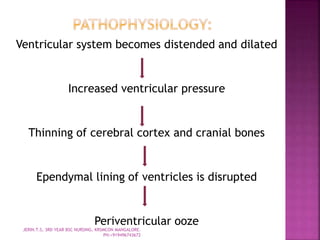
JERIN.T.S, 3RD YEAR BSC NURSING, KRSMCON
MANGALORE. PH:+919496743672
 Hydrocephalus is characterized by dilation
of the cerebral ventricles, increased CSF
(predominantly of about 80-90%) formed by
the choroid plexus of the lateral, third or
fourth ventricles by an active transport
process across the endothelium of
capillaries in the villus process of the
choroid plexus. The arachnoid villus is the
primary site of CSF absorption.
JERIN.T.S, 3RD YEAR BSC NURSING, KRSMCON MANGALORE.
PH:+919496743672
 Hydrocephalus is the abnormal
accumulation of cerebrospinal fluid in
the intracranial spaces. It occurs due
to imbalance between production or
absorption of CSF or due to
obstruction of the CSF pathways. It
results in the dilatation of the
cerebral ventricles and enlargement of
head.
JERIN.T.S, 3RD YEAR BSC NURSING, KRSMCON MANGALORE.
PH:+919496743672
 Increased production (obstructive
hydrocephalus) eg. Pseudo tumor cerebri,
Choroid plexus papilloma.
 Obstruction to the flow (communication
hydrocephalus) eg. Inflammatory Adhesions,
developmental obstructive lesions.
 Interference with absorption eg. Cavernous
sinus thrombosis
JERIN.T.S, 3RD YEAR BSC NURSING, KRSMCON MANGALORE.
PH:+919496743672
Clinically the causes are:
1. Congenital hydrocephalus:
i. Arnold-chiari malformation: In which there
is a displacement of the brain stem and
cerebellum through foramen magnum, into
upper cervical part of the spine. It is generally
associated with spina bifida and
meningomyelocele.
ii. Dandy-walker anomaly: In which
congenital septa or membrane block the
outlet of the fourth ventricle.
iii. Malformations or stenotic lesions of
aqueduct cerebri.
iv. Malformations of arachnoid villi.
JERIN.T.S, 3RD YEAR BSC NURSING, KRSMCON MANGALORE.
PH:+919496743672
 2. Acquired hydrocephalus:
 i. Inflammatory: Meningitis occasionally
encephalitis in first few months of life.
 ii. Traumatic: Birth trauma, head injury,
intracranial hemorrhage.
 iii. Neoplastic: Space occupying lesions like
tuberculoma, subdural hematoma or abscess
gliomas etc.
 iv. Chemical: Hypervitaminosis A
 v. Connective tissue disorders:
Achondroplasia.
JERIN.T.S, 3RD YEAR BSC NURSING, KRSMCON MANGALORE.
PH:+919496743672
 1. Congenital hydrocephalus:
It is present right at birth or becomes
apparent in the first few months of life.
 2. Acquired hydrocephalus:
Develops later in association with or as a
sequele to the causative factor.
 Types depending on the site of obstructions
a. Communicating hydrocephalus:
The ventricular system is patent and the
site of block is in the basal Subarachnoid
JERIN.T.S, 3RD YEAR BSC NURSING, KRSMCON MANGALORE.
PH:+919496743672
Sulci or at the arachnoid villi. Meningitis is the
most common cause of Communicating
hydrocephalus.
b. Non-communicating hydrocephalus:
There is a blockage of CSF pathway at or
proximal to the outlet foramina of the Fourth
ventricle the cause is usually congenital.
c. Arrested hydrocephalus:
A large proportion of both congenital and
acquired hydrocephalus may undergo
spontaneous arrest and may not require active
surgical intervention.
JERIN.T.S, 3RD YEAR BSC NURSING, KRSMCON MANGALORE.
PH:+919496743672
Ventricular system becomes distended and dilated
Increased ventricular pressure
Thinning of cerebral cortex and cranial bones
Ependymal lining of ventricles is disrupted
Periventricular ooze
JERIN.T.S, 3RD YEAR BSC NURSING, KRSMCON MANGALORE.
PH:+919496743672
Subependymal edema occurs and white matter
is compressed
Downward bulging of third ventricle
compresses the optic nerves and hypophysis
cerebri with dilation of sella turcica
Atrophy of choroid plexus and cortex occurs
JERIN.T.S, 3RD YEAR BSC NURSING, KRSMCON MANGALORE.
PH:+919496743672
These depend on age at onset, duration and
severity.
Early onset (0-2 years):
 50% of children may be asymptomatic.
 Vomiting, drowsiness, failure to thrive, shrill cry.
 Delayed motor milestones are common symptoms
 Progressive increase in head circumference
 Head shape is abnormal and overshadows face
 Frontal bossing and triangular facias
 The skin of scalp is shiny and tense with dilated
veins and sparse hair
 The anterior fontanelle is open, large and non-
pulsatile
JERIN.T.S, 3RD YEAR BSC NURSING, KRSMCON MANGALORE.
PH:+919496743672
 Open squamo-parietal suture beyond the first month is
an early sign of hydrocephalus
 Ocular signs such as 6th nerve palsy
 Sunset sign, Ptosis and Nystagmus may be present
 Spasticity of lower limbs due to compression of
periventricular white matter may develop
 Pseudo bulsar palsy may be present and results in
regurgitation, dysphonia and stridor
 Late onset (2-10 years):
 Papilledema, headache and vomiting are usual
presenting features
 Increasing head size is present in only 605
 McEwen’s sign (crack pot resonance on percussion of
the skull)
 Psychomotor retardation and gait anomaly in 30% and
epilepsy in 20%
JERIN.T.S, 3RD YEAR BSC NURSING, KRSMCON MANGALORE.
PH:+919496743672
 Physical examination and neurological assessment
 Accurate serial recording of the head circumference
 Increase in head circumference in first 3 months of
life more than 1 cm every fortnight should arouse
suspicion of hydrocephalus
 Persistent widening of parietal sutures
 Positive Trans illumination of infant head,
 Typical cracked pot sound (Macewen’s sign) of the
skull bone
 Ophthalmoscopy
 MRI, CT scan, cranial ultrasonography and X-ray skull -
site of obstruction and in congenital hydrocephalus to
identify associated malformations.
JERIN.T.S, 3RD YEAR BSC NURSING, KRSMCON MANGALORE.
PH:+919496743672
 Carbonic anhydrase inhibitors like
Acetazolamide (Diamox) in doses of
50mg/kg/day to reduce CSF production in
slow progressive hydrocephalus.
 Oral glycerol and Isosoribide is also used
to reduce CSF production.
JERIN.T.S, 3RD YEAR BSC NURSING, KRSMCON MANGALORE.
PH:+919496743672
 Hydrocephalus can be treated through a variety
of surgical procedures including direct operation
on the lesion causing the obstruction, such as
tumor.
 Intracranial shunts for selected cases of non-
communicating hydrocephalus to divert fluid
from the obstructed segment of the ventricular
system to the subarachnoid space.
 Extracranial shunts (most common) to divert
fluid from the ventricular system to extracranial
compartments, frequently the peritoneum or
right atrium.
JERIN.T.S, 3RD YEAR BSC NURSING, KRSMCON MANGALORE.
PH:+919496743672
 Ventriculo peritoneal shunt (V-P shunt): lateral
ventricle or the spinal subarachnoid space to the
peritoneal cavity.
 2. Ventriculo atrial shunt (V-A shunt): dilated
lateral ventricle through a burr hole in the parietal
region, ear and into a vein down to a point where it
discharges into the right atrium or superior
venacava
 3. Ventriculo pleural shunt:
Diverts CSF to the pleural cavity, when the V-P or V-A
route cannot be used.
 4. Ventricle-gall bladder shunt:
Diverts CSF to the common bile duct
JERIN.T.S, 3RD YEAR BSC NURSING, KRSMCON MANGALORE.
PH:+919496743672
 Need for shunt revision frequently occurs
because of occlusion, infection or malfunction.
 Shunt revision may be necessary because of
growth of the child, newer models, however
include coiled tubing to allow the shunt to grow
with the child.
 Shunt dependency frequently occurs, the child
rapidly manifests symptoms of increased
intracranial pressure of the shunt does not
function optimally.
 Children with V-A shunt may experience
endocardial contusions and clotting, leading to
bacterial endocarditis, bacteremia and
ventriculitis or thromboembolism and
corpulmonale.JERIN.T.S, 3RD YEAR BSC NURSING, KRSMCON MANGALORE.
PH:+919496743672
 Infants:
 Assess head circumference – point out largest
circumference
 Palpate fontanelle for bulging and tenderness
 Assess pupillary response
 Assess level of consciousness
 Evaluate breathing patterns and effectiveness
 Assess feeding patterns and patterns of emesis
 Assess motor activity
 Determine attainment of developmental
milestones.
JERIN.T.S, 3RD YEAR BSC NURSING, KRSMCON MANGALORE.
PH:+919496743672
 Older child:
 Measure vital signs for signs of increased ICP
 Assess patterns of headache, emesis
 Determine pupillary response
 Evaluate the level of consciousness
 Assess motor function
 Evaluate attainment of mile stones, school
performance
 Obtain parents report of recent behaviour.
JERIN.T.S, 3RD YEAR BSC NURSING, KRSMCON MANGALORE.
PH:+919496743672
 Impaired cerebral tissue perfusion related to
increased ICP.
 Impaired nutritional status less than body requirement
related to reduced oral intake and vomiting
 Risk for impaired skin integrity related to enlarged
head.
 Anxiety related to abnormal condition and surgical
interventions.
 Risk for infection related to introduction of infecting
organism through the shunt.
 Risk for fluid volume deficit related to CSF drainage.
 Ineffective family coping related to life threatening
problem of infant.
JERIN.T.S, 3RD YEAR BSC NURSING, KRSMCON MANGALORE.
PH:+919496743672
 Observe for the signs of increased ICP i.e. for altered mental state,
vomiting, strabismus, slowed respiration, decreased pulse or increased
pulse.
 Measure the occipital frontal circumference daily
 Daily palpate the fontanelles for bulging, size and tension.
 Record vital signs
 Small and frequent feeds are given to child to prevent vomiting.
 Give the feeds only when the child is relaxed and calm.
 After every feed burp the baby and place him on his right side with head
elevated to prevent aspiration.
 While holding the baby, avoid pressure on the neck by supporting the head.
 Position of the baby is changed every 2 hourly to prevent the occurrence of
pressure sores on the scalp and to prevent hypostatic pneumonia.
 A pad of cotton can be placed under the head.
 The infant must be kept clean and dry especially around the creases of the
neck.
 While moving the baby proper should be given to the head.
 Give psychological support and detailed information to parents regarding
each investigation and about the condition of the child.
 Involve the parents in daily care of the child.
JERIN.T.S, 3RD YEAR BSC NURSING, KRSMCON MANGALORE.
PH:+919496743672
 The vital signs are checked every 15-30 mins.
 Observe for increased ICP and if present it should be reported.
 If shunt is done, the catheter tract is checked for swelling and
tenderness.
 The shunt is tested for patency.
 The infant is positioned on the operative site to prevent pressure on
the shunt valve.
 If there is no vomiting, place the baby on his back to prevent pressure
on one side of the scalp.
 Observe the fontanelles; if it is depressed lower the head end of the
bed.
 Mild analgesics are given for pain.
 Administration of prophylactic antibiotics is done to prevent infection.
 Maintain the oral hygiene and give skin care daily.
 Allow proper drainage of CSF, through the shunt, pump the shunt, and
carefully compress the valve for instructed timing.
 If there is any feeding difficulty, report to the surgeon maintains the
fluid and electrolyte balance.
 Explain to the parents regarding the conditions of the child and also
about the procedures to be carried out for the child.JERIN.T.S, 3RD YEAR BSC NURSING, KRSMCON MANGALORE.
PH:+919496743672
 Seizures
 Herniation of brain
 Persistent increased ICP
 Developmental delay
 Infections
 Neurological deficits
 Motor and intellectual handicaps
 Visual problems
 Aggressive and delinquent behaviour.
 Shunt complications are found as shunt revision due to
shunt occlusion, malfunction, infection and physical
growth of the child.
 Ventriculoatrial (V-A) shunt may be complicated with
endocarditis, bacteremia, thromboembolism,
ventriculitis, corpulmonale etc.JERIN.T.S, 3RD YEAR BSC NURSING, KRSMCON MANGALORE.
PH:+919496743672

Hydrocephalus

  • 1.
    JERIN.T.S, 3RD YEARBSC NURSING, KRSMCON MANGALORE. PH:+919496743672
  • 2.
     Hydrocephalus ischaracterized by dilation of the cerebral ventricles, increased CSF (predominantly of about 80-90%) formed by the choroid plexus of the lateral, third or fourth ventricles by an active transport process across the endothelium of capillaries in the villus process of the choroid plexus. The arachnoid villus is the primary site of CSF absorption. JERIN.T.S, 3RD YEAR BSC NURSING, KRSMCON MANGALORE. PH:+919496743672
  • 3.
     Hydrocephalus isthe abnormal accumulation of cerebrospinal fluid in the intracranial spaces. It occurs due to imbalance between production or absorption of CSF or due to obstruction of the CSF pathways. It results in the dilatation of the cerebral ventricles and enlargement of head. JERIN.T.S, 3RD YEAR BSC NURSING, KRSMCON MANGALORE. PH:+919496743672
  • 4.
     Increased production(obstructive hydrocephalus) eg. Pseudo tumor cerebri, Choroid plexus papilloma.  Obstruction to the flow (communication hydrocephalus) eg. Inflammatory Adhesions, developmental obstructive lesions.  Interference with absorption eg. Cavernous sinus thrombosis JERIN.T.S, 3RD YEAR BSC NURSING, KRSMCON MANGALORE. PH:+919496743672
  • 5.
    Clinically the causesare: 1. Congenital hydrocephalus: i. Arnold-chiari malformation: In which there is a displacement of the brain stem and cerebellum through foramen magnum, into upper cervical part of the spine. It is generally associated with spina bifida and meningomyelocele. ii. Dandy-walker anomaly: In which congenital septa or membrane block the outlet of the fourth ventricle. iii. Malformations or stenotic lesions of aqueduct cerebri. iv. Malformations of arachnoid villi. JERIN.T.S, 3RD YEAR BSC NURSING, KRSMCON MANGALORE. PH:+919496743672
  • 6.
     2. Acquiredhydrocephalus:  i. Inflammatory: Meningitis occasionally encephalitis in first few months of life.  ii. Traumatic: Birth trauma, head injury, intracranial hemorrhage.  iii. Neoplastic: Space occupying lesions like tuberculoma, subdural hematoma or abscess gliomas etc.  iv. Chemical: Hypervitaminosis A  v. Connective tissue disorders: Achondroplasia. JERIN.T.S, 3RD YEAR BSC NURSING, KRSMCON MANGALORE. PH:+919496743672
  • 7.
     1. Congenitalhydrocephalus: It is present right at birth or becomes apparent in the first few months of life.  2. Acquired hydrocephalus: Develops later in association with or as a sequele to the causative factor.  Types depending on the site of obstructions a. Communicating hydrocephalus: The ventricular system is patent and the site of block is in the basal Subarachnoid JERIN.T.S, 3RD YEAR BSC NURSING, KRSMCON MANGALORE. PH:+919496743672
  • 8.
    Sulci or atthe arachnoid villi. Meningitis is the most common cause of Communicating hydrocephalus. b. Non-communicating hydrocephalus: There is a blockage of CSF pathway at or proximal to the outlet foramina of the Fourth ventricle the cause is usually congenital. c. Arrested hydrocephalus: A large proportion of both congenital and acquired hydrocephalus may undergo spontaneous arrest and may not require active surgical intervention. JERIN.T.S, 3RD YEAR BSC NURSING, KRSMCON MANGALORE. PH:+919496743672
  • 9.
    Ventricular system becomesdistended and dilated Increased ventricular pressure Thinning of cerebral cortex and cranial bones Ependymal lining of ventricles is disrupted Periventricular ooze JERIN.T.S, 3RD YEAR BSC NURSING, KRSMCON MANGALORE. PH:+919496743672
  • 10.
    Subependymal edema occursand white matter is compressed Downward bulging of third ventricle compresses the optic nerves and hypophysis cerebri with dilation of sella turcica Atrophy of choroid plexus and cortex occurs JERIN.T.S, 3RD YEAR BSC NURSING, KRSMCON MANGALORE. PH:+919496743672
  • 11.
    These depend onage at onset, duration and severity. Early onset (0-2 years):  50% of children may be asymptomatic.  Vomiting, drowsiness, failure to thrive, shrill cry.  Delayed motor milestones are common symptoms  Progressive increase in head circumference  Head shape is abnormal and overshadows face  Frontal bossing and triangular facias  The skin of scalp is shiny and tense with dilated veins and sparse hair  The anterior fontanelle is open, large and non- pulsatile JERIN.T.S, 3RD YEAR BSC NURSING, KRSMCON MANGALORE. PH:+919496743672
  • 12.
     Open squamo-parietalsuture beyond the first month is an early sign of hydrocephalus  Ocular signs such as 6th nerve palsy  Sunset sign, Ptosis and Nystagmus may be present  Spasticity of lower limbs due to compression of periventricular white matter may develop  Pseudo bulsar palsy may be present and results in regurgitation, dysphonia and stridor  Late onset (2-10 years):  Papilledema, headache and vomiting are usual presenting features  Increasing head size is present in only 605  McEwen’s sign (crack pot resonance on percussion of the skull)  Psychomotor retardation and gait anomaly in 30% and epilepsy in 20% JERIN.T.S, 3RD YEAR BSC NURSING, KRSMCON MANGALORE. PH:+919496743672
  • 13.
     Physical examinationand neurological assessment  Accurate serial recording of the head circumference  Increase in head circumference in first 3 months of life more than 1 cm every fortnight should arouse suspicion of hydrocephalus  Persistent widening of parietal sutures  Positive Trans illumination of infant head,  Typical cracked pot sound (Macewen’s sign) of the skull bone  Ophthalmoscopy  MRI, CT scan, cranial ultrasonography and X-ray skull - site of obstruction and in congenital hydrocephalus to identify associated malformations. JERIN.T.S, 3RD YEAR BSC NURSING, KRSMCON MANGALORE. PH:+919496743672
  • 14.
     Carbonic anhydraseinhibitors like Acetazolamide (Diamox) in doses of 50mg/kg/day to reduce CSF production in slow progressive hydrocephalus.  Oral glycerol and Isosoribide is also used to reduce CSF production. JERIN.T.S, 3RD YEAR BSC NURSING, KRSMCON MANGALORE. PH:+919496743672
  • 15.
     Hydrocephalus canbe treated through a variety of surgical procedures including direct operation on the lesion causing the obstruction, such as tumor.  Intracranial shunts for selected cases of non- communicating hydrocephalus to divert fluid from the obstructed segment of the ventricular system to the subarachnoid space.  Extracranial shunts (most common) to divert fluid from the ventricular system to extracranial compartments, frequently the peritoneum or right atrium. JERIN.T.S, 3RD YEAR BSC NURSING, KRSMCON MANGALORE. PH:+919496743672
  • 16.
     Ventriculo peritonealshunt (V-P shunt): lateral ventricle or the spinal subarachnoid space to the peritoneal cavity.  2. Ventriculo atrial shunt (V-A shunt): dilated lateral ventricle through a burr hole in the parietal region, ear and into a vein down to a point where it discharges into the right atrium or superior venacava  3. Ventriculo pleural shunt: Diverts CSF to the pleural cavity, when the V-P or V-A route cannot be used.  4. Ventricle-gall bladder shunt: Diverts CSF to the common bile duct JERIN.T.S, 3RD YEAR BSC NURSING, KRSMCON MANGALORE. PH:+919496743672
  • 17.
     Need forshunt revision frequently occurs because of occlusion, infection or malfunction.  Shunt revision may be necessary because of growth of the child, newer models, however include coiled tubing to allow the shunt to grow with the child.  Shunt dependency frequently occurs, the child rapidly manifests symptoms of increased intracranial pressure of the shunt does not function optimally.  Children with V-A shunt may experience endocardial contusions and clotting, leading to bacterial endocarditis, bacteremia and ventriculitis or thromboembolism and corpulmonale.JERIN.T.S, 3RD YEAR BSC NURSING, KRSMCON MANGALORE. PH:+919496743672
  • 18.
     Infants:  Assesshead circumference – point out largest circumference  Palpate fontanelle for bulging and tenderness  Assess pupillary response  Assess level of consciousness  Evaluate breathing patterns and effectiveness  Assess feeding patterns and patterns of emesis  Assess motor activity  Determine attainment of developmental milestones. JERIN.T.S, 3RD YEAR BSC NURSING, KRSMCON MANGALORE. PH:+919496743672
  • 19.
     Older child: Measure vital signs for signs of increased ICP  Assess patterns of headache, emesis  Determine pupillary response  Evaluate the level of consciousness  Assess motor function  Evaluate attainment of mile stones, school performance  Obtain parents report of recent behaviour. JERIN.T.S, 3RD YEAR BSC NURSING, KRSMCON MANGALORE. PH:+919496743672
  • 20.
     Impaired cerebraltissue perfusion related to increased ICP.  Impaired nutritional status less than body requirement related to reduced oral intake and vomiting  Risk for impaired skin integrity related to enlarged head.  Anxiety related to abnormal condition and surgical interventions.  Risk for infection related to introduction of infecting organism through the shunt.  Risk for fluid volume deficit related to CSF drainage.  Ineffective family coping related to life threatening problem of infant. JERIN.T.S, 3RD YEAR BSC NURSING, KRSMCON MANGALORE. PH:+919496743672
  • 21.
     Observe forthe signs of increased ICP i.e. for altered mental state, vomiting, strabismus, slowed respiration, decreased pulse or increased pulse.  Measure the occipital frontal circumference daily  Daily palpate the fontanelles for bulging, size and tension.  Record vital signs  Small and frequent feeds are given to child to prevent vomiting.  Give the feeds only when the child is relaxed and calm.  After every feed burp the baby and place him on his right side with head elevated to prevent aspiration.  While holding the baby, avoid pressure on the neck by supporting the head.  Position of the baby is changed every 2 hourly to prevent the occurrence of pressure sores on the scalp and to prevent hypostatic pneumonia.  A pad of cotton can be placed under the head.  The infant must be kept clean and dry especially around the creases of the neck.  While moving the baby proper should be given to the head.  Give psychological support and detailed information to parents regarding each investigation and about the condition of the child.  Involve the parents in daily care of the child. JERIN.T.S, 3RD YEAR BSC NURSING, KRSMCON MANGALORE. PH:+919496743672
  • 22.
     The vitalsigns are checked every 15-30 mins.  Observe for increased ICP and if present it should be reported.  If shunt is done, the catheter tract is checked for swelling and tenderness.  The shunt is tested for patency.  The infant is positioned on the operative site to prevent pressure on the shunt valve.  If there is no vomiting, place the baby on his back to prevent pressure on one side of the scalp.  Observe the fontanelles; if it is depressed lower the head end of the bed.  Mild analgesics are given for pain.  Administration of prophylactic antibiotics is done to prevent infection.  Maintain the oral hygiene and give skin care daily.  Allow proper drainage of CSF, through the shunt, pump the shunt, and carefully compress the valve for instructed timing.  If there is any feeding difficulty, report to the surgeon maintains the fluid and electrolyte balance.  Explain to the parents regarding the conditions of the child and also about the procedures to be carried out for the child.JERIN.T.S, 3RD YEAR BSC NURSING, KRSMCON MANGALORE. PH:+919496743672
  • 23.
     Seizures  Herniationof brain  Persistent increased ICP  Developmental delay  Infections  Neurological deficits  Motor and intellectual handicaps  Visual problems  Aggressive and delinquent behaviour.  Shunt complications are found as shunt revision due to shunt occlusion, malfunction, infection and physical growth of the child.  Ventriculoatrial (V-A) shunt may be complicated with endocarditis, bacteremia, thromboembolism, ventriculitis, corpulmonale etc.JERIN.T.S, 3RD YEAR BSC NURSING, KRSMCON MANGALORE. PH:+919496743672