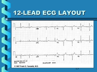
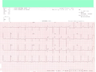
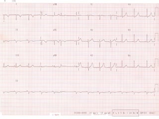
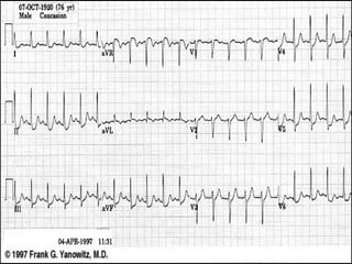
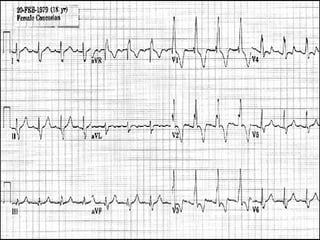
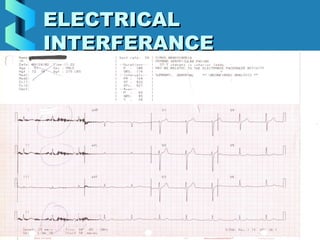
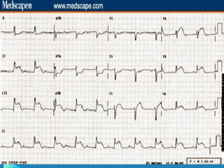
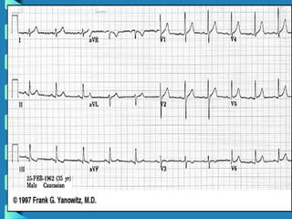
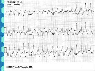
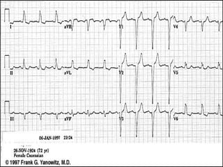
MAKING ECG’S EASYMAKING ECG’S EASY
APPLICATION FOR THEAPPLICATION FOR THE
ECGECG
EVALUATING THE ECGEVALUATING THE ECG
2
Upon completion one willUpon completion one will
be able to:be able to:
• Describe what an ECG is.Describe what an ECG is.
• Describe the proper hook-upDescribe the proper hook-up
procedure for a 12-Lead ECGprocedure for a 12-Lead ECG
• Identify basic normal ECGIdentify basic normal ECG
waveform morphology.waveform morphology.
• Distinguish between basic ECGDistinguish between basic ECG
arrhythmia and artifact.arrhythmia and artifact.
3
QUICK REVIEW OFQUICK REVIEW OF
HEARTHEART
PurposePurpose
• Pumps bloodPumps blood
Basic AnatomyBasic Anatomy
• 4 chambers4 chambers
• 2 sides2 sides
• 4 valves4 valves
4
THE CONDUCTINGYTHE CONDUCTINGY
SYSTEMSYSTEM
• SA NodeSA Node
• Inter-nodalInter-nodal
pathwaypathway
• AV NodeAV Node
• Bundle of HISBundle of HIS
• Bundle BranchesBundle Branches
• Purkinje FibersPurkinje Fibers
5
RELATIONSHIPRELATIONSHIP
6
CARDIAC CYCLESCARDIAC CYCLES
7
12 LEAD ECG12 LEAD ECG
Learning objectives:Learning objectives:
Describe the correctDescribe the correct
placement of all electrodesplacement of all electrodes
Proper Skin preparationProper Skin preparation
Care of the ECG machine, wiresCare of the ECG machine, wires
and electrodesand electrodes
8
Principles ofPrinciples of
ElectrocardiographElectrocardiograph
• Electrocardiograph – is theElectrocardiograph – is the
instrument that records theinstrument that records the
electrical activity of the heartelectrical activity of the heart
• Electrocardiogram (ECG) is theElectrocardiogram (ECG) is the
record of that activityrecord of that activity
9
HOOKING UP THEHOOKING UP THE
12-LEAD ECG12-LEAD ECG
• Proper skin prepProper skin prep
• Placement of the limbPlacement of the limb
electrodeselectrodes
• Placement of the chestPlacement of the chest
electrodeselectrodes
10
SKIN PREPARATIONSKIN PREPARATION
• REASON FOR SKINREASON FOR SKIN
PREPARATIONPREPARATION
• 5 STEP METHOD5 STEP METHOD
• 2 STEP METHOD2 STEP METHOD
11
ELECTRODEELECTRODE
PLACEMENTPLACEMENT
• LIMB LEADSLIMB LEADS
• CHEST LEADCHEST LEAD
12
LIMB LEADSLIMB LEADS
• Bipolar leadsBipolar leads
I II IIII II III
• Augment leadsAugment leads
Avr Avl AvfAvr Avl Avf
 
13
CHEST LEADSCHEST LEADS
• 6 UNIPOLAR6 UNIPOLAR
LEADSLEADS
• V1V1
• V2V2
• V3V3
• V4V4
• V5V5
• V6V6
14
LETS REVIEWLETS REVIEW
• What is an ECGWhat is an ECG
• What are the limb leads?What are the limb leads?
• What are the chest leads?What are the chest leads?
• Why do skin prep?Why do skin prep?
15
12-LEAD ECG LAYOUT12-LEAD ECG LAYOUT
16
INFORMATION ON THEINFORMATION ON THE
ECGECG
• Patients demographicsPatients demographics
• Heart rate and measurementsHeart rate and measurements
• Speed ECG is recorded atSpeed ECG is recorded at
• Voltage ECG is recorded atVoltage ECG is recorded at
• What the filter is set onWhat the filter is set on
17
INFORMATION ON THEINFORMATION ON THE
12-LEAD ECG12-LEAD ECG
18
WHAT YOU NEED TOWHAT YOU NEED TO
LOOK FORLOOK FOR
• Are the limb leads hooked upAre the limb leads hooked up
correctly?correctly?
• Are the chest leads hooked upAre the chest leads hooked up
correctly?correctly?
• Is the ECG free of artifact.Is the ECG free of artifact.
• Is this ECG a Critical ValueIs this ECG a Critical Value
19
IS the ECG HOOKED UPIS the ECG HOOKED UP
CORRECTLY?CORRECTLY?
LIMB LEADSLIMB LEADS
Normal 12-leadNormal 12-lead
• AVR – alwaysAVR – always
negativenegative
• Lead I – alwaysLead I – always
positivepositive
• Lead II, III –Lead II, III –
positive orpositive or
biphasicbiphasic
CHEST LEADSCHEST LEADS
COLUMN IIICOLUMN III
• R wave progressionR wave progression
• Small to TallSmall to Tall
COLUMN IVCOLUMN IV
• R wave progressionR wave progression
• Tall to SmallTall to Small
20
RULING OUT LIMB LEADRULING OUT LIMB LEAD
REVERSALREVERSAL
• Avr is always negativeAvr is always negative
• Lead I is always positiveLead I is always positive
• Lead II and III positive for the PLead II and III positive for the P
wave and usually the QRSwave and usually the QRS
complexcomplex
21
CHEST LEADSCHEST LEADS
COLUMN IIICOLUMN III
• R wave progressionR wave progression
• Small to TallSmall to Tall
COLUMN IVCOLUMN IV
• R wave progressionR wave progression
• Tall to SmallTall to Small
22
LIMB LEADSLIMB LEADS
23
CHEST LEADSCHEST LEADS
24
25
26
27
28
OTHER PROBLEMSOTHER PROBLEMS
WITH THE ECGWITH THE ECG
• ArtifactArtifact
• Electrical interferenceElectrical interference
• Somatic tremorSomatic tremor
• Wandering baselineWandering baseline
29
EINTHOVENS TRIANGLEEINTHOVENS TRIANGLE
30
ARTIFACT ON THE ECGARTIFACT ON THE ECG
31
FIND THE ARTEFACTFIND THE ARTEFACT
32
WANDERIN BASELINEWANDERIN BASELINE
33
SOMATIC TREMORSOMATIC TREMOR
34
ELECTRICALELECTRICAL
INTERFERANCEINTERFERANCE
35
BASIC CRITICALBASIC CRITICAL
VALUESVALUES
• Bradycardia – HRBradycardia – HR << 40bpm40bpm
• Tachycardia HRTachycardia HR >> 120bpm120bpm
• PVC’s - 4 or more in a rowPVC’s - 4 or more in a row
• ST ElevationST Elevation
36
LOOKING AT THELOOKING AT THE
RHYTHMRHYTHM
• Evaluate theEvaluate the
rhythm strip atrhythm strip at
the bottom ofthe bottom of
the 12-lead forthe 12-lead for
the followingthe following
• Is the rhythmIs the rhythm
regular orregular or
irregular?irregular?
• Is there a PIs there a P
wave beforewave before
every QRSevery QRS
complexcomplex
• Are they anyAre they any
abnormal beats.abnormal beats.
37
FINDING THE HEARTFINDING THE HEART
RATERATE
• 3 METHODS3 METHODS
• RATE RULERRATE RULER
• COUNTINGCOUNTING
• BOX METHODBOX METHOD
38
BOX METHODBOX METHOD
39
40
41
42
43
ECTOPIC BEATSECTOPIC BEATS
44
ECTOPIC BEATSECTOPIC BEATS
45
LETS SUMMARIZELETS SUMMARIZE
How do we produce an excellentHow do we produce an excellent
12-lead ECG?12-lead ECG?
Proper skin prepProper skin prep
Correct electrode placementCorrect electrode placement
Recognize and know how toRecognize and know how to
correct problemscorrect problems
Recognize basic critical valuesRecognize basic critical values

Ecg made easy

  • 1.
    MAKING ECG’S EASYMAKINGECG’S EASY APPLICATION FOR THEAPPLICATION FOR THE ECGECG EVALUATING THE ECGEVALUATING THE ECG
  • 2.
    2 Upon completion onewillUpon completion one will be able to:be able to: • Describe what an ECG is.Describe what an ECG is. • Describe the proper hook-upDescribe the proper hook-up procedure for a 12-Lead ECGprocedure for a 12-Lead ECG • Identify basic normal ECGIdentify basic normal ECG waveform morphology.waveform morphology. • Distinguish between basic ECGDistinguish between basic ECG arrhythmia and artifact.arrhythmia and artifact.
  • 3.
    3 QUICK REVIEW OFQUICKREVIEW OF HEARTHEART PurposePurpose • Pumps bloodPumps blood Basic AnatomyBasic Anatomy • 4 chambers4 chambers • 2 sides2 sides • 4 valves4 valves
  • 4.
    4 THE CONDUCTINGYTHE CONDUCTINGY SYSTEMSYSTEM •SA NodeSA Node • Inter-nodalInter-nodal pathwaypathway • AV NodeAV Node • Bundle of HISBundle of HIS • Bundle BranchesBundle Branches • Purkinje FibersPurkinje Fibers
  • 5.
  • 6.
  • 7.
    7 12 LEAD ECG12LEAD ECG Learning objectives:Learning objectives: Describe the correctDescribe the correct placement of all electrodesplacement of all electrodes Proper Skin preparationProper Skin preparation Care of the ECG machine, wiresCare of the ECG machine, wires and electrodesand electrodes
  • 8.
    8 Principles ofPrinciples of ElectrocardiographElectrocardiograph •Electrocardiograph – is theElectrocardiograph – is the instrument that records theinstrument that records the electrical activity of the heartelectrical activity of the heart • Electrocardiogram (ECG) is theElectrocardiogram (ECG) is the record of that activityrecord of that activity
  • 9.
    9 HOOKING UP THEHOOKINGUP THE 12-LEAD ECG12-LEAD ECG • Proper skin prepProper skin prep • Placement of the limbPlacement of the limb electrodeselectrodes • Placement of the chestPlacement of the chest electrodeselectrodes
  • 10.
    10 SKIN PREPARATIONSKIN PREPARATION •REASON FOR SKINREASON FOR SKIN PREPARATIONPREPARATION • 5 STEP METHOD5 STEP METHOD • 2 STEP METHOD2 STEP METHOD
  • 11.
  • 12.
    12 LIMB LEADSLIMB LEADS •Bipolar leadsBipolar leads I II IIII II III • Augment leadsAugment leads Avr Avl AvfAvr Avl Avf  
  • 13.
    13 CHEST LEADSCHEST LEADS •6 UNIPOLAR6 UNIPOLAR LEADSLEADS • V1V1 • V2V2 • V3V3 • V4V4 • V5V5 • V6V6
  • 14.
    14 LETS REVIEWLETS REVIEW •What is an ECGWhat is an ECG • What are the limb leads?What are the limb leads? • What are the chest leads?What are the chest leads? • Why do skin prep?Why do skin prep?
  • 15.
  • 16.
    16 INFORMATION ON THEINFORMATIONON THE ECGECG • Patients demographicsPatients demographics • Heart rate and measurementsHeart rate and measurements • Speed ECG is recorded atSpeed ECG is recorded at • Voltage ECG is recorded atVoltage ECG is recorded at • What the filter is set onWhat the filter is set on
  • 17.
    17 INFORMATION ON THEINFORMATIONON THE 12-LEAD ECG12-LEAD ECG
  • 18.
    18 WHAT YOU NEEDTOWHAT YOU NEED TO LOOK FORLOOK FOR • Are the limb leads hooked upAre the limb leads hooked up correctly?correctly? • Are the chest leads hooked upAre the chest leads hooked up correctly?correctly? • Is the ECG free of artifact.Is the ECG free of artifact. • Is this ECG a Critical ValueIs this ECG a Critical Value
  • 19.
    19 IS the ECGHOOKED UPIS the ECG HOOKED UP CORRECTLY?CORRECTLY? LIMB LEADSLIMB LEADS Normal 12-leadNormal 12-lead • AVR – alwaysAVR – always negativenegative • Lead I – alwaysLead I – always positivepositive • Lead II, III –Lead II, III – positive orpositive or biphasicbiphasic CHEST LEADSCHEST LEADS COLUMN IIICOLUMN III • R wave progressionR wave progression • Small to TallSmall to Tall COLUMN IVCOLUMN IV • R wave progressionR wave progression • Tall to SmallTall to Small
  • 20.
    20 RULING OUT LIMBLEADRULING OUT LIMB LEAD REVERSALREVERSAL • Avr is always negativeAvr is always negative • Lead I is always positiveLead I is always positive • Lead II and III positive for the PLead II and III positive for the P wave and usually the QRSwave and usually the QRS complexcomplex
  • 21.
    21 CHEST LEADSCHEST LEADS CHESTLEADSCHEST LEADS COLUMN IIICOLUMN III • R wave progressionR wave progression • Small to TallSmall to Tall COLUMN IVCOLUMN IV • R wave progressionR wave progression • Tall to SmallTall to Small
  • 22.
  • 23.
  • 24.
  • 25.
  • 26.
  • 27.
  • 28.
    28 OTHER PROBLEMSOTHER PROBLEMS WITHTHE ECGWITH THE ECG • ArtifactArtifact • Electrical interferenceElectrical interference • Somatic tremorSomatic tremor • Wandering baselineWandering baseline
  • 29.
  • 30.
    30 ARTIFACT ON THEECGARTIFACT ON THE ECG
  • 31.
  • 32.
  • 33.
  • 34.
  • 35.
    35 BASIC CRITICALBASIC CRITICAL VALUESVALUES •Bradycardia – HRBradycardia – HR << 40bpm40bpm • Tachycardia HRTachycardia HR >> 120bpm120bpm • PVC’s - 4 or more in a rowPVC’s - 4 or more in a row • ST ElevationST Elevation
  • 36.
    36 LOOKING AT THELOOKINGAT THE RHYTHMRHYTHM • Evaluate theEvaluate the rhythm strip atrhythm strip at the bottom ofthe bottom of the 12-lead forthe 12-lead for the followingthe following • Is the rhythmIs the rhythm regular orregular or irregular?irregular? • Is there a PIs there a P wave beforewave before every QRSevery QRS complexcomplex • Are they anyAre they any abnormal beats.abnormal beats.
  • 37.
    37 FINDING THE HEARTFINDINGTHE HEART RATERATE • 3 METHODS3 METHODS • RATE RULERRATE RULER • COUNTINGCOUNTING • BOX METHODBOX METHOD
  • 38.
  • 39.
  • 40.
  • 41.
  • 42.
  • 43.
  • 44.
  • 45.
    45 LETS SUMMARIZELETS SUMMARIZE Howdo we produce an excellentHow do we produce an excellent 12-lead ECG?12-lead ECG? Proper skin prepProper skin prep Correct electrode placementCorrect electrode placement Recognize and know how toRecognize and know how to correct problemscorrect problems Recognize basic critical valuesRecognize basic critical values

Editor's Notes

  • #4 In an adult with a healthy heart, the heart rate is usually about 72 beats per minute. The excitatory and electrical conduction system of the heart is responsible for the contraction and relaxation of the heart muscle. The heart is divided into four chambers, but it functions as a two sided pump. Top is the right and left atria Bottom is the right and left ventricles The right side of the heart receives and pumps venous blood to the lungs. The left side of the heart receives (from the lungs) and pumps arterial blood to the body.
  • #5 The sinoatrial node (SA node) is the pacemaker where the electrical impulse is generated. This node is located along the posterior wall of the right atrium right beneath the opening of the superior vena cava. It is crescent shaped and about 3 mm wide and 1 cm long. The impulse travels from the SA node through the internodal pathways to the atrioventricular node (AV node). The AV node is responsible for conduction of the impulse from the atria to the ventricles. The impulse is delayed slightly at this point to allow complete emptying of the atria before the ventricles contract. The impulse continues through the AV bundle and down the left and right bundle branches of the Purkinje fibers. The Purkinje fibers conduct the impulse to all parts of the ventricles, causing contraction (Guyton, 1982).
  • #6 Lets look at how the conduction system related to what we record on the ECG. P wave: the sequential activation (depolarization) of the right and left atria QRS complex: right and left ventricular depolarization (normally the ventricles are activated simultaneously) ST-T wave ventricular repolarization U wave: origin for this wave is not clear - but probably represents &amp;quot;afterdepolarizations&amp;quot; in the ventricles  PR interval: time interval from onset of atrial depolarization (P wave) to onset of ventricular depolarization (QRS complex)QRS duration: duration of ventricular muscle depolarizationQT interval: duration of ventricular depolarization and repolarizationRR interval: duration of ventricular cardiac cycle (an indicator of ventricular rate)PP interval: duration of atrial cycle (an indicator of atrial rate)
  • #7 Recorded on graph paper. Time is measured across Voltage is measured up and down. Small squares 1mm high 0.04 secs wide 5 small squares = 1 large square 1 large square is 5mm high and 0.20 sec wide
  • #11 Both re-usable and disposable electrodes rely on the electrolyte in the electrodes to make an effective connection between the machines electrode and the patient’s electro-physiological signals.   These signals are not picked up just off the surface of the skin, where dead or dry skin, oils and hair all prevent the signals from being detected. The signals are under this surface, and thus we have to prepare the patient so that the electrolyte can reach the signals beneath.   This is achieved by: Removing oil, greasy and dirt from the skins sites with alcohol  
  • #12 ELECTRODE PLACEMENT The standard 12-Lead ECG is a collect of tracings of electrical activity occurring in the heart. Each lead provides a tracing, which is characteristic of a different view of the same electrical activity. Other words we take 12 different angles (pictures) of the same activity. In a standard 12-lead ECG there are 6 limb leads with a three-electrode connection made to the Right arm, Left arm and Left leg. The right leg is also hooked up to the machine but this is your ground lead and does not generate a signal on the ECG.
  • #14 The chest electrodes are labelled “V” and are numbered from 1 to 6. The placement of these electrodes needs to be exact to give the optimum information as possible. If the electrodes are placed incorrectly on the chest, the tracing will reveal duplication of some information, while other areas will not be represented properly. Incorrect placement of the electrodes can lead to serious errors of interpretation.  There are six chest leads: V1, V2,V3,V4, V5 and V6. V1 at the fourth intercostal space, at the right margin of the sternum V2at the fourth intercostal space, at the left margin of the sternum V3midway between the position of leads V2 and V4 (in a straight line) V4at the fifth intercostals space at the junction of the left midclavicular line V5 midway between the position of leads V4 and V6 (straight down from the axillary Line on the same horizontal position as V4 and V6 V6at the horizontal position of V4, at the left of the midaxillary line.
  • #23 Lets look at this ECG for the limb leads? Is lead I positive? Is AVR negative We have reversed our limb electrodes we have the right arm on the left arm and reverse.
  • #24 We need to have R wave progression In column III the R wave(first positive wave of the QRS) goes from short to tall V1 shortest R wave V2 R wave taller than in V1 and V3 has the tallest R wave. In column IV the R wave is always the shortest in V6.
  • #26 Is this ECG hooked up correctly for the limb leads? Is it for the chest leads?
  • #27 Lets evaluate this ECG to see if the Limb leads and Chest Leads are hooked up correctly?
  • #28 Is this ECG HOOKED UP CORRECTLY FOR LIMB AND CHEST?
  • #30 Lead I = right arm and left arm Lead II = right arm and left Leg Lead III = left arm and Left Leg.
  • #39 Look at the number of large boxes before two R waves and then using this method find the HR . I just remember that 2 ½ large boxes or less is critical value for tachycardia 7 or more large boxes is critical for bradycardia
  • #40 Look at the rhythm strip is it regular or irregular. How many large boxes are between two R waves = 5 = 60bpm
  • #41 Is the rhythm regular? What is the heart rate?