This document contains 17 questions about ECG interpretations with corresponding answers. Each question provides an ECG reading and clinical scenario, and the answers analyze the ECG findings and provide a diagnosis. Some of the conditions addressed include atrial fibrillation, atrial flutter, ventricular tachycardia, STEMI, non-STEMI, Brugada syndrome, WPW syndrome, hyperkalemia, pulmonary embolism, pericarditis, hypothermia, and more. The document serves as a teaching aid with examples of ECG interpretations for various cardiac conditions and presentations.
This document discusses paroxysmal supraventricular tachycardia (PSVT), which represents a subset of supraventricular tachycardias (SVTs) characterized by abrupt onset and termination of a regular, rapid tachycardia. The main types of PSVT are atrioventricular nodal reentrant tachycardia (AVNRT) and atrioventricular reentrant tachycardia (AVRT) involving an accessory pathway. The document provides details on the mechanisms, clinical presentations, evaluations and management of these arrhythmias. Vagal maneuvers and adenosine are first-line treatment options that can terminate the tachycardias by slowing conduction through the at
Ventricular tachycardia can occur due to various causes like acute myocardial infarction, chronic infarction, dilated cardiomyopathy, etc. It is classified as sustained, non-sustained, monomorphic, polymorphic, etc. based on characteristics. Diagnosis involves ECG, echocardiogram, and monitoring. Treatment depends on hemodynamic stability and includes electrical cardioversion, antiarrhythmic drugs like amiodarone, lidocaine, ablation, and ICD implantation in selected cases. Recurrence risk is high in structurally abnormal hearts and prevention involves controlling triggers, antiarrhythmics, and ICDs.
This document contains a series of lectures from Professor Dr. Md Toufiqur Rahman on the topic of Q waves on electrocardiograms (ECGs). The professor defines a Q wave as any negative deflection preceding an R wave, representing normal left-to-right depolarization of the interventricular septum. Small, normal Q waves can be seen in specific left-sided leads. Larger or abnormal Q waves may indicate myocardial infarction or other conditions like cardiomyopathy. Examples of pathological Q waves and their associations with ST segments and T waves are shown from different leads.
This document provides a template for systematically interpreting electrocardiograms (ECGs). It outlines 13 sections to analyze, including ECG type and recording, rate, rhythm and axis, P wave, PR interval, QRS complex, ST segment, T wave, and U wave. Additional features like delta waves are also addressed. Various abnormalities are defined and associated diagnoses are provided. The template aims to help clinicians make accurate ECG interpretations through a standardized approach.
ST segment elevations can be seen in acute myocardial infarction (AMI) but also have other causes. Non-AMI causes of ST elevation include left bundle branch block, left ventricular hypertrophy, pericarditis, Brugada syndrome, and early repolarization. The morphology, distribution, and magnitude of ST elevations, as well as other ECG features, can help differentiate AMI from other causes of ST elevation. It can be challenging to diagnose AMI using ECG criteria alone, as around half of AMI cases present without typical ST elevation patterns.
This document provides an outline for a module on ventricular tachyarrhythmias. It begins with module objectives of differentiating types of ventricular tachycardias using ECGs. The outline then describes ventricular tachyarrhythmias, discusses characteristics like mechanisms and sustained vs nonsustained types. It classifies ventricular tachyarrhythmias as monomorphic or polymorphic, and provides subcategories within each with descriptions and ECG recognition details. Specific arrhythmias discussed include idiopathic VT, bundle branch reentry, ventricular flutter, fibrillation, and Torsades de Pointes.
This document provides information on evaluating and diagnosing chest pain, including differential diagnoses and case scenarios. It outlines objectives of establishing a differential diagnosis for chest pain and knowing how to diagnose conditions like myocardial infarction (MI), pulmonary embolism (PE), pneumothorax, and aortic dissection. Common etiologies of chest pain are described. Case scenarios provide examples of applying history, physical exam findings, and test results to arrive at probable diagnoses for various patient presentations of chest pain. Key investigations and management strategies for conditions like MI are also reviewed.
This document provides an overview of ECG interpretation including:
- The anatomy of the heart's conduction system and how ECG leads are attached
- How to read an ECG strip and calculate heart rate
- Normal P, QRS, and T waves along with intervals like PR and QT
- Abnormalities that can indicate conditions like blocks, arrhythmias, and hypertrophy
- Electrolyte imbalances that can affect the ECG tracing
It concludes with examples of ECG strips and questions to test the reader's understanding.
This document discusses supraventricular tachycardias (SVT). It defines different types of SVT including paroxysmal SVT, which is common in emergency rooms. Quality of life is often poor for those with paroxysmal SVT. The document discusses mechanisms of SVT including reentry circuits, enhanced automaticity, and triggered activity. It provides details on differentiating AV nodal reentrant tachycardia from AV reentrant tachycardia using electrocardiogram findings. Treatment options discussed include carotid sinus massage, adenosine, and catheter ablation.
The document discusses electrocardiograms (ECGs) in the context of acute coronary syndrome. It begins by describing the normal conduction system and the 12 standard ECG leads. It then explains how ECGs are recorded and the positioning of limb and precordial leads. The document discusses ST segments, T waves, and how to evaluate for ST elevations. It defines acute coronary syndrome and describes the classifications of ST-elevation MI, non-ST-elevation MI, and unstable angina based on ECG and cardiac enzyme findings. Specific ECG patterns for lateral, inferior, septal, and posterior wall MIs are also shown.
This document provides an overview of atrial fibrillation (AF), including its pathogenesis, types, diagnosis, and management. Some key points:
- AF is the most common cardiac arrhythmia, affecting around 6% of those over 65. It increases the risk of stroke.
- It occurs when the normal sinus rhythm is overridden by disorganized electrical impulses, usually originating in the lungs.
- Types include paroxysmal, persistent, and permanent. Symptoms range from none to palpitations, dyspnea, chest pain, and neurological issues.
- Diagnosis is made via ECG showing irregular rhythm without P waves. Workup evaluates for underlying causes and stroke risk factors.
Atrial fibrillation and atrial flutter are types of arrhythmia where the heart beats irregularly. Atrial fibrillation occurs when rapid, irregular electrical signals cause the heart's upper chambers (atria) to beat very fast and irregularly. Atrial flutter is similar but the heart beats fast in a regular pattern. These conditions are diagnosed through electrocardiograms which detect abnormal heart rhythms. Holter monitors and event recorders can also detect arrhythmias over longer periods of time when symptoms occur. Complications include stroke and heart failure, so treatment focuses on rate or rhythm control and preventing clots.
Atrial tachycardias can originate from different sites in the atria and have various mechanisms. Common sites include the right atrial appendage, coronary sinus ostium, and crista terminalis. Mechanisms include focal automaticity, triggered activity, microreentry, and macroreentry. Macroreentry is the most common mechanism and can involve single or double loop circuits around anatomical barriers or scar tissue. Diagnosis involves electrocardiographic localization of the origin and electrophysiological testing including pacing maneuvers to evaluate for entrainment. Catheter ablation is often curative by targeting the arrhythmia origin site or critical portions of reentrant circuits.
ventricular premature complexes and idioventricular rhythm identification is important in the ICU ..they may run into arryhthmias..look over my seminar...
any queries...
Ventricular tachycardia is a fast heart rhythm originating from the ventricles with a rate over 100 bpm. It is classified based on duration (sustained vs non-sustained), morphology (monomorphic, polymorphic, sinusoidal), and symptoms. Causes include structural heart disease, electrolyte abnormalities, drugs, and prolonged QT interval. Diagnosis involves ECG criteria showing ventricular origin. Treatment depends on hemodynamic stability and may include antiarrhythmic drugs, implantable cardioverter-defibrillator, catheter ablation, or surgery. Recurrent ventricular tachycardia is managed long term with devices, drugs, and treatment of underlying causes.
Sick sinus syndrome describes dysfunction of the heart's sinoatrial node, which can cause abnormal heart rhythms like bradycardia, tachycardia, and alternating slow and fast rhythms. It is usually caused by non-specific degeneration of the conduction system in older adults. Various types of heart block exist that interfere with conduction in the heart, from first degree involving prolonged PR intervals to third degree or complete heart block where no impulses reach the ventricles. Bundle branch blocks occur when the left or right bundle branch is blocked, delaying conduction and causing characteristic ECG patterns.
1) The document defines wide complex tachycardia as a rhythm with a QRS duration ≥120ms and heart rate >100 bpm.
2) The main causes listed are ventricular tachycardia (80% of cases) and supraventricular tachycardia with aberrancy.
3) Key features that can help differentiate the underlying rhythm include QRS duration, axis, morphology, and the presence or absence of AV dissociation on electrocardiogram.
crème de la crème basics to understand electrocardiographic analysis in an easy & simple way with some specifications to its use in Emergency medicine/clinical toxicology practice.
This document discusses the stages of ECG changes seen in acute pericarditis. It notes that stage I shows ST elevation in most leads with PR depression and upright T waves. Stage II shows resolving ST elevation and flattened T waves over days. Stage III shows isolated T wave inversions when ST has returned to normal. Stage IV shows complete normalization of ST and T waves, which can take weeks to months. Variations from these stages are common and PR depression is an early sign. Differential diagnoses include early repolarization and STEMI.
The document provides guidance on performing and interpreting 12-lead electrocardiograms (ECGs). It outlines the proper procedure for applying electrodes, including skin preparation and placement of limb and chest leads. Key aspects that must be checked include verifying the leads are attached correctly, the ECG is free of artifact, and identifying any critical findings such as arrhythmias or ST segment changes. Interpreting the ECG requires evaluating the rhythm, measuring the heart rate, identifying normal and abnormal waveforms, and relating findings to the patient's condition.
This document provides a summary of basics of electrocardiography (ECG/EKG). It discusses the history and development of ECG technology. It describes the components of a normal ECG waveform including the P, QRS, and T waves. It explains how to determine heart rate from an ECG and identify different arrhythmias based on the waveform. Key anatomical structures involved in heart's electrical conduction system are also outlined.
This patient presented with progressive chest pain on exertion and shortness of breath. A physical exam revealed a systolic murmur and echocardiogram showed aortic stenosis with a mean gradient of 32mm Hg and valve area of 0.88cm^2. A cardiac catheterization showed severe aortic stenosis with a peak gradient of 68mm Hg and valve area of 0.83cm^2. Given her symptoms and severity of stenosis, surgical aortic valve replacement is recommended. Coronary angiography will also be performed to assess for need for concomitant CABG prior to surgery.
This is a comprehensive approach to a hypertensive patient presenting to the emergency department.
Discussing:-
- Hypertensive emergency
- Hypertensive Urgency
- Hypertensive Crisis
- Hypertensive encephalopathy and retinopathy
- Accelerated Hypertension
- Malignant hypertension
This document contains 6 ECG readings from patients presenting with various chest complaints. For each reading, it provides the clinical interpretation of the ECG findings and recommends next steps in management, such as administering thrombolytics, referring for angiography, or implanting a pacemaker. Common presentations included prior heart attacks, heart block, acute myocardial infarction, and supraventricular tachycardia. The recommended treatments were aimed at addressing the underlying cardiac issues and providing symptom relief.
This document contains 16 medical cases involving electrocardiogram (ECG) readings and interpretations. Each case provides an ECG image and description of a patient's symptoms or medical history, and asks the reader to identify the diagnosis or next step. The document also includes explanations of various ECG patterns and conditions, such as R on T phenomenon, hyperkalemia, Brugada syndrome, lead reversals, and long QT interval with T-wave alternans. The goal is to teach readers how to properly analyze ECGs and apply that analysis to diagnosing cardiac conditions.
This document provides an overview of cardiac physiology concepts including preload, afterload, contractility and the phases of the cardiac cycle. It then links these concepts to ECG patterns and discusses how electrolyte imbalances can affect the ECG. Specific conditions like atrial fibrillation, myocardial infarction and ventricular tachycardia are examined. A case study example is presented of a patient experiencing a potential inferior wall MI. Key aspects of the ECG like the ST segment and T wave are discussed in relation to evaluating for cell injury during a heart attack. Finally, a short quiz is provided to test recognition of ECG components.
A 28-year-old school teacher and mother of 3 children presented with chest pain and palpitations. Her ECG showed normal sinus rhythm. A 53-year-old male with a history of pontine hemorrhage presented with chest pain. His two sets of cardiac enzymes were normal and his ECG showed NSR with LVH and LV strain pattern. A 65-year-old male with a history of recurrent heart failure and non-ischemic cardiomyopathy was admitted to the CCU and his ECG showed NSR with LBBB.
The document contains ECG readings from multiple patients. It discusses cases of supraventricular tachycardia, anterolateral ischemia, atrial fibrillation with complete heart block, right bundle branch block, ventricular extra systoles, anterior myocardial infarction, hypokalemia, acute anterolateral myocardial infarction, and chronic obstructive pulmonary disease. Diagnoses are provided for each case.
Topik 6 - ECG in clinical practica (Advanced ECG).pdfcarolussiahaan1
This document presents 8 clinical cases involving ECG interpretations by a junior doctor supervised by an expert. Each case provides background on the patient and their symptoms, the junior doctor's interpretation, and the expert's analysis of ECG abnormalities and recommended actions. The expert identifies issues such as lead misplacement, arrhythmias mimicking STEMI, and subtle signs of ACS. Through these cases, the expert aims to teach the junior doctor to carefully examine ECGs, consider alternative diagnoses, and treat concerning abnormalities promptly while continuing to improve skills in ECG interpretation.
The rhythm is best analyzed by looking at a rhythm strip.
On a 12 lead ECG this is usually a 10 second recording from Lead II.
Confirm or corroborate any findings in this lead by checking the other leads.
A longer rhythm strip, recorded perhaps recorded at a slower speed, may be helpful.
1. The document describes 4 ECG findings from patients presenting with various symptoms. The first case shows ventricular bigeminy in a patient with chest pain. The second case shows sinus tachycardia with S1Q3T3 pattern in a bedridden patient with breathlessness, indicating pulmonary embolism. The third case shows ventricular tachycardia in a patient recently diagnosed with myocardial infarction. The fourth case provides the Brugada criteria used to diagnose Brugada syndrome.
- The document discusses electrocardiography (ECG), including what an ECG is, how to perform one, and how to interpret the results. Key aspects include placing 10 electrodes on the patient and using them to form 12 leads that examine the heart from different angles. The ECG traces the heart's electrical activity through waves like the P, QRS, and T waves. Interpreting the ECG involves checking various parameters like rate, rhythm, intervals, and amplitudes to identify any abnormalities.
Atypical stemi patterns and stemi equivalentsDr Varun Patel
This presentation highlights all Atypical STEMI patterns and STEMI equivalents. It includes conditions like Isolated posterior STEMI, De WInter's sign, Isolated ST depressions in aVL, ST Elevations in lead aVR, Presumed new LBBB, Sgarbossa Criteria, etc
This document summarizes an ECG review presentation given by Dr. Eric Hockstad. It begins with an overview of ECG basics including components like the P wave, PR interval, QRS complex, ST segment, T wave, and QT interval. It then covers various cardiac conditions and how they present on ECG such as heart block, arrhythmias, bundle branch blocks, ST segment changes, and more. Examples are provided of ECGs demonstrating STEMI and cath lab images. Clinical cases are also presented and summarized with ECG findings, treatment outcomes, and teaching points.
An electrocardiogram (ECG or EKG) records the electrical activity of the heart. It is a painless test that provides important information about heart function and any problems with heart rhythm or electrical conduction. The ECG traces the heart's electrical signals as waves on paper or a computer screen. Abnormal wave patterns or intervals can indicate conditions like arrhythmias, heart attacks, or damage to the heart muscle. The ECG is a very commonly used test that provides valuable data to diagnose cardiac issues.
The document provides information about various cardiac conditions that can be identified on ECGs, echocardiograms, chest x-rays and other cardiac tests. It describes the findings and diagnoses for 12 different clinical cases, including polymorphic atrial tachycardia, preexcited atrial fibrillation, AV nodal reentrant tachycardia, Brugada syndrome, R-on-T phenomenon, hypokalemia, catecholaminergic polymorphic ventricular tachycardia, atrial lead dislodgment, atrial flutter, pulmonary and aortic pressures in ventricular septal defect, and intra-aortic balloon pump positioning.
Arrhythmias refer to abnormalities in the cardiac rhythm. There are two main types: bradycardia where the heart rate is slow, and tachycardia where the heart rate is fast. Specific arrhythmias include sinus bradycardia, various types of heart block, atrial fibrillation, atrial flutter, AV nodal re-entry tachycardia, ventricular tachycardia, and ventricular fibrillation. Diagnosis involves electrocardiography and other tests. Treatment depends on the type of arrhythmia but may include medications, catheter ablation, pacemaker implantation, or cardioversion. Lifestyle modifications and avoiding arrhythmia triggers can also help management.
This document reviews STEMI (ST elevation myocardial infarction) recognition and treatment. It defines a STEMI as elevated ST segments on an ECG due to blocked coronary arteries. Imposters like left bundle branch block can mimic STEMIs. The anatomy, ECG interpretation rules, and signs of STEMI versus no STEMI are described. Treatment includes aspirin, nitroglycerin, oxygen, and morphine or dilaudid for pain management in the pre-hospital setting. Recognizing true STEMIs amid imposters like left bundle branch block is a critical skill for emergency responders.
The document provides information on basics of EKG, including:
1) Anatomy and physiology of the cardiac conduction cycle, graphic representation of the cardiac cycle, and anatomy of normal sinus rhythm.
2) Common arrhythmias including locations they stem from and typical ones seen.
3) The EKG procedure including patient preparation and lead placement.
4) Typical cardiac medications and cardiac labs. Abnormal EKG patterns are also described like myocardial infarction, ventricular fibrillation, and more.
The document provides information on basics of EKG, including:
1) Anatomy and physiology of the cardiac conduction cycle, how the electrical movement is represented graphically, and anatomy of normal sinus rhythm.
2) Common arrhythmias like premature ventricular complexes, junctional rhythms, and types of heart block.
3) The EKG procedure and patient preparation.
4) Typical cardiac medications and labs used to diagnose cardiac conditions.
Basic ECG Introductory Course Final 2018-converted (3).pptxHishamDarayseh
This document provides an introduction and objectives for an ECG interpretation course. It covers normal ECG patterns, how to read an ECG, identifying arrhythmias, conduction abnormalities, myocardial infarction, and metabolic effects. Key points are emphasized on recognizing P waves, QRS complexes, calculating heart rate, axis determination, and criteria for diagnosing conditions from the ECG. Common arrhythmias, blocks, and infarction signs are outlined. The document concludes with references for further practice and examples.
This ECG shows ventricular tachycardia in a middle-aged patient presenting with palpitations and dizziness. Key findings include a regular broad complex tachycardia with northwest axis and an atypical RBBB pattern in V1 with a taller left "rabbit ear", indicating ventricular tachycardia rather than supraventricular tachycardia with aberrant conduction. Immediate treatment is needed given the life-threatening nature of sustained ventricular tachycardia.
The document discusses several types of pediatric heart disease. It describes congenital heart defects as the most common type, affecting about 8 in 1,000 births. Some specific congenital defects mentioned include heart valve disorders, holes in the heart walls, and tetralogy of Fallot. The document also discusses acquired conditions like atherosclerosis, arrhythmias, Kawasaki disease, heart murmurs, pericarditis, rheumatic heart disease, viral infections of the heart, cardiomyopathy, pulmonary hypertension, and myocarditis. It provides details on symptoms, causes, and treatments for each condition.
Sudden cardiac death is the sudden, unexpected death caused by loss of heart function, most often due to abnormal heart rhythms. It accounts for about half of heart disease deaths and can result from structural heart issues or heart attacks. While generally unpredictable, factors like high blood pressure, smoking, inactivity, and family history can increase risk. For those at high risk, screening and interventions like Implantable cardioverter-defibrillators may help reduce risk or treat abnormal heart rhythms. Lifestyle changes to reduce risk factors and treatment of underlying conditions may also help decrease risk of sudden cardiac death.
I. The document discusses how to interpret timing on electrocardiograms (ECGs) and determine heart rates. Each large square on an ECG represents 0.2 seconds and is divided into 5 smaller squares of 0.04 seconds each.
II. It also provides methods for assessing regularity of heart rhythms using ECG tracings and distinguishing between bradycardias and tachycardias, as well as narrow and broad complex rhythms.
III. Specific arrhythmias discussed include ventricular tachycardia, polymorphic ventricular tachycardia, torsades de pointes, and broad complex rhythms that can originate from the atria like atrial fibrillation.
This document provides guidance for reporting electrocardiogram (ECG) results in a thorough yet concise manner. It recommends including key details such as the patient's identity and date, ventricular rate, rhythm regularity, sinus rhythm status, cardiac axis, PR and QRS interval measurements, P wave, QRS complex, ST segment and T wave assessments, and differential diagnosis in the report. The goal is to have a routine structure for consistently analysing and communicating ECG investigation findings to colleagues.
A pigtail catheter is a medical device used in procedures like angiography. It has a curved tip that prevents the catheter from slipping out of place. The document discusses the use of pigtail catheters but provides little detail on the specific procedure or context for its use.
Mumps is a contagious viral infection that is transmitted through saliva or mucus from an infected person and causes swelling of the salivary glands. Common symptoms include fever, headache, and swelling of the parotid glands located below the ears. While mumps is usually mild and self-limiting, complications can include meningitis, pancreatitis, or orchitis in males. Vaccination with two doses of the MMR vaccine is highly effective at preventing mumps.
How long does the brain stay active after deathαямαи мαℓιк
The brain can remain active for up to six minutes after the heart stops. CPR within this window may help the brain survive the lack of oxygen. Different types of memory are stored at different levels in the brain. Short-term memory encoded as current neuronal firing and short-term potentiation decay immediately or within minutes after loss of consciousness. Long-term memory stored as synaptic weights and neural wiring can remain intact longer after brain death but are not accessible once a large number of neurons have died.
1. Myocardial infarction occurs when blood flow decreases or stops to the heart muscle, depriving it of oxygen and causing cell death.
2. The ECG, blood tests of cardiac enzymes like troponin, and symptoms are used to diagnose a myocardial infarction.
3. Different coronary artery blockages can cause infarcts in specific heart regions, shown by distinct ST segment changes on ECG - inferior infarcts raise ST in II, III, AVF, etc.
Endocarditis is an inflammation of the inner lining of the heart that is usually caused by a bacterial infection. Common symptoms include fever, chills, heart murmurs, and muscle or joint pain. It can be diagnosed through echocardiograms, blood tests, and electrocardiograms. Treatment involves intravenous antibiotics for weeks and may require heart valve surgery to repair damage. Complications can include abnormal heart rhythms, blood clots, and embolisms that spread the infection to other organs like the kidneys, lungs, and brain.
Echocardiography is a technique used to assess the heart's function and structures. It uses ultrasound to create moving images of the heart and detect any abnormalities that may be present. The document provides an introduction to echocardiography and its basic principles and applications.
The document describes various ultrasound views used to image the heart, including the parasternal long axis and short axis views from the sternum, apical views from the apex of the heart, subcostal views from below the ribs, and suprasternal views from above the sternum. For each view, it indicates the transducer placement and orientation and the heart structures that can be seen from that view.
The document discusses drugs used to treat erectile dysfunction (ED). There are five oral phosphodiesterase type 5 inhibitors (PDE5Is) including sildenafil, tadalafil, vardenafil, avanafil, and alprostadil. PDE5Is work by inhibiting the PDE5 enzyme and increasing blood flow to the penis to facilitate erection. Alprostadil is a prostaglandin E1 injection or suppository that works locally in the penis. All drugs have similar side effects like headaches and flushing but differ in how long their effects last and if they interact with food. Proper use and understanding the mechanisms and pharmacokinetics of these drugs is important for
I. A coronary stent is a mesh tube placed in arteries to treat heart disease and keep arteries open. It is implanted via angioplasty.
II. Stents are implanted by threading a balloon catheter into the heart and inflating the balloon to expand the stent against the artery wall.
III. While generally safe, potential risks of stents include blood clots, infection, and re-narrowing of the artery known as restenosis.
A coronary angiogram is an x-ray imaging procedure used to evaluate coronary artery disease. It involves inserting a catheter into an artery and using contrast dye and fluoroscopy to visualize the arteries. It can identify blockages and determine the appropriate treatment such as medications, angioplasty, stenting, or bypass surgery. The procedure takes 20-30 minutes and is usually not very uncomfortable with local anesthesia and sedation. Risks include bleeding, infection, contrast reactions, and damage to arteries.
This document provides information about behavioral sciences and the biopsychosocial model of health care. It defines behavioral sciences as a branch of science that deals with human and animal behavior through systematic observation and experimentation. It notes key differences between behavioral sciences and social sciences in their level of analysis. The document also describes the biopsychosocial model, which views health as influenced by biological, psychological, and social factors rather than just biological ones. It discusses implications and challenges of this model in medicine.
The biopsychosocial model considers biological, psychological, and social factors that influence health. It aims to treat the whole person rather than just symptoms. This leads doctors to consider relationships, lifestyle, environment, and preventative care alongside medical treatment. However, challenges include poverty, behaviors like misusing antibiotics, and lack of access to healthcare.
The document discusses several species of trematodes (parasitic flatworms) that infect humans, including their life cycles, transmission, sites of infection, and associated diseases. It focuses on the important trematodes Schistosoma species (which cause schistosomiasis), Clonorchis sinensis (liver fluke), Paragonimus westermani (lung fluke), Fasciola hepatica, and Fasciolopsis buski. It provides details on the life cycles, pathogenesis, clinical findings, and diagnosis of schistosomiasis. It also briefly discusses Fasciola hepatica (sheep liver fluke) and Fasciolopsis buski, which infect the bile
This document discusses two types of tissue nematodes: Wuchereria bancrofti and Onchocerca volvulus. W. bancrofti causes filariasis and elephantiasis when transmitted via mosquitoes. It causes inflammation and obstruction of lymphatic vessels leading to edema. O. volvulus causes onchocerciasis (river blindness) when transmitted via blackflies. It causes inflammation, nodules, and microfilariae that migrate to the eyes and skin, potentially causing blindness, skin thickening, and dermatitis. Both are diagnosed via blood smears (W. bancrofti) or skin biopsies (O. volvulus)
This document discusses Plasmodium and Toxoplasma, which cause malaria and toxoplasmosis respectively. It covers their life cycles, transmission, pathogenesis, clinical findings, and laboratory diagnosis. Plasmodium is transmitted by mosquitoes and causes malaria, while Toxoplasma is transmitted through contact with cat feces or undercooked meat and causes toxoplasmosis including congenital infection. Both involve asymptomatic infection in healthy individuals but can cause severe disease in immunocompromised patients.
This document summarizes information about four pathogens - Leishmania donovani, Leishmania tropica, Trypanosoma cruzi, and Trypanosoma gambiense/rhodesiense. It describes their life cycles, which involve transmission between hosts via sandfly or tsetse fly vectors. It also discusses the diseases they cause, including visceral leishmaniasis, cutaneous leishmaniasis, Chagas disease, and sleeping sickness. Signs and symptoms are provided for each disease's acute and chronic stages. Methods for laboratory diagnosis focus on identifying the pathogens in blood, tissue, or spinal fluid samples via microscopy or serology.
- Video recording of this lecture in English language: https://youtu.be/kqbnxVAZs-0
- Video recording of this lecture in Arabic language: https://youtu.be/SINlygW1Mpc
- Link to download the book free: https://nephrotube.blogspot.com/p/nephrotube-nephrology-books.html
- Link to NephroTube website: www.NephroTube.com
- Link to NephroTube social media accounts: https://nephrotube.blogspot.com/p/join-nephrotube-on-social-media.html
These lecture slides, by Dr Sidra Arshad, offer a quick overview of the physiological basis of a normal electrocardiogram.
Learning objectives:
1. Define an electrocardiogram (ECG) and electrocardiography
2. Describe how dipoles generated by the heart produce the waveforms of the ECG
3. Describe the components of a normal electrocardiogram of a typical bipolar lead (limb II)
4. Differentiate between intervals and segments
5. Enlist some common indications for obtaining an ECG
6. Describe the flow of current around the heart during the cardiac cycle
7. Discuss the placement and polarity of the leads of electrocardiograph
8. Describe the normal electrocardiograms recorded from the limb leads and explain the physiological basis of the different records that are obtained
9. Define mean electrical vector (axis) of the heart and give the normal range
10. Define the mean QRS vector
11. Describe the axes of leads (hexagonal reference system)
12. Comprehend the vectorial analysis of the normal ECG
13. Determine the mean electrical axis of the ventricular QRS and appreciate the mean axis deviation
14. Explain the concepts of current of injury, J point, and their significance
Study Resources:
1. Chapter 11, Guyton and Hall Textbook of Medical Physiology, 14th edition
2. Chapter 9, Human Physiology - From Cells to Systems, Lauralee Sherwood, 9th edition
3. Chapter 29, Ganong’s Review of Medical Physiology, 26th edition
4. Electrocardiogram, StatPearls - https://www.ncbi.nlm.nih.gov/books/NBK549803/
5. ECG in Medical Practice by ABM Abdullah, 4th edition
6. Chapter 3, Cardiology Explained, https://www.ncbi.nlm.nih.gov/books/NBK2214/
7. ECG Basics, http://www.nataliescasebook.com/tag/e-c-g-basics
Promoting Wellbeing - Applied Social Psychology - Psychology SuperNotesPsychoTech Services
A proprietary approach developed by bringing together the best of learning theories from Psychology, design principles from the world of visualization, and pedagogical methods from over a decade of training experience, that enables you to: Learn better, faster!
share - Lions, tigers, AI and health misinformation, oh my!.pptxTina Purnat
• Pitfalls and pivots needed to use AI effectively in public health
• Evidence-based strategies to address health misinformation effectively
• Building trust with communities online and offline
• Equipping health professionals to address questions, concerns and health misinformation
• Assessing risk and mitigating harm from adverse health narratives in communities, health workforce and health system
Rasamanikya is a excellent preparation in the field of Rasashastra, it is used in various Kushtha Roga, Shwasa, Vicharchika, Bhagandara, Vatarakta, and Phiranga Roga. In this article Preparation& Comparative analytical profile for both Formulationon i.e Rasamanikya prepared by Kushmanda swarasa & Churnodhaka Shodita Haratala. The study aims to provide insights into the comparative efficacy and analytical aspects of these formulations for enhanced therapeutic outcomes.
8 Surprising Reasons To Meditate 40 Minutes A Day That Can Change Your Life.pptxHolistified Wellness
We’re talking about Vedic Meditation, a form of meditation that has been around for at least 5,000 years. Back then, the people who lived in the Indus Valley, now known as India and Pakistan, practised meditation as a fundamental part of daily life. This knowledge that has given us yoga and Ayurveda, was known as Veda, hence the name Vedic. And though there are some written records, the practice has been passed down verbally from generation to generation.
2. Question1
• A 35 year old man presents with
palpitations. He has been drinking
heavily with friends over the weekend.
This is his ECG. Present your findings
and give a diagnosis.
3.
4. Answer
• Rate100 – 150
• Rhythm Irregularly irregular
• AxisNormal
• PR/P-wave No p-wave seen. Fibrillating base line
• QRSNarrow
• ST/T-wave Normal
• QTc/other Normal
• Diagnosis:
• This ECG shows atrial fibrillation (AF) with a fast ventricular response. With this history the
underlying diagnosis would fit with a ‘holiday heart’ syndrome.
5. Question2
• A 45 year old business man presents with
a feeling that his heart is racing. He also
has some shortness of breath. This is his
ECG. Present your findings and give a
diagnosis.
6.
7. Answer
• Rate150
• Rhythm Regular
• AxisNormal
• PR/P-wave No p-waves. Seesaw baseline
• QRSNarrow
• QTc/other Normal
• Diagnosis:
• This is atrial flutter. The atria contract at 300 beats per minute causing a ‘seesaw’ baseline.
Beats are transmitted with a 2:1, 3:1 or 4:1 block, leading to ventricular rates of 150, 100 and 75
BPM respectively.
8. Question 3
• A 75 year old man with a history of
COPD presents with fever and increased
sputum production. An ECG is taken in
the emergency department. What does it
show?
9.
10. Answer
• Rate100 – 150
• Rhythm Irregularly irregular
• AxisNormal
• PR/P wave Polymorphic p-waves (see arrows)
• QRSNarrow
• ST/T wave Normal
• QTc/other Normal
• Diagnosis:
• This is polymorphic atrial tachycardia. It occurs in respiratory disease and reflects an aberrant foci
of atrial excitation. The morphology of the p-waves is therefore variable but all p-waves are
transmitted via the bundle of His and therefore the QRS complexes are all the same.
11. Question4
• A 65 year old man is found
unresponsive. He has no central pulse
and is making no respiratory effort.
Surprisingly someone has done an ECG.
What would you do?
12.
13. Answer
I. We will not go through the ECG as the most important information is in the clinical
history.
II. This is pulseless electrical activity (PEA). It is the most extreme example of why you
should look at the patient in conjunction with the ECG! There are no specific ECG
changes in PEA – the most important thing is to recognize that this patient is in cardiac
arrest and to start chest compressions and Advanced Life Support (ALS) immediately.
III. However, the ECG may help you ascertain the underlying pathology. In this case there
are low voltage QRS complexes which may simply due to large body habitus or could
indicate pathology ‘interrupting’ the signal between the heart and the electrode. This
can include pericardial fluid or pneumothorax. This is worth thinking about as
tamponade and tension pneumothorax are both reversible causes of PEA.
14. Question5
•A fit and well 31 year old man
presents for a routine insurance
medical. This is his ECG. Present
your findings and give the diagnosis.
15.
16. Answer
• Rate85 ............ Rhythm Regular
• AxisNormal
• PR/P-wave Normal
• QRSNarrow
• ST/T-wave Normal
• QTc/other Normal
• Diagnosis:
• This is a normal ECG. There are many variants of normal and it is worth looking at as many ECGs
as possible to get exposed to the common variants. It is crucial to remember that a very sick patient
can have a normal ECG so always use all the information available to you and don’t rely on the ECG
alone.
17. Question6
• A 65 year old man with a history of
ischaemic heart disease is found
unresponsive. He has no central pulse and is
making no respiratory effort. This is his
ECG. What is the diagnosis and what will
you do?
18.
19. Answer
• Rate 150
• Rhythm Regular
• Axis Left axis deviation
• PR/P wave Not visible
• QRS Wide
• ST/T wave Unable to assess
• QTc/other Unable to assess
• Diagnosis:
• This is ventricular tachycardia (VT) and in this case the patient is in cardiac arrest as they have no central pulse. He should
be treated as per ALS guidelines with chest compressions beginning immediately. This is a shockable rhythm and should be
treated using the ALS algorithm with DC cardioversion and adrenaline.
• If the patient was conscious the ALS algorithm would not be necessary and management depends on symptoms. If acutely
symptomatic urgent DC cardioversion is indicated. If there were no symptoms of decompensation (e.g. shortness of breath,
chest pain, shock, confusion, syncope) he could be managed pharmaceutically in the first instance.
20. Question7
• A 40 year old lady comes to the emergency
department from her husband’s funeral with
a sensation of ‘fluttering’ in her chest. She is
feeling very anxious. An ECG is performed.
What is the diagnosis?
21.
22. Answer
• Rate 160 .............. Rhythm Regular
• Axis Normal
• PR/P wave Not visible
• QRS Narrow
• ST/T wave Slight lateral ST depression
• QTc/other Normal
• Diagnosis:
• The history makes a sinus tachycardia secondary to anxiety seem likely. However, sinus rhythm rarely goes above 120
BPM and in this case there are no p-waves visible. This is therefore a junctional supraventricular tachycardia (SVT): a
narrow-complex tachycardia originating from the AV node. Treatment includes vagal manoeuvres followed by
adenosine.
• Atrial flutter would be a reasonable differential as the rate is regular and close to 150. However, there is no variation in
the baseline and not a hint of sawtooth appearance so this is less likely than SVT.
23. Question8
• A 58 year old man who attends the
emergency department with chest pain loses
consciousness whilst he is having his initial
ECG. He has no central pulse and is taking
occasional deep breaths. What is going on?
24.
25. Answer
• Rate Initially 100, then 300
• Rhythm Initially regular, then irregular
• PR/P wave Initially present, then unable to visualise
• QRSInitially narrow, then wide
• ST/T wave Initially massive ST elevation in II III and aVF with reciprocal depression in I and
aVL. Then unable to visualise
• QTc/other Unable to assess
• Diagnosis:
• This is ECG initially shows an inferior STEMI, which then deteriorates into ventricular
fibrillation (VF). The breaths described are agonal breaths – this does not represent normal
respiratory effort and resuscitation for cardiac arrest with CPR should be started immediately.
26. Question9
•A 72 year old lady presents
with collapse. This is her ECG.
Present your findings. How
would you proceed?
27.
28. Answer
• Rate50 bpm .......Rhythm Regular
• AxisNormal
• PR/P wave Normal
• QRSNarrow
• ST/T wave Normal
• QTc/other Normal
• Diagnosis:
• This is sinus bradycardia. In a young fit person this rate may be normal. However, in the
context of a more elderly person and presenting with collapse it should be further
investigated. A medication review, blood tests including thyroid function, repeat ECGs, chest
x-ray, echocardiogram and 24-hour tape would be reasonable first-line investigations.
29. Question 10
•A 60 year old man presents with
tight central chest pain radiating to
his left shoulder. This is his initial
ECG. Present your findings and give
a diagnosis.
30.
31. Answer
• Rate 90 ...... Rhythm Regular
• AxisNormal .......... PR/P wave Normal
• QRSNarrow
• ST/T wave Grossly elevated in V2, V3, V4, V5 and V6. Reciprocal depression in II, III and
aVF.
• QTc/other Normal
• Diagnosis:
• This patient has ST elevation in the anterior and lateral leads. This is therefore an
anterolateral ST elevation MI (STEMI). This dramatic ST elevation is also referred to as
‘tombstone’ ST elevation, both for its resemblance to a tombstone and as a reflection on the
poor prognosis without rapid intervention.
32. Question11
• A 55 year old renal dialysis patient presents
to the emergency department having missed
his last session of dialysis due to feeling dizzy
and unwell. This is his ECG. Present your
findings and give a diagnosis.
33.
34. Answer
• Rate 100 – 150 .............. Rhythm Irregular
• Axis Unable to establish ...................... PR/P wave Not visible
• QRS Widened ..................... ST/T wave Merged with QRS
• QTc/other Unable to assess
• Diagnosis:
• This is the classic sine wave ECG pattern of severe hyperkalaemia. It can quickly deteriorate into
ventricular fibrillation (VF). There are three main ECG changes in hyperkalaemia:
I. 1. In the early stages of you may only see tenting or peaking of the t-waves.
II. 2. Later changes involve a decrease in height of the p-wave and increase in length of the PR interval as
conduction is slowed through the atrial myocardium.
III. 3. This is later accompanied by widening of the QRS and merging of the QRS complex and the t-wave.
This pattern eventually deteriorates to the sine wave pattern seen above.
35. Question12
• A 65 year old woman presents with chest
pain radiating to her jaw and down her left
arm. It feels like her ‘normal’ angina but on
this occasion it has not eased with GTN
spray. This is her ECG. Present your findings
and give the diagnosis.
36.
37. Answer
• Rate60 ................ Rhythm Normal
• AxisNormal .................... PR/P wave Normal
• QRSNormal
• ST/T wave T wave inverted in II III and aVF , V4 – V5. ST elevation in aVR>1mm
• QTc/other Normal
• Diagnosis:
• On initial inspection this looks like an inferolateral NSTEMI. There is (we assume new) t-wave
inversion in consecutive leads which fit with an anatomical territory (inferolateral) and most
importantly there is ongoing ischaemic sounding chest pain not eased by GTN. However,
note the ST elevation in aVR. As such, this is more suggestive of critical left main stem
38. Question13
• A 25 year old man presents with a
collapse which occurred as he was
playing in a football match. He has
suffered episodes of fainting in the past.
This is his ECG. What is the diagnosis?
39.
40. Answer
• Rate60 ....................... Rhythm Regular
• AxisNormal ................... PR/P wave Shortened PR interval
• QRS‘Slurred’ upstroke on QRS ........... ST/T wave Normal
• QTc/other Normal
• Diagnosis:
• This picture of shortened PR interval and slurred QRS upstroke – also know as a ‘delta wave’
– are typical of Wolff-Parkinson White (WPW) syndrome. These changes represent
transmission through an accessory pathway. The history of collapse in this case is concerning
as these episodes could be due to re-entrant tachycardias which can be fatal. Other features
not seen here which may be present in WPW include a dominant R wave in V1 and T wave
inversion in the anterior chest leads.
41. Question14
•An 18 year old man signs up to
join the army. He is fit and well.
This is his ECG taken at his
medical examination. Is it
42.
43. Answer
• Rate60 ................ Rhythm Regular
• AxisNormal ............. PR/P wave Prolonged PR interval
• QRSWide in the inferior lateral leads
• ST/T wave Abnormal in V1, V2 and V3 with unusually-shaped ‘coved’ ST elevation
• QTc/other Normal
• Diagnosis:
• No it is certainly not normal. This ECG is characteristic of Brugada Syndrome (Type 1). In
leads V1 – V3 there is >2mm ST elevation, the T waves are inverted and the ST segment has a
characteristic ‘coved’ shape. This condition has a high risk of sudden death from ventricular
fibrillation (VF). Treatment is with an implantable cardioverter-defibillator (ICD).
44. Question15
•A 58 year old smoker presents with
tight epigastric pain. He looks sweaty
and unwell. One of the nurses shows
you his routine ECG. What is the
diagnosis?
45.
46. Answer
• Rate45 ............ Rhythm Regular
• AxisNormal ............... QRS Narrow
• ST/T wave Dramatic ST depression in V1 – V3
• Diagnosis:
• This is acute posterior MI. What we see in the anterior leads is the equivalent of ‘upside
down’ ST elevation. Imagine flipping the ECG paper over and looking at it from behind or
looking at the ECG in a mirror held along the inferior border. You would see ST elevation (the
deep ST depression reversed), t-wave inversion (upright t-waves seen upside down) and this
represents what is going on in the posterior region of the heart. Another clue is the
bradycardia seen in this case: the vessels supplying the posterior of the heart also supply the
‘pacemaker’ region of the SA node.
47. Question16
•A 29 year old presents with central
chest pain. She has a history of
recent flu-like illness but no
significant past medical history. This
is her ECG. What is the diagnosis?
48.
49. Answer
• Rate60
• Rhythm Regular
• AxisNormal
• PR/P wave PR segment depression
• QRSNarrow
• ST/T wave Widespread ST elevation (saddle shaped)
• QTc/other Normal
• Diagnosis:
• The diagnosis is pericarditis. Pericarditis often presents in young people after a history of
viral illness. He you can see the characteristic widespread saddle-shaped ST elevation and PR
depression.
50. Question17
• A 70 year old woman presents with
sudden onset of chest pain. The pain is
crushing in nature and radiates up to
her jaw. This is her ECG. Present your
findings and give the diagnosis.
51.
52. Answer
• Rate100 ....... Rhythm Regular
• AxisNormal
• PR/P wave Every p-wave followed by a QRS
• QRSNarrow
• ST/T wave ST elevation in II III and aVF
• QTc/other Normal
• Diagnosis:
• This ECG shows ST elevation in the inferior region of the heart. This patient should be
assessed and treated urgently for a STEMI, ideally with primary angioplasty. Immediate
management also includes aspirin, clopidogrel, heparin, nitrites, morphine and controlled
oxygen.
53. Question18
• A 45 woman has just stepped off a flight
from Japan when she develops severe
pleuritic chest pain and shortness of breath.
On examination her chest is clear. Present
your findings. What is the most likely
diagnosis?
54.
55. Answer
• Rate100 ............. Rhythm Regular
• AxisRight axis deviation ................ PR/P wave Normal
• QRSWide – right bundle branch block (RBBB)
• ST/T wave T wave inversion in lead III ............. QTc/other Normal
• Diagnosis:
• Given the history, examination and ECG findings, pulmonary embolism (PE) is the most
likely diagnosis. In PE the constellation of ECG findings of ‘S1Q3T3’ is classically described. It
refers to a deep S wave in lead I, pathological Q wave in lead III and inverted T in V3 (and
other anterior leads). However, though it may be classical it is extremely rare in clinical
practice! The most commonly observed ECG abnormality in PE is a sinus tachycardia. There
may also be RBBB or a RV strain pattern with T wave inversion in V1 to V4.
56. Question19
• It is early January and a middle-aged
man is found lying in a park. He is
surrounded by bottles of Buckfast and
has a GCS of 9. An ECG is performed in
the ambulance. What is going on?
57.
58. Answer
• Rate50 ........... Rhythm Regular
• AxisNormal ................. PR/P wave Normal
• QRSNarrow ................ ST/T wave Normal
• QTc/other J wave visible after the QRS
• Diagnosis:
• This patient is hypothermic. The positive deflection after the QRS but before the t-wave is an
Osbourne J-wave; these can also be seen in subarachnoid haemorrhage (SAH) and
hypercalcaemia. Classically a hypothermic patient is bradycardic and their ECG will show J-
waves. Treatment in this case would be with gentle rewarming provided there was no
immediate risk to life from an arrhythmia.
59. Question20
• A 61 year old woman presents to the
emergency department with diarrhoea and
vomiting. She has recently been started on
furosemide by her GP for hypertension.
What has happened?
60.
61. Answer
• AnswerRate 85 ............. Rhythm Regular
AxisLeft axis (may be normal) ........... PR/P wave Normal
QRSNarrow .............. ST/T wave Normal
• QTc/other Prolonged QTc
Diagnosis:
This ECG shows changes consistent with hypokalaemia. This has likely be precipitated by the
new loop diuretic. Note also that furosemide is not a first-line treatment for hypertension.
Classically hypokalaemia causes t-wave flattening with ST depression. In severe cases you
may see a U-wave. This is a positive deflection following the t-wave but preceding the p-wave.
These are found in hypokalaemia but also in hypercalcaemia and thyrotoxicosis.
62. Question21
• An 18 year old lady is found collapsed at
home. When you see her she has a GCS
of 10 and you notice that her pupils are
dilated. This is her ECG. Present your
findings and give the diagnosis.
63.
64. Answer
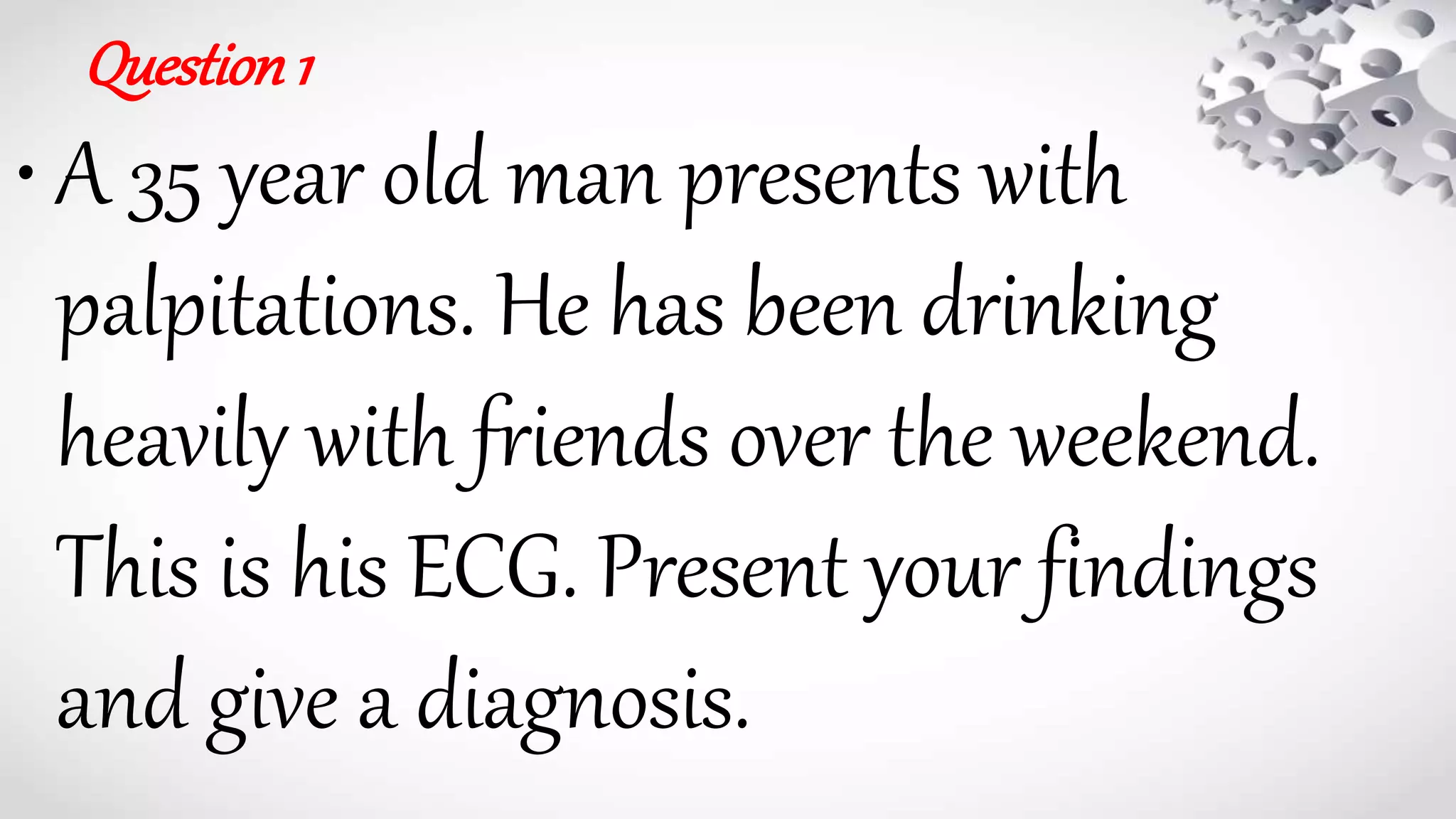
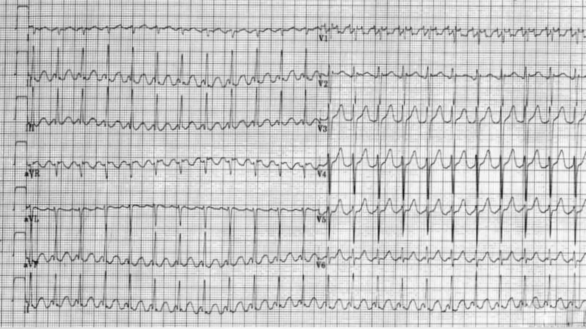
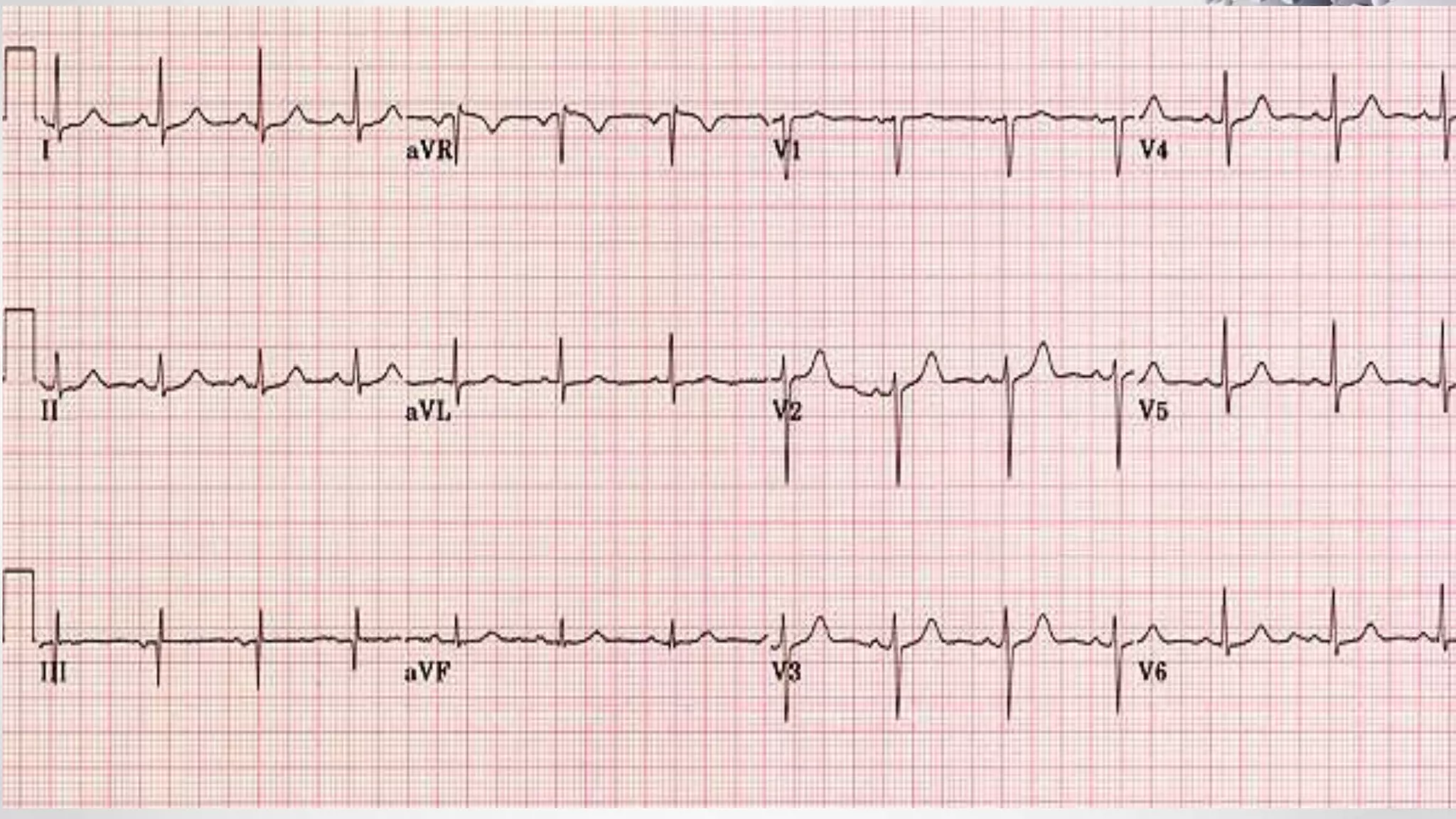
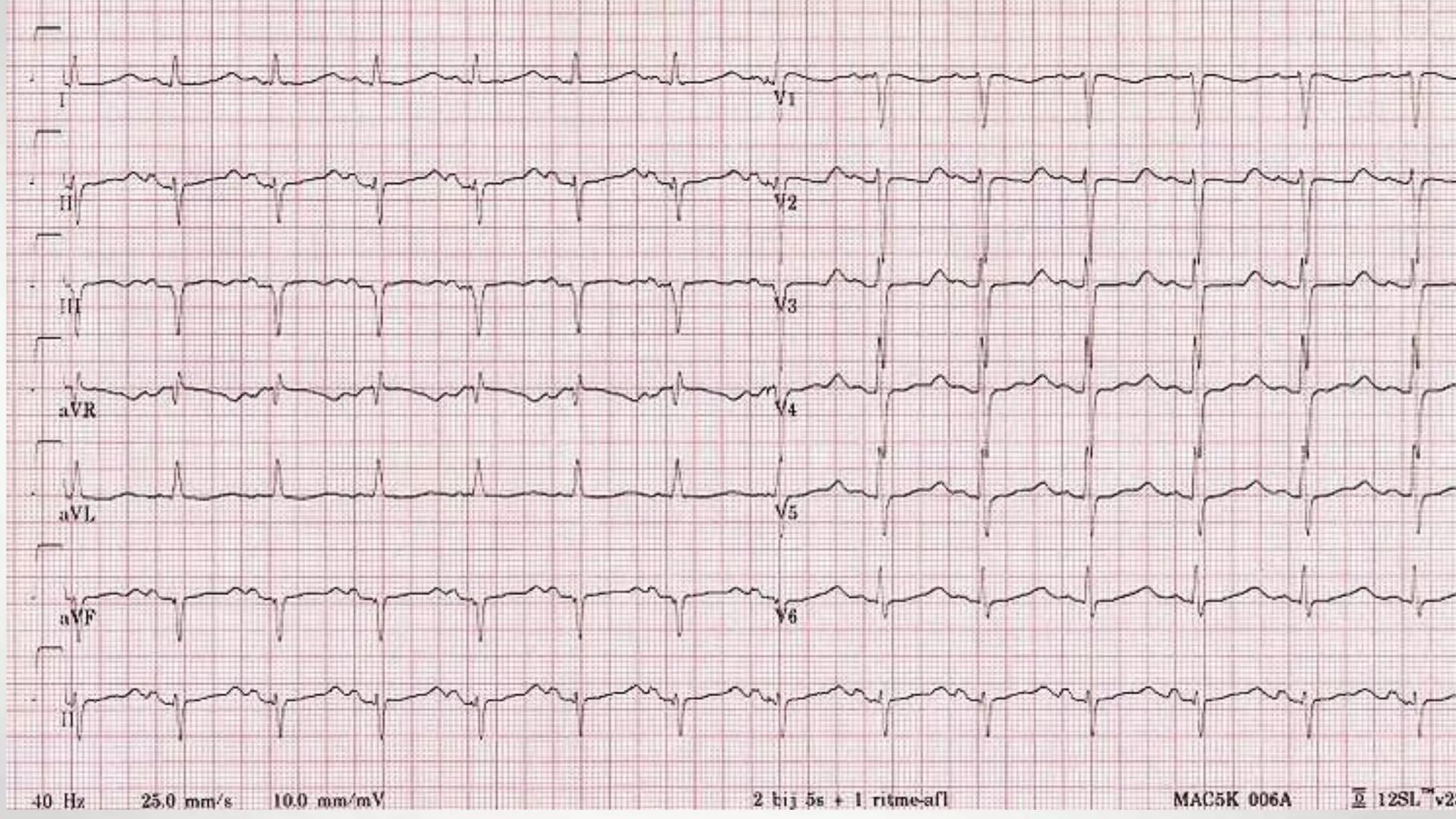
• Rate 85 .... Rhythm Regular ........ PR/P wave Unable to assess ............... QRS Wide
• ST/T wave Wide ............... QTc/other Prolonged
• Diagnosis:
1. The diagnosis is tricyclic antidepressant overdose. This causes widening of the QRS complex and
lengthening of the QT interval due to blockade of sodium channels. 2. [/toggle_item]
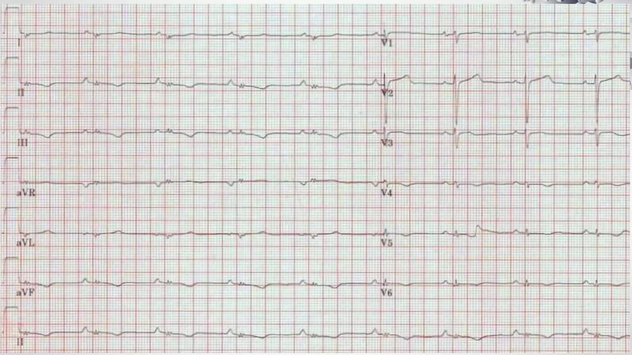
3. [toggle_item title=”What would you do?” active=”false”].......4. A,B,C,D,E (ventilation may be required)
5. Bloods including paracetamol level; ABG (likely metabolic acidosis) .6. Activated charcoal if within 8hrs of
ingestion
7. Sodium bicarbonate (50ml of 8.4%).. 8. Give if any arrhythmia or QRS>110
Further options:
If ventricular tachycardia: lignocaine (avoid beta blockers, amiodarone and calcium blockers)
If seizures: benzodiazepines
65. Question22
•A 45 year old man is found
collapsed at home. There is no
history available. This is his ECG.
What is the diagnosis?
66.
67. Answer
• RateHighly variable – up to 300 bpm .... Rhythm Irregular
• AxisUnable to assess ............ PR/P wave Absent during episodes of extreme
tachycardia
. QRSWide ............. ST/T wave Unable to assess ............. QTc/otherUnable to assess
• Diagnosis:
• This is a difficult case and shows runs of polymorphic VT or Torsades de pointes (literally
translated as twisting of the points). It has a number of causes including medications
(especially psychotropics) and electrolyte imbalance. Essentially any cause of long QT can
precipitate polymorphic VT.
• Management in the first instance is magnesium 2g IV, independent of serum magnesium
concentration before treating any other cause of long QT.
68. Question23
• A 50 year old man presents with collapse. He has
been unwell recently with a chest infection for
which he has been prescribed clarithromycin
from his GP. He also takes medication for his
hayfever at this time of year. What is most
concerning here?
69.
70. Answer
• Rate60 ............. RhythmRegular ............ Axis Normal
• PR/P wave Normal ... QRS Narrow ........ ST/T wave Normal
• QTc/other Prolonged QT interval
• Diagnosis:
• This patient has a prolonged QT interval and a cause for this should be sought. Medications
are the likely culprits in this case: both clarithromycin and the antihistamine
diphenhydramine can cause prolonged QT interval.
• The normal length of the QT varies with heart rate and there is a formula that is applied to
correct for this. ECG machines automatically provide you with this ‘corrected QT’ (QTc).
Normal QTc is generally under 480ms. As a rule of thumb, if the end of the QT interval is
over over half way to the next QRS then consider long QT.