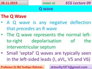
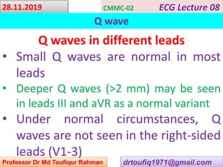
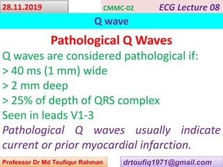
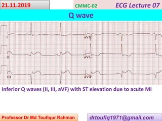
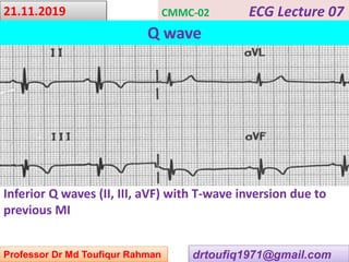
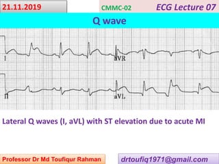
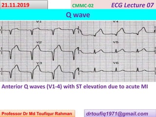
This document contains a series of lectures from Professor Dr. Md Toufiqur Rahman on the topic of Q waves on electrocardiograms (ECGs). The professor defines a Q wave as any negative deflection preceding an R wave, representing normal left-to-right depolarization of the interventricular septum. Small, normal Q waves can be seen in specific left-sided leads. Larger or abnormal Q waves may indicate myocardial infarction or other conditions like cardiomyopathy. Examples of pathological Q waves and their associations with ST segments and T waves are shown from different leads.
Biatrial enlargement is diagnosed when criteria for both right and left atrial enlargement are present on the same ECG.
The diagnosis of biatrial enlargement requires criteria for LAE and RAE to be met in either lead II, lead V1 or a combination of leads.
Case-1:
A 23 years old medical student presented with occasional palpitation, shortness of breath and chest discomfort. He had the following ECG.
A 53 years old gentleman presented with palpitations for last 5 hours. He is smoker, diabetic, dyslipidemic and hypertensive. He had exertional chest discomfort for last 5 years and did coronary angiogram 3 years back and CAG revealed TVD and advised for revascularization. But he refused and was irregular in medication and reluctant for life style modification. He came to emergency department with this ECG.
crème de la crème basics to understand electrocardiographic analysis in an easy & simple way with some specifications to its use in Emergency medicine/clinical toxicology practice.
Wolff–Parkinson–White syndrome (WPW) is one of several disorders of the conduction system of the heart that are commonly referred to as pre-excitation syndromes. WPW is caused by the presence of an abnormal accessory electrical conduction pathway between the atria and the ventricles. Electrical signals traveling down this abnormal pathway (known as the bundle of Kent) may stimulate the ventricles to contract prematurely, resulting in a unique type of supra-ventricular tachycardia referred to as an atrio-ventricular reciprocating tachycardia.
Wolff–Parkinson–White syndrome (WPW) is one of several disorders of the conduction system of the heart that are commonly referred to as pre-excitation syndromes. WPW is caused by the presence of an abnormal accessory electrical conduction pathway between the atria and the ventricles. Electrical signals travelling down this abnormal pathway (known as the bundle of Kent) may stimulate the ventricles to contract prematurely, resulting in a unique type of supraventricular tachycardia referred to as an atrioventricular reciprocating tachycardia.The incidence of WPW is between 0.1% and 0.3% in the general population.Sudden cardiac death in people with WPW is rare (incidence of less than 0.6%), and is usually caused by the propagation of an atrial tachydysrhythmia (rapid and abnormal heart rate) to the ventricles by the abnormal accessory pathway.
ECG Lecture: Sinus arrest, sinoatrial exit block, AV block and escape rhythmsMichael-Joseph Agbayani
Simple ECG lecture about sinus arrest, sinoatrial exit block, AV block and escape rhythms. Slideshow was made with an audience of medical professionals in mind.
A 45 years old lady presented with generalized weakness and palpitations. She is a diagnosed case of chronic renal failure with Diabetes mellitus and Hypertension. Her serum K+ level is 6.8 meq/L. She had the following ECG.
Case; A 54 years old gentleman complained of chest discomfort on exertion for the last 5 months. He is smoker for 10 years, diabetic for 5 years and hypertensive for 3 years. He had the following ECG.
Case: A 25 years old gentleman presented with chest pain and fever .He was normotensive, non-smoker and non-diabetic. His pulse 128b/min and BP-130/80 mm Hg. Troponin I was normal.
Case: A 58 years old gentleman complained of severe central chest pain with excessive sweating 5 days back. He is smoker for 7 years, diabetic for 5 years and hypertensive for 4 years. His BP-90/70 mm Hg. He had the following ECG.
Case-1: A 45 years old lady presented with sudden severe chest discomfort with excessive sweating for last 2 hours. She was diabetic and dyslipidemic and hypertensive. She had history of taking oral contraceptive pills (OCP).. She had the following ECG.
Case: A 34 years old lady presented with shortness of breath , chest discomfort, palpitations , cough, fever , joint pain and skin rash. Her CXR showed nodular lesion in lung field and cardiomegaly. Her serum BNP level was raised. Her echocardiography showed dilated cardiomyopathy with low ejection fraction. She had the following ECG.
Case: A 51 years old gentleman presented with occasional chest discomfort. He was diabetic and smoker. He had a history of myocardial infarction 6 weeks back. He had the following ECG.
The QT interval is the time from the start of the Q wave to the end of the T wave.
It represents the time taken for ventricular depolarisation and repolarisation, effectively the period of ventricular systole from ventricular isovolumetric contraction to isovolumetric relaxation.
Case-1: A 23 years old gentleman presented with generalized weakness and fatigue , 2 hours after several bouts of vomiting and diarrhea. His serum potassium was 2.3 mEq/L and he had the following ECG.
Survival in patients with advanced heart failure (AHF) has improved over the last 2 decades. An increasing number of patients however, are dying with progressive heart failure over the same duration. Optimal utilization of medical therapies and devices like implantable defibrillators and biventricular pacemakers are the likely reasons patients are surviving longer albeit with progressive HF.
Evolution in mechanical circulatory support (MCS) devices has occurred over the same period, such that they can now be rapidly instituted providing support for pump failure, often percutaneously, with timely restitution of physiologic and metabolic derangements with fewer complications.
MCS devices can be classified as Short term and Long term. Short term devices such as Intraaortic balloon pumps (IABP), Impella ®, TandemHeart® or Venoarterial extracorporeal membrane oxygenation (VA – ECMO) using a Cardiohelp® device, are usually employed as ‘Bridge to Recovery’(BTR) or Bridge to Decision’(BTD), usually in acute settings. Long term devices such as implantable left ventricular assist devices (LVADs) e.g. Heartmate II® & 3®, Heart ware HVAD® are implanted as ‘Bridge to transplant’ (BTT) or ‘Destination therapy’ (DT) usually in patients ‘sliding’ on inotropes when they are transplant eligible (BTT) or ineligible (DT) respectively.
Ventricular assist devices have traditionally been developed for left ventricular support in case of severe left heart or biventricular dysfunction. Historically, right ventricular (RV) dysfunction following LVAD implantation or as a component of biventricular dysfunction was managed with either medical therapy, temporary VADs (i.e. ECMO configuration with continuous flow centrifugal pumps like CentriMag®, Rotaflow ®) or occasionally with LVADs placed on the right side. Recently the Impella RP® and ProtekDuo®, percutaneously placed pumps with inflow in the inferior vena cava & right atrium respectively and outflow in pulmonary artery, have become available as less invasive options, for short term RV support.
The Syncardia® is the only approved total artificial heart system currently in use; however various biventricular, total heart systems (e.g. BiVACOR®) in development show promise.
Mechanical circulatory devices provide attractive, viable, physiologically plausible ventricular support options that can be used effectively in carefully selected patients.
Case: A 45 years old presented with chest discomfort and excessive sweating for last 2 hours. He was diabetic, smoker and dyslipidemic. His pulse 68b/min and BP-130/80 mm Hg. In emergency department he had the following ECG.
Case: A 25 years old gentleman presented with chest pain and fever .He was normotensive, non-smoker and non-diabetic. His pulse 128b/min and BP-130/80 mm Hg. Troponin I was normal.
Case: A 54 years old lady presented with chest discomfort and excessive sweating for last 4 hours. She was diabetic, hypertensive and dyslipidemic. Her pulse 62b/min and BP-160/90 mm Hg. In emergency department she had the following ECG.
Wellens’ Syndrome: Exception to the Rule: One Referral at a Time!asclepiuspdfs
We describe two patients with Wellens’ syndrome. In these patients, the electrocardiogram changes must be recognized promptly and accurately. These cases are managed aggressively and early invasive treatment approach is recommended to avoid myocardial infarction and death.
Pulmonary hypertension (PH) is a complex and progressive
condition characterised by high blood pressure in the lungs, leading
to significant health challenges. This book is dedicated to unravelling
the intricacies of PH, encompassing its pathophysiology, diagnosis,
management and emerging research trends. It is designed to serve
as a comprehensive guide for clinicians, researchers and students
in the field of cardiology and respiratory medicine, as well as a
valuable resource for patients and their families seeking to deepen
their understanding of this condition.
The chapters of this book are structured to provide a detailed
insight into the various facets of PH. Starting with the basic
pathophysiology and classification systems, I delve into the
clinical presentation, diagnostic criteria and the nuances of
managing this condition, including both pharmacological and
non-pharmacological approaches. Special attention is given to
the unique challenges posed by pediatric PH, PH in pregnancy
and the management of co-morbidities and complications.
Recognizing the rapid advancements in the field, this book also
dedicates a significant portion to discussing current research
trends, future therapeutic targets and evolving diagnostic
techniques. Real-world case studies and patient testimonies are
included to provide a practical perspective, highlighting the
impact of PH on patients’ lives and the importance of a patient-
centered approach to care.
The field of pulmonary hypertension is one of dynamic change
and I growing understanding. Through this book, we aim to
provide a thorough and up-to-date resource that reflects the
current state of knowledge and practice in the field of PH, while
also offering a glimpse into the future directions of research and
treatment. It is my hope that this book will not only enhance the
understanding of PH among healthcare professionals but also offer
support and information to patients and their families navigating
this challenging condition.
I wish to express our heartfelt gratitude to the following
individuals whose unwavering support and contributions have
played a pivotal role in the creation of this book "Innovations in
Cardiology: From Fundamentals to Frontiers – Short Notes in
Cardiology," My sincere thanks go to: Professor Sufia Rahman,
Professor Abdullah Al Shafi Majumder, Professor dr. Abduz
Zaher, Professor Syed Azizul Haque, Professor Dr Nurunnahar
Fatema Begum; Professor Md. Atahar Ali, Professor Dr. Afzalur
Rahman, Professor Fazila- Tun- Nessa Malik, Professor Kh.
Qamrul Islam; Professor Dr. GM Faruque, Professor M.
Maksumul Haq,Professo Dr. Sajal Krisna Banerjee; Professor
Dr. STM Abu Azam; Professor Mir Jamal Uddin, Professor
Mohammad Abdur Rashid, Professor Dr. AKM Fazlur Rahman,
Professor Dr. Abdul Kader Akanda, Professor Dr. AQM Reza,
Professor Dr. Saiful Islam; Dr. Shams Munwar; Professor Dr.
Chaudhury Meshkat Ahmed, Professor Dr. Khaled Mohsin,
Professor Abdul Wadud Chowdhury, Professor Razia Sultana
Mahmud,Professor Dr. M Touhidul Haque; Professor Dr. Md.
Sahabuddin, Professor Prabir Kumar Das, Professor Dr. Baren
Chakraborty, Professor Dr. Amirul Khusru, Dr. Kaiser Nasrullah
Khan, Professor Ashok Dutta, Professor Md. Khalequzzaman,
Dr. Abdullah Al Jamil, Professor Dr. Amal Kumar Choudhury,
Professor Mostafa Zaman Babul, Professor Dr Dipal Krishna
Adhikary, Professor Dr. Dipankar Chandra Nag professor Dr.
Moeen Uddin Ahmed, professor Mir nesar Uddin; Brig. Gen. Dr.
Syeda Aleya Sultana,Professor Dr Syed Nasir Uddin; Professor
Dr Mohsin Hossain; Dr. Sm Shahidul Haque; Professor Dr
Tawfiq Shahriar Huq; Dr. SM Quamrul Haque; Professor Dr.
Mamunur Rashid Sizar, Professor Dr. Mohsin Ahmed, Professor
Dr. Zillur Rahman; Professor Dr. Tanjima Parveen; Professor Dr.
Harisul hoque, Dr. Reyan Anis, Dr. Ashish Dey, Dr. Mohammad
Ullah firoz, Professor Dr. Udoy Shankar Roy; Dr. Nuruddin
Tareq; Dr. Md. Towhiduzzaman, Dr. Kh. Asaduzzaman, Dr.
AKM Monwarul Islam, Dr. Abdul Momen, Dr. Md. Shafiqur
Rahman Patwary, Dr. Md, Zulfiker Ali Lenin; Dr. Mahbub
Mansur, Dr. CM Shaheen Kabir, Dr. Rumi Alam, Dr. Farah
ii
Tasneem Mowmi, Dr. Rashid Ahmed, Dr. Mohammad Anowar
Hossain, Dr. Mohammad Nasimul Gani,Professor Dr abu Tarek
Iqbal, Dr. Husnayen Nanna, Dr. Abdul Malek, Dr, Ajoy Kumar
Datta, Dr. Nur Alam; Dr. Sahela Nasrin; Dr. Haripada sarker, Dr.
Anisul Awal, Dr. Shaila Nabi; Professor Dr. Umme Salma Khan;
Dr SM Ahsan Habib; Professor Dr Solaiman Hossain; Dr. Bijoy
Dutta,Dr. Shahana Zaman; Dr. Ishrat Jahan shimu, Dr. Ibrahim
Khalil. Dr. Chayan Kumar Singha, Dr. Kazi Nazrul Islam, Dr.
Kamal pasha; professor Dr. Liakat Hossain Tapan, Professor Dr.
Mamun Iqbal, Professor Dr. MG Azam, Dr. Lima Asrin Sayami,
Dr. Smita Kanungo; Dr. Sadequl Islam Shamol; Dr. Swadesh
chakraborty; Dr. Md. Rasul Amin Shepon; Dr. Saqif shahriar;
Your collective wisdom, expertise and commitment to the field
of cardiology have enriched the content of this book. Your
mentorship and guidance have been invaluable in shapi
Definition: Cardiac arrhythmias refer to abnormal heart rhythms, where the heartbeat may be too slow (bradycardia), too fast (tachycardia), or irregular.
These irregularities disrupt the normal electrical signaling in the heart.
In a world where hearts beat free and bold,
A silent foe creeps, its story untold,
Rheumatic whispers, in hushed refrain,
A tale of love's struggle, of heartache and pain.
A childhood song, innocent and sweet,
Takes a tragic turn, hearts skip a beat,
Rheumatic winds blow, fierce and unseen,
Leaving scars on hearts that once danced so keen.
Valves that should open, a rhythmic embrace,
Now bear the weight of this silent chase,
Rheumatic echoes, a haunting refrain,
Leaving imprints of sorrow, of loss and of pain.
But amidst the shadows, there's hope that glows,
A symphony of care, compassion bestows,
With knowledge and love, we stand side by side,
To mend these hearts, to be a healing guide.
Rheumatic battles, we'll face them anew,
A united front, a relentless crew,
For every heart deserves freedom's embrace,
And in the face of rheumatic storms, we'll find grace.
So let's raise our voices, let the world hear,
The fight against rheumatic pain, we hold dear,
With courage and faith, we'll rewrite the verse,
A tale of triumph, of hearts that converse.
"Rheumatic fever reminds us that our body is a delicate symphony, and neglecting even the slightest discord can lead to profound consequences." -
"In the battle against rheumatic fever, awareness and early intervention are our most potent allies."
"Rheumatic fever teaches us the vital lesson that the heart, both physical and emotional, must be nurtured with care and vigilance." -
"Every case of rheumatic fever avoided is a triumph of knowledge, compassion, and the will to protect our most vital instrument, the heart." -
"Rheumatic fever serves as a reminder that even the strongest fortresses need vigilant guardians to shield against the unseen enemies within." -
Case Scenario: You're presenting research findings on hypertension prevalence in
different regions. What Excel chart type would best visualize the variation in
prevalence across regions?
Options: A) Line chart B) Pie chart C) Bar chart D) Scatter plot E) Radar chart Answer:
C) Bar chart
Explanation: A bar chart effectively compares values across different categories,
making it ideal for visualizing the variation in hypertension prevalence across different
regions.
Case Scenario: You're analyzing patient demographics, and you want to find the
most common blood type among your patients. What Excel function would help
you identify the mode of the blood types?
Options: A) MEDIAN B) MODE C) COUNTIF D) AVERAGE E) SUM Answer: B)
MODE
Explanation: The MODE function in Excel helps you find the most frequently occurring
value in a range, making it suitable for identifying the most common blood type among
patients.
Case Scenario: You're conducting a study on the effects of exercise on blood
pressure. What Excel tool would you use to create a summary table showing
average blood pressure before and after exercise?
Options: A) Goal Seek B) PivotTable C) Data Validation D) Filter E) Sort Answer: B)
PivotTable
Explanation: A PivotTable in Excel can summarize data and calculate averages,
making it suitable for creating a summary table showing average blood pressure before
and after exercise.
Case Scenario: You're managing patient records and need to categorize patients
into age groups for analysis. What Excel function would you use to assign each
patient to a specific age category?
Options: A) VLOOKUP B) IF C) COUNTIF D) INDEX E) MATCH Answer: B)
IF
Explanation: The IF function in Excel allows you to apply conditional logic. It's useful
for categorizing patients into age groups based on their ages.
Case Scenario: You're analyzing the effectiveness of a new drug on reducing
cholesterol levels in patients. Which Excel function would you use to calculate
the percentage reduction in cholesterol for each patient?
Options: A) SUMIF B) AVERAGEIF C) MEDIAN D) COUNTIF E) IF Answer: E) IF
Explanation: The IF function in Excel allows you to apply conditional logic. It's useful
for calculating the percentage reduction in cholesterol levels based on the original and
post-treatment values.
Case Scenario: You're preparing a presentation on global prevalence rates of
different heart diseases. What Excel chart type would best display the proportion
of each disease in relation to the whole?
Options: A) Line chart B) Scatter plot C) Bar chart D) Pie chart E) Area chart Answer:
D) Pie chart
Explanation: A pie chart effectively displays proportions and percentages, making it
ideal for showcasing the proportion of each heart disease in relation to the total.
Case Scenario: You're managing a database of medical research papers, including
titles, authors, and publication years. What Excel tool can you use to quickly find
papers published between cert
5. A 5 years old boy presents with fever & swelling of knee and ankle joint for 3 weeks. Write down 3 important D/D. Discuss the treatment of acute rheumatic fever with carditis. (DU-09Ju)
Three important differential diagnoses of a 5-year-old boy presenting with fever and joint swelling for 3 weeks include:
Septic arthritis: This is an acute bacterial infection of a joint that causes similar symptoms to rheumatic fever but is usually monoarticular and associated with more severe pain, redness, and tenderness of the affected joint. Septic arthritis requires urgent drainage and antibiotics.
Juvenile idiopathic arthritis: This is a group of chronic autoimmune disorders that can present with fever, joint swelling, and stiffness. The diagnosis is based on clinical features, laboratory tests, and imaging studies. The treatment may include nonsteroidal anti-inflammatory drugs, disease-modifying antirheumatic drugs, and biologic agents.
Reactive arthritis: This is an inflammatory joint disease that can occur after an infection, especially with certain bacteria such as Chlamydia, Salmonella, or Shigella. Reactive arthritis usually affects the lower limb joints, such as knees, ankles, and feet, and may be associated with skin rash, eye inflammation, or urethritis. The treatment may include antibiotics, nonsteroidal anti-inflammatory drugs, and corticosteroids.
Assuming the diagnosis of acute rheumatic fever with carditis, the treatment usually involves a combination of antibiotics and anti-inflammatory drugs. The antibiotics aim to eradicate the streptococcal infection and prevent further rheumatic fever recurrences, while the anti-inflammatory drugs aim to reduce the inflammation and symptoms of carditis. The specific regimen may vary depending on the severity of carditis, the presence of other complications, and the patient's age and weight. In general, the following principles apply:
Antibiotics: A 10-day course of oral or intramuscular penicillin is the first-line antibiotic for acute rheumatic fever, as it is effective against most strains of streptococci and has low toxicity. Alternative antibiotics may be used for patients who are allergic to penicillin or have recurrent rheumatic fever despite adequate penicillin therapy. Long-term prophylaxis with penicillin is recommended to prevent recurrences, usually until the age of 21 years or for 10 years after the last episode of rheumatic fever, whichever is longer.
Anti-inflammatory drugs: High-dose aspirin or nonsteroidal anti-inflammatory drugs (NSAIDs) such as ibuprofen or naproxen are usually given for the first 2-3 weeks of acute rheumatic fever to control fever, pain, and inflammation. Corticosteroids such as prednisone or methylprednisolone may be used in severe cases of carditis or when other therapies are not effective or contraindicated. The duration and dose of anti-inflammatory drugs should be tailored to the patient's response and adverse effects, such as gastric
The Cardiovascular System: Life's Vital Transport System
The cardiovascular system, comprising the heart, blood vessels, and blood, is a fundamental physiological network in the body.
It facilitates the circulation of oxygen, nutrients, hormones, and immune cells while eliminating waste products.
Essential for maintaining tissue function, energy production, and overall homeostasis.
Defining the Cardiovascular System
The cardiovascular system, also known as the circulatory system, is a complex network responsible for circulating vital substances throughout the body.
Components of the Cardiovascular System
Heart: A muscular organ that pumps blood, generating the force required to propel blood through the blood vessels.
Blood Vessels: A network of tubes that carry blood to and from various body tissues.
Blood: A specialized fluid containing red and white blood cells, platelets, and plasma, essential for nutrient and gas exchange.
Exploring Applied Physiology of the Cardiovascular System
The cardiovascular system is a cornerstone of human health, regulating the circulation of vital nutrients, oxygen, and waste products throughout the body.
Understanding the applied physiology of this system is essential for healthcare professionals to provide effective medical care and interventions.
Importance of Applied Cardiovascular Physiology
Effective healthcare requires a deep comprehension of how the cardiovascular system functions under various conditions.
Applied physiology knowledge empowers healthcare practitioners to make informed decisions, diagnose disorders, and formulate targeted treatment plans.
Focus on Practical Applications in Healthcare
This presentation delves into the practical aspects of cardiovascular physiology that directly impact clinical practice.
We will explore how physiological concepts are translated into real-world medical scenarios and interventions.
By grasping the applied physiology of the cardiovascular system, healthcare providers can optimize patient care, enhance diagnostics, and improve treatment outcomes.
Throughout this presentation, we'll bridge the gap between theoretical understanding and its practical implications in the field of healthcare.
Understanding the Components
The cardiovascular system comprises three crucial components: the heart, blood vessels, and blood.
Heart: A muscular organ that pumps blood, ensuring a continuous flow throughout the body.
Blood Vessels: A network of tubes that transport blood to and from various tissues.
Blood: A specialized fluid that carries nutrients, oxygen, hormones, and removes waste products.
Role in Oxygen and Nutrient Delivery
Oxygen from the lungs and nutrients from the digestive system are transported to body tissues through the bloodstream.
These essential components are required for cellular metabolism and energy production.
Peripartum cardiomyopathy (PPCM) is a type of heart disease that affects women during the last month of pregnancy or in the first few months after delivery. It is characterized by a weakened and enlarged heart muscle, which makes it difficult for the heart to pump blood efficiently to the rest of the body. The exact cause of PPCM is unknown, but it is believed to be related to the hormonal changes and increased demands on the heart that occur during pregnancy. Symptoms of PPCM can include shortness of breath, fatigue, chest pain, swelling in the legs and feet, and palpitations. Treatment for PPCM usually involves medications to improve heart function and supportive care to manage symptoms. In severe cases, advanced treatments such as implantable devices or heart transplantation may be necessary. With early diagnosis and treatment, most women with PPCM can recover completely and go on to lead healthy lives.during pregnancy.
The diagnosis of PPCM is based on clinical symptoms, such as shortness of breath, fatigue, chest pain, and edema, along with imaging studies, such as echocardiography. Treatment for PPCM usually involves medications to improve heart function and supportive care to manage symptoms. These medications can include beta-blockers, ACE inhibitors, diuretics, and inotropic agents. In severe cases, advanced treatments such as mechanical circulatory support or heart transplantation may be necessary.
The prognosis for PPCM varies depending on the severity of the disease and the presence of underlying comorbidities. However, with early diagnosis and appropriate treatment, most women with PPCM can recover completely and go on to lead healthy lives. The recurrence rate of PPCM in subsequent pregnancies is approximately 20%, and women who have had PPCM are advised to avoid future pregnancies or undergo careful monitoring and management during pregnancy.
There are still many unanswered questions about PPCM, including its exact cause, optimal diagnostic and treatment strategies, and long-term outcomes. Further research is needed to better understand this complex and potentially life-threatening condition.
In conclusion, PPCM is a rare but serious form of heart disease that can occur during or after pregnancy. Early recognition and management of this condition are critical in preventing complications and improving outcomes for both the mother and the baby. Future research will continue to shed light on the pathophysiology and optimal management of PPCM.
Cardiac rehabilitation is a comprehensive program that aims to improve the health and quality of life of individuals with cardiovascular disease. This review article provides an overview of current evidence-based practices and the benefits of cardiac rehabilitation. The article discusses the components of cardiac rehabilitation, including medical evaluation, physical activity and exercise training, nutrition counseling and education, psycho social support and counseling, cardiac risk factor management, medication management, and tobacco cessation counseling. The article also discusses the effectiveness of cardiac rehabilitation in reducing mortality rates, improving functional capacity, and reducing the risk of future cardiovascular events. Additionally, the article explores the future directions of cardiac rehabilitation, including personalized medicine, technology integration, home-based programs, expanded target populations, and a multidisciplinary approach. Healthcare providers play a crucial role in encouraging and referring eligible patients to cardiac rehabilitation programs as part of their treatment plan. The review concludes that cardiac rehabilitation is an essential aspect of the management of cardiovascular disease and highlights the need for further research and development in this dynamic field.
Outline of CPR manual
I. Introduction
A. Definition of CPR
1. Explanation of what CPR stands for
2. Definition of CPR as a life-saving technique
B. Importance of CPR
1. Statistics on cardiac arrest and survival rates
2. Explanation of why CPR is crucial for saving lives
C. Objective of the manual
1. Explanation of what readers will learn from the manual
2. Statement of the manual's purpose
II. Getting Started with CPR
A. Assessing the situation
1. Importance of assessing the situation before starting CPR
2. Factors to consider when assessing the situation
B. Checking for responsiveness
1. Explanation of how to check for responsiveness
2. Importance of checking for responsiveness
C. Activating the emergency response system
1. Explanation of when to activate the emergency response system
2. Step-by-step guide to activating the emergency response system
III. Basic Life Support Techniques
A. Key components of basic life support
1. Explanation of the components of basic life support
2. Importance of each component
B. The ABCs of CPR
1. Explanation of the ABCs of CPR
2. Importance of each step in the ABCs of CPR
C. Performing chest compressions
1. Explanation of how to perform chest compressions
2. Importance of proper chest compression technique
D. Delivering rescue breaths
1. Explanation of how to deliver rescue breaths
2. Importance of proper rescue breath technique
E. Utilizing an automated external defibrillator (AED)
1. Explanation of what an AED is and how it works
2. Step-by-step guide to using an AED
F. Administering medications during CPR
1. Explanation of medications used during CPR
2. Dosages and administration guidelines for each medication
IV. Advanced Life Support Techniques
A. Advanced airway management
1. Explanation of advanced airway management techniques
2. Importance of advanced airway management in CPR
B. Advanced monitoring techniques
1. Explanation of advanced monitoring techniques
2. Importance of advanced monitoring in CPR
C. Invasive interventions
1. Explanation of invasive interventions
2. Importance of invasive interventions in CPR
D. Extracorporeal membrane oxygenation (ECMO)
1. Explanation of ECMO
2. Importance of ECMO in CPR
V. Improving Outcomes in CPR
A. Factors influencing CPR outcomes
1. Explanation of factors that influence CPR outcomes
2. Importance of understanding these factors
B. Strategies for improving CPR outcomes
1. Explanation of strategies for improving CPR outcomes
2. Importance of implementing these strategies
C. The role of high-quality CPR in improving outcomes
1. Explanation of what high-quality CPR is
2. Importance of performing high-quality CPR
VI. Special Considerations in CPR
A. CPR in special populations
1. Explanation of special populations that require unique CPR techniques
2. Importance of understanding these unique CPR techniques
B. CPR in special settings
1. Explanation of special settings that require unique CPR techniques
2. Importance of understanding these unique CPR techniques
C.
I. Introduction
A. Brief explanation of World Hypertension Day
B. Importance of addressing hypertension as a global health issue
C. Overview of the objectives of the presentation
II. Understanding Hypertension
A. Definition and classification of hypertension
B. Prevalence and global burden of hypertension
C. Risk factors and causes of hypertension
D. Health implications and complications associated with hypertension
III. World Hypertension Day 2023
A. Background and significance of World Hypertension Day
B. Theme and key messages for World Hypertension Day 2023
C. Activities and events organized worldwide to raise awareness
IV. Goals and Objectives
A. Key goals set for World Hypertension Day 2023
B. Promoting prevention and early detection of hypertension
C. Encouraging healthy lifestyle modifications
D. Enhancing public knowledge about hypertension management
V. Initiatives and Campaigns
A. Overview of global initiatives and campaigns
B. Collaborations with international organizations, NGOs, and healthcare professionals
C. Campaign materials and resources available for public use
VI. Strategies for Hypertension Prevention and Control
A. Implementing population-level interventions
B. Screening and diagnosis strategies
C. Lifestyle modifications (diet, physical activity, stress management)
D. Pharmacological management and treatment guidelines
VII. Public Awareness and Education
A. Importance of raising public awareness about hypertension
B. Educational campaigns and resources for the general public
C. Role of healthcare professionals in educating patients
VIII. Impact and Achievements
A. Highlighting the impact of previous World Hypertension Day campaigns
B. Success stories and achievements in hypertension prevention and control
C. Lessons learned and areas for improvement
IX. Conclusion
A. Recap of the key points discussed
B. Call to action for individuals, communities, and policymakers
C. Encouragement to spread awareness and take steps towards hypertension prevention
. Introduction
A. Definition and prevalence of hypertension in the elderly
B. Importance of managing hypertension in this population
II. Risk Factors and Complications
A. Common risk factors for hypertension in the elderly
B. Potential complications associated with uncontrolled hypertension
III. Diagnostic Process
A. Blood pressure measurement techniques and guidelines
B. Target blood pressure goals for elderly patients
C. Identification of secondary causes of hypertension
IV. Non-Pharmacological Management
A. Lifestyle modifications
1. Dietary recommendations (e.g., DASH diet, sodium reduction)
2. Weight management and physical activity
3. Smoking cessation and alcohol moderation
B. Stress management and relaxation techniques
V. Pharmacological Management
A. First-line antihypertensive medications
B. Considerations for drug selection in the elderly
1. Drug interactions and comorbidities
2. Adverse effects and tolerability
C. Individualized treatment approach based on patient characteristics
VI. Monitoring and Follow-Up
A. Frequency of blood pressure monitoring
B. Importance of medication adherence
C. Adjusting treatment based on patient response
D. Collaborative care and involvement of healthcare professionals
VII. Special Considerations
A. Polypharmacy and medication management
B. Management of hypertension in frail and institutionalized elderly
C. Cognitive impairment and medication adherence
VIII. Controversies and Challenges
A. Blood pressure targets and guidelines in the elderly
B. Conflicting evidence on specific antihypertensive agents
C. Adherence issues and barriers to effective management
IX. Conclusion
A. Summary of key points discussed
B. Importance of comprehensive management in elderly patients
C. Future directions in hypertension management for the elderly
I. Introduction
A. Definition of CPR
1. Explanation of what CPR stands for
2. Definition of CPR as a life-saving technique
B. Importance of CPR
1. Statistics on cardiac arrest and survival rates
2. Explanation of why CPR is crucial for saving lives
C. Objective of the manual
1. Explanation of what readers will learn from the manual
2. Statement of the manual's purpose
II. Getting Started with CPR
A. Assessing the situation
1. Importance of assessing the situation before starting CPR
2. Factors to consider when assessing the situation
B. Checking for responsiveness
1. Explanation of how to check for responsiveness
2. Importance of checking for responsiveness
C. Activating the emergency response system
1. Explanation of when to activate the emergency response system
2. Step-by-step guide to activating the emergency response system
III. Basic Life Support Techniques
A. Key components of basic life support
1. Explanation of the components of basic life support
2. Importance of each component
B. The ABCs of CPR
1. Explanation of the ABCs of CPR
2. Importance of each step in the ABCs of CPR
C. Performing chest compressions
1. Explanation of how to perform chest compressions
2. Importance of proper chest compression technique
D. Delivering rescue breaths
1. Explanation of how to deliver rescue breaths
2. Importance of proper rescue breath technique
E. Utilizing an automated external defibrillator (AED)
1. Explanation of what an AED is and how it works
2. Step-by-step guide to using an AED
F. Administering medications during CPR
1. Explanation of medications used during CPR
2. Dosages and administration guidelines for each medication
IV. Advanced Life Support Techniques
A. Advanced airway management
1. Explanation of advanced airway management techniques
2. Importance of advanced airway management in CPR
B. Advanced monitoring techniques
1. Explanation of advanced monitoring techniques
2. Importance of advanced monitoring in CPR
C. Invasive interventions
1. Explanation of invasive interventions
2. Importance of invasive interventions in CPR
D. Extracorporeal membrane oxygenation (ECMO)
1. Explanation of ECMO
2. Importance of ECMO in CPR
V. Improving Outcomes in CPR
A. Factors influencing CPR outcomes
1. Explanation of factors that influence CPR outcomes
2. Importance of understanding these factors
B. Strategies for improving CPR outcomes
1. Explanation of strategies for improving CPR outcomes
2. Importance of implementing these strategies
C. The role of high-quality CPR in improving outcomes
1. Explanation of what high-quality CPR is
2. Importance of performing high-quality CPR
VI. Special Considerations in CPR
A. CPR in special populations
1. Explanation of special populations that require unique CPR techniques
2. Importance of understanding these unique CPR techniques
B. CPR in special settings
1. Explanation of special settings that require unique CPR techniques
2. Importance of understanding these unique CPR techniques
C. CPR during a pandemic
1
HOT NEW PRODUCT! BIG SALES FAST SHIPPING NOW FROM CHINA!! EU KU DB BK substit...GL Anaacs
Contact us if you are interested:
Email / Skype : kefaya1771@gmail.com
Threema: PXHY5PDH
New BATCH Ku !!! MUCH IN DEMAND FAST SALE EVERY BATCH HAPPY GOOD EFFECT BIG BATCH !
Contact me on Threema or skype to start big business!!
Hot-sale products:
NEW HOT EUTYLONE WHITE CRYSTAL!!
5cl-adba precursor (semi finished )
5cl-adba raw materials
ADBB precursor (semi finished )
ADBB raw materials
APVP powder
5fadb/4f-adb
Jwh018 / Jwh210
Eutylone crystal
Protonitazene (hydrochloride) CAS: 119276-01-6
Flubrotizolam CAS: 57801-95-3
Metonitazene CAS: 14680-51-4
Payment terms: Western Union,MoneyGram,Bitcoin or USDT.
Deliver Time: Usually 7-15days
Shipping method: FedEx, TNT, DHL,UPS etc.Our deliveries are 100% safe, fast, reliable and discreet.
Samples will be sent for your evaluation!If you are interested in, please contact me, let's talk details.
We specializes in exporting high quality Research chemical, medical intermediate, Pharmaceutical chemicals and so on. Products are exported to USA, Canada, France, Korea, Japan,Russia, Southeast Asia and other countries.
These simplified slides by Dr. Sidra Arshad present an overview of the non-respiratory functions of the respiratory tract.
Learning objectives:
1. Enlist the non-respiratory functions of the respiratory tract
2. Briefly explain how these functions are carried out
3. Discuss the significance of dead space
4. Differentiate between minute ventilation and alveolar ventilation
5. Describe the cough and sneeze reflexes
Study Resources:
1. Chapter 39, Guyton and Hall Textbook of Medical Physiology, 14th edition
2. Chapter 34, Ganong’s Review of Medical Physiology, 26th edition
3. Chapter 17, Human Physiology by Lauralee Sherwood, 9th edition
4. Non-respiratory functions of the lungs https://academic.oup.com/bjaed/article/13/3/98/278874
ARTIFICIAL INTELLIGENCE IN HEALTHCARE.pdfAnujkumaranit
Artificial intelligence (AI) refers to the simulation of human intelligence processes by machines, especially computer systems. It encompasses tasks such as learning, reasoning, problem-solving, perception, and language understanding. AI technologies are revolutionizing various fields, from healthcare to finance, by enabling machines to perform tasks that typically require human intelligence.
These lecture slides, by Dr Sidra Arshad, offer a quick overview of physiological basis of a normal electrocardiogram.
Learning objectives:
1. Define an electrocardiogram (ECG) and electrocardiography
2. Describe how dipoles generated by the heart produce the waveforms of the ECG
3. Describe the components of a normal electrocardiogram of a typical bipolar leads (limb II)
4. Differentiate between intervals and segments
5. Enlist some common indications for obtaining an ECG
Study Resources:
1. Chapter 11, Guyton and Hall Textbook of Medical Physiology, 14th edition
2. Chapter 9, Human Physiology - From Cells to Systems, Lauralee Sherwood, 9th edition
3. Chapter 29, Ganong’s Review of Medical Physiology, 26th edition
4. Electrocardiogram, StatPearls - https://www.ncbi.nlm.nih.gov/books/NBK549803/
5. ECG in Medical Practice by ABM Abdullah, 4th edition
6. ECG Basics, http://www.nataliescasebook.com/tag/e-c-g-basics
Report Back from SGO 2024: What’s the Latest in Cervical Cancer?bkling
Are you curious about what’s new in cervical cancer research or unsure what the findings mean? Join Dr. Emily Ko, a gynecologic oncologist at Penn Medicine, to learn about the latest updates from the Society of Gynecologic Oncology (SGO) 2024 Annual Meeting on Women’s Cancer. Dr. Ko will discuss what the research presented at the conference means for you and answer your questions about the new developments.
Tom Selleck Health: A Comprehensive Look at the Iconic Actor’s Wellness Journeygreendigital
Tom Selleck, an enduring figure in Hollywood. has captivated audiences for decades with his rugged charm, iconic moustache. and memorable roles in television and film. From his breakout role as Thomas Magnum in Magnum P.I. to his current portrayal of Frank Reagan in Blue Bloods. Selleck's career has spanned over 50 years. But beyond his professional achievements. fans have often been curious about Tom Selleck Health. especially as he has aged in the public eye.
Follow us on: Pinterest
Introduction
Many have been interested in Tom Selleck health. not only because of his enduring presence on screen but also because of the challenges. and lifestyle choices he has faced and made over the years. This article delves into the various aspects of Tom Selleck health. exploring his fitness regimen, diet, mental health. and the challenges he has encountered as he ages. We'll look at how he maintains his well-being. the health issues he has faced, and his approach to ageing .
Early Life and Career
Childhood and Athletic Beginnings
Tom Selleck was born on January 29, 1945, in Detroit, Michigan, and grew up in Sherman Oaks, California. From an early age, he was involved in sports, particularly basketball. which played a significant role in his physical development. His athletic pursuits continued into college. where he attended the University of Southern California (USC) on a basketball scholarship. This early involvement in sports laid a strong foundation for his physical health and disciplined lifestyle.
Transition to Acting
Selleck's transition from an athlete to an actor came with its physical demands. His first significant role in "Magnum P.I." required him to perform various stunts and maintain a fit appearance. This role, which he played from 1980 to 1988. necessitated a rigorous fitness routine to meet the show's demands. setting the stage for his long-term commitment to health and wellness.
Fitness Regimen
Workout Routine
Tom Selleck health and fitness regimen has evolved. adapting to his changing roles and age. During his "Magnum, P.I." days. Selleck's workouts were intense and focused on building and maintaining muscle mass. His routine included weightlifting, cardiovascular exercises. and specific training for the stunts he performed on the show.
Selleck adjusted his fitness routine as he aged to suit his body's needs. Today, his workouts focus on maintaining flexibility, strength, and cardiovascular health. He incorporates low-impact exercises such as swimming, walking, and light weightlifting. This balanced approach helps him stay fit without putting undue strain on his joints and muscles.
Importance of Flexibility and Mobility
In recent years, Selleck has emphasized the importance of flexibility and mobility in his fitness regimen. Understanding the natural decline in muscle mass and joint flexibility with age. he includes stretching and yoga in his routine. These practices help prevent injuries, improve posture, and maintain mobilit
MANAGEMENT OF ATRIOVENTRICULAR CONDUCTION BLOCK.pdfJim Jacob Roy
Cardiac conduction defects can occur due to various causes.
Atrioventricular conduction blocks ( AV blocks ) are classified into 3 types.
This document describes the acute management of AV block.
Couples presenting to the infertility clinic- Do they really have infertility...Sujoy Dasgupta
Dr Sujoy Dasgupta presented the study on "Couples presenting to the infertility clinic- Do they really have infertility? – The unexplored stories of non-consummation" in the 13th Congress of the Asia Pacific Initiative on Reproduction (ASPIRE 2024) at Manila on 24 May, 2024.
Factory Supply Best Quality Pmk Oil CAS 28578–16–7 PMK Powder in Stockrebeccabio
Factory Supply Best Quality Pmk Oil CAS 28578–16–7 PMK Powder in Stock
Telegram: bmksupplier
signal: +85264872720
threema: TUD4A6YC
You can contact me on Telegram or Threema
Communicate promptly and reply
Free of customs clearance, Double Clearance 100% pass delivery to USA, Canada, Spain, Germany, Netherland, Poland, Italy, Sweden, UK, Czech Republic, Australia, Mexico, Russia, Ukraine, Kazakhstan.Door to door service
Hot Selling Organic intermediates
Factory Supply Best Quality Pmk Oil CAS 28578–16–7 PMK Powder in Stock
ECG-Q wave
1. MBBS, FCPS, MD, FACC, FESC, FRCP, FSCAI,
FCCP,FAPSC, FAPSIC, FAHA,FACP,FASE
Professor and Head of Cardiology
Colonel Malek Medical College , Manikganj.
For under-graduates
drtoufiq19711@yahoo.com21/11/2019
Under graduate version 2019
2. drtoufiq1971@gmail.com
28.11.2019 CMMC-02 ECG Lecture 09
Q wave
Professor Dr Md Toufiqur Rahman
The Q Wave
• A Q wave is any negative deflection
that precedes an R wave
• The Q wave represents the normal left-
to-right depolarisation of the
interventricular septum
• Small ‘septal’ Q waves are typically seen
in the left-sided leads (I, aVL, V5 and V6)
4. drtoufiq1971@gmail.com
28.11.2019 CMMC-02 ECG Lecture 08
Q wave
Professor Dr Md Toufiqur Rahman
Q waves in different leads
• Small Q waves are normal in most
leads
• Deeper Q waves (>2 mm) may be seen
in leads III and aVR as a normal variant
• Under normal circumstances, Q
waves are not seen in the right-sided
leads (V1-3)
5. drtoufiq1971@gmail.com
28.11.2019 CMMC-02 ECG Lecture 08
Q wave
Professor Dr Md Toufiqur Rahman
Pathological Q Waves
Q waves are considered pathological if:
> 40 ms (1 mm) wide
> 2 mm deep
> 25% of depth of QRS complex
Seen in leads V1-3
Pathological Q waves usually indicate
current or prior myocardial infarction.
6. drtoufiq1971@gmail.com
21.11.2019 CMMC-02 ECG Lecture 07
Q wave
Professor Dr Md Toufiqur Rahman
Differential Diagnosis/ Causes of Q waves
• Myocardial infarction
• Cardiomyopathies —
Hypertrophic (HCM), infiltrative myocardial
disease
• Rotation of the heart — Extreme clockwise
or counter-clockwise rotation
• Lead placement errors — e.g. upper limb
leads placed on lower limbs
7. drtoufiq1971@gmail.com
21.11.2019 CMMC-02 ECG Lecture 07
Q wave
Professor Dr Md Toufiqur Rahman
Loss of normal Q waves
• The absence of small septal Q
waves in leads V5-6 should be
considered abnormal.
• Absent Q waves in V5-6 is most
commonly due to LBBB.