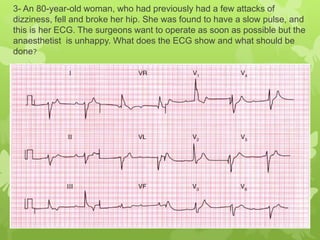
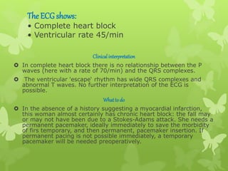
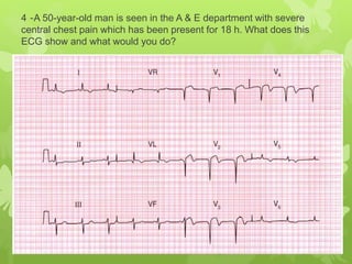
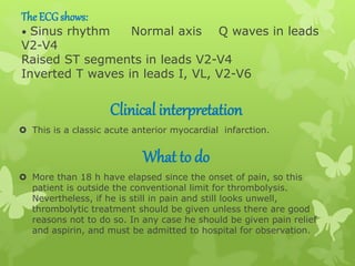
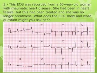
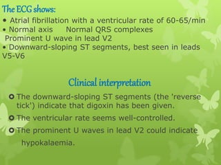
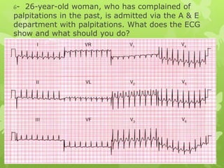
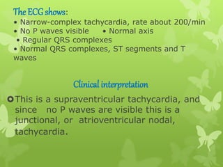
2- A 60-year-old man was seen as an out-patient, complaining of rather
vague central chest pain on exertion. He had never had pain at rest.
What does this ECG show and what would you do next
The ECG shows:
• Sinus rhythm Normal axis Small Q waves in leads II, III, VF
• Biphasic T waves in leads II, V6; inverted T waves in leads III, VF
Markedly peaked T waves in leads V1-V2
Clinicalinterpretation
 The Q waves in the inferior leads, together
with inverted T waves, point to an old inferior
myocardial infarction.
 While symmetrically peaked T waves in the anterior leads can be
due to hyper-kalaemia, or to ischaemia , they are frequently a
normal variant.
What to do
 The patient seems to have had a myocardial infarction at some
point in the past, and by implication his vague chest pain may be
due to cardiac ischaemia.
 Attention must be paid to risk factors (smoking, blood
pressure, plasma cholesterol), and he probably needs long-
term treatment with aspirin and a statin.
 An exercise test will be the best way of deciding whether he has
coronary disease that merits angiography
3- An 80-year-old woman, who had previously had a few attacks of
dizziness, fell and broke her hip. She was found to have a slow pulse, and
this is her ECG. The surgeons want to operate as soon as possible but the
anaesthetist is unhappy. What does the ECG show and what should be
done?
The ECG shows:
• Complete heart block
• Ventricular rate 45/min
Clinicalinterpretation
 In complete heart block there is no relationship between the P
waves (here with a rate of 70/min) and the QRS complexes.
 The ventricular 'escape‘ rhythm has wide QRS complexes and
abnormal T waves. No further interpretation of the ECG is
possible.
Whatto do
 In the absence of a history suggesting a myocardial infarction,
this woman almost certainly has chronic heart block: the fall may
or may not have been due to a Stokes-Adams attack. She needs a
permanent pacemaker, ideally immediately to save the morbidity
of firs temporary, and then permanent, pacemaker insertion. If
permanent pacing is not possible immediately, a temporary
pacemaker will be needed preoperatively.
4 -A 50-year-old man is seen in the A & E department with severe
central chest pain which has been present for 18 h. What does this
ECG show and what would you do?
The ECGshows:
• Sinus rhythm Normal axis Q waves in leads
V2-V4
Raised ST segments in leads V2-V4
Inverted T waves in leads I, VL, V2-V6
Clinical interpretation
 This is a classic acute anterior myocardial infarction.
What to do
 More than 18 h have elapsed since the onset of pain, so this
patient is outside the conventional limit for thrombolysis.
Nevertheless, if he is still in pain and still looks unwell,
thrombolytic treatment should be given unless there are good
reasons not to do so. In any case he should be given pain relief
and aspirin, and must be admitted to hospital for observation.
5 - This ECG was recorded from a 60-year-old woman
with rheumatic heart disease. She had been in heart
failure, but this had been treated and she was no
longer breathless. What does the ECG show and what
question might you ask her?
The ECG shows:
• Atrial fibrillation with a ventricular rate of 60-65/min
• Normal axis Normal QRS complexes
Prominent U wave in lead V2
• Downward-sloping ST segments, best seen in leads
V5-V6
Clinical interpretation
 The downward-sloping ST segments (the 'reverse
tick') indicate that digoxin has been given.
 The ventricular rate seems well-controlled.
 The prominent U waves in lead V2 could indicate
hypokalaemia.
What to do?
 Ask the patient about her appetite: the
earliest symptom of digoxin toxicity is
appetite loss, followed by nausea and
vomiting.
 If the patient is being treated with
diuretics, check the serum potassium level
- a low potassium level potentiates the
effects of digoxin. If in doubt, the serum
digoxin level is easily measured
6- 26-year-old woman, who has complained of
palpitations in the past, is admitted via the A & E
department with palpitations. What does the ECG
show and what should you do?
The ECG shows:
• Narrow-complex tachycardia, rate about 200/min
• No P waves visible • Normal axis
• Regular QRS complexes
• Normal QRS complexes, ST segments and T
waves
Clinical interpretation
This is a supraventricular tachycardia, and
since no P waves are visible this is a
junctional, or atrioventricular nodal,
tachycardia.
What to do
 Junctional tachycardia is the commonest form of
paroxysmal tachycardia in young people, and
presumably explains her previous episodes of
palpitations.
 Attacks of junctional tachycardia may be terminated
by any of the manoeuvres that lead to vagal
stimulation - Valsalva's manoeuvre, carotid sinus
pressure, or immersion of the face in cold water. If
these are unsuccessful, intravenous adenosine should
be given by bolus injection. Adenosine has a very
short half-life, but can cause flushing and occasionally
asthma.
 If adenosine proves unsuccessful, verapamil 5-10 mg
given by bolus injection will usually restore sinus
rhythm. Otherwise, DC cardioversion is indicated
14 A 50-year-old man is admitted to hospital as an
emergency, having had chest pain characteristic of a
myocardial infarction for 4 h. Apart from the features
associated with pain there are no abnormal physical findings.
What does this ECG show and what would you do?
The ECG shows:
• Sinus rhythm • Normal axis • Small Q waves
in lead III but not elsewhere
• Elevated ST segments in leads II, III, VF, with
upright T waves
• T wave inversion in lead VL
• Suggestion of ST segment depression in leads
V2-V3
Clinical interpretation
A classic ECG of an acute inferior myocardial
infarction, with lead VL indicating ischaemia.
The rate of development of Q waves is very variable
What to do
 Pain relief must take priority. In the
absence of contraindications (i.e. risk of
bleeding from any important site), the
patient should be given aspirin and then a
thrombolytic agent
Ecg case
Ecg case

Ecg case

  • 2.
    2- A 60-year-oldman was seen as an out-patient, complaining of rather vague central chest pain on exertion. He had never had pain at rest. What does this ECG show and what would you do next
  • 3.
    The ECG shows: •Sinus rhythm Normal axis Small Q waves in leads II, III, VF • Biphasic T waves in leads II, V6; inverted T waves in leads III, VF Markedly peaked T waves in leads V1-V2 Clinicalinterpretation  The Q waves in the inferior leads, together with inverted T waves, point to an old inferior myocardial infarction.  While symmetrically peaked T waves in the anterior leads can be due to hyper-kalaemia, or to ischaemia , they are frequently a normal variant.
  • 4.
    What to do The patient seems to have had a myocardial infarction at some point in the past, and by implication his vague chest pain may be due to cardiac ischaemia.  Attention must be paid to risk factors (smoking, blood pressure, plasma cholesterol), and he probably needs long- term treatment with aspirin and a statin.  An exercise test will be the best way of deciding whether he has coronary disease that merits angiography
  • 5.
    3- An 80-year-oldwoman, who had previously had a few attacks of dizziness, fell and broke her hip. She was found to have a slow pulse, and this is her ECG. The surgeons want to operate as soon as possible but the anaesthetist is unhappy. What does the ECG show and what should be done?
  • 6.
    The ECG shows: •Complete heart block • Ventricular rate 45/min Clinicalinterpretation  In complete heart block there is no relationship between the P waves (here with a rate of 70/min) and the QRS complexes.  The ventricular 'escape‘ rhythm has wide QRS complexes and abnormal T waves. No further interpretation of the ECG is possible. Whatto do  In the absence of a history suggesting a myocardial infarction, this woman almost certainly has chronic heart block: the fall may or may not have been due to a Stokes-Adams attack. She needs a permanent pacemaker, ideally immediately to save the morbidity of firs temporary, and then permanent, pacemaker insertion. If permanent pacing is not possible immediately, a temporary pacemaker will be needed preoperatively.
  • 7.
    4 -A 50-year-oldman is seen in the A & E department with severe central chest pain which has been present for 18 h. What does this ECG show and what would you do?
  • 8.
    The ECGshows: • Sinusrhythm Normal axis Q waves in leads V2-V4 Raised ST segments in leads V2-V4 Inverted T waves in leads I, VL, V2-V6 Clinical interpretation  This is a classic acute anterior myocardial infarction. What to do  More than 18 h have elapsed since the onset of pain, so this patient is outside the conventional limit for thrombolysis. Nevertheless, if he is still in pain and still looks unwell, thrombolytic treatment should be given unless there are good reasons not to do so. In any case he should be given pain relief and aspirin, and must be admitted to hospital for observation.
  • 9.
    5 - ThisECG was recorded from a 60-year-old woman with rheumatic heart disease. She had been in heart failure, but this had been treated and she was no longer breathless. What does the ECG show and what question might you ask her?
  • 10.
    The ECG shows: •Atrial fibrillation with a ventricular rate of 60-65/min • Normal axis Normal QRS complexes Prominent U wave in lead V2 • Downward-sloping ST segments, best seen in leads V5-V6 Clinical interpretation  The downward-sloping ST segments (the 'reverse tick') indicate that digoxin has been given.  The ventricular rate seems well-controlled.  The prominent U waves in lead V2 could indicate hypokalaemia.
  • 11.
    What to do? Ask the patient about her appetite: the earliest symptom of digoxin toxicity is appetite loss, followed by nausea and vomiting.  If the patient is being treated with diuretics, check the serum potassium level - a low potassium level potentiates the effects of digoxin. If in doubt, the serum digoxin level is easily measured
  • 12.
    6- 26-year-old woman,who has complained of palpitations in the past, is admitted via the A & E department with palpitations. What does the ECG show and what should you do?
  • 13.
    The ECG shows: •Narrow-complex tachycardia, rate about 200/min • No P waves visible • Normal axis • Regular QRS complexes • Normal QRS complexes, ST segments and T waves Clinical interpretation This is a supraventricular tachycardia, and since no P waves are visible this is a junctional, or atrioventricular nodal, tachycardia.
  • 14.
    What to do Junctional tachycardia is the commonest form of paroxysmal tachycardia in young people, and presumably explains her previous episodes of palpitations.  Attacks of junctional tachycardia may be terminated by any of the manoeuvres that lead to vagal stimulation - Valsalva's manoeuvre, carotid sinus pressure, or immersion of the face in cold water. If these are unsuccessful, intravenous adenosine should be given by bolus injection. Adenosine has a very short half-life, but can cause flushing and occasionally asthma.  If adenosine proves unsuccessful, verapamil 5-10 mg given by bolus injection will usually restore sinus rhythm. Otherwise, DC cardioversion is indicated
  • 15.
    14 A 50-year-oldman is admitted to hospital as an emergency, having had chest pain characteristic of a myocardial infarction for 4 h. Apart from the features associated with pain there are no abnormal physical findings. What does this ECG show and what would you do?
  • 16.
    The ECG shows: •Sinus rhythm • Normal axis • Small Q waves in lead III but not elsewhere • Elevated ST segments in leads II, III, VF, with upright T waves • T wave inversion in lead VL • Suggestion of ST segment depression in leads V2-V3 Clinical interpretation A classic ECG of an acute inferior myocardial infarction, with lead VL indicating ischaemia. The rate of development of Q waves is very variable
  • 17.
    What to do Pain relief must take priority. In the absence of contraindications (i.e. risk of bleeding from any important site), the patient should be given aspirin and then a thrombolytic agent