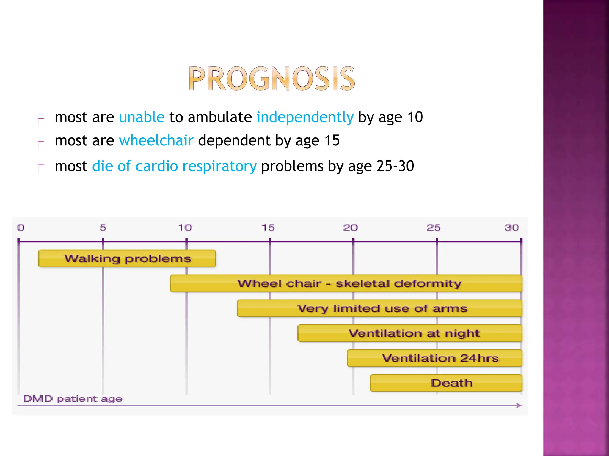
BY ANIEDU, UGOCHUKWU I.
(B.Sc, MD(in view)
History
Introduction
Genetics
Pathogenesis
Clinical features/Diagnosis
Prognosis
Treatment
Summary and Conclusion
References
The disease was first described by the Neapolitan
physician Giovanni Semmola in 1834 and Gaetano
Conte in 1836
DMD is named after the French neurologist Guillaume
Benjamin Amand Duchenne
In an 1861 publication, Duchenne established the
diagnostic criteria that are still used
William Richard Gowers was the first to deduce the
genetic basis for the disease
In 1986, Louis M. Kunkel provided molecular genetic
confirmation of the X-linked recessive inheritance
pattern
The muscular dystrophies are a group of
genetically determined, progressive diseases of
skeletal muscle
They are non-inflammatory and have no
neurological cause
Duchenne muscular dystrophy (DMD) is the most
common muscular dystrophy affecting 1 in 3500
males born worldwide.
Seen in males only (expect in females with
TURNER’S SYNDROME)
DMD is inherited in an X-linked recessive
pattern(defect at Xp21 locus)
Females will typically be carriers for
the disease while males will be
affected
The son of a carrier mother has a 50%
chance of inheriting the defective gene
from his mother.
The daughter of a carrier mother has a
50% chance of being a carrier or having
two normal copies of the gene.
The disorder is caused by a mutation in
the dystrophin gene, the largest gene
located on the human X chromosome which
codes for the protein dystrophin
Without dystrophin, muscles are susceptible
to mechanical injury and undergo repeated
cycles of necrosis and regeneration.
Ultimately, regenerative capabilities are
exhausted or inactivated
Dystrophin is responsible for connecting
the cytoskeleton of each muscle fiber to the
underlying basal lamina
The absence of dystrophin permits
excess calcium to penetrate the sarcolemma
leading to mitochondrial dysfunction
mitochondrial dysfunction gives rise to an
amplification of stress-induced cytosolic calcium
signals and an amplification of stress-
induced reactive-oxygen species (ROS) production.
Increased oxidative stress within the cell damages the sarcolemma and
eventually results in the death of the cell.
Muscle fibers undergo necrosis and are ultimately replaced
with adipose and connective tissue
Age of onset is between 2-6 years of age
Stage 1 – Presymptomatic
Creatine kinase usually elevated
Positive family history
Stage 2- Early ambulatory
clumsy & Waddling gait, manifesting in children aged
2-6 years; secondary to hip girdle muscle weakness
Inexorable progressive weakness in the proximal
musculature, initially in the lower extremities, but
later involving the neck flexors, shoulders, and arms
Meryon’s sign
child slips through examiner’s grasp when lifted under
arms
Possible toe-walking
Can climb stairs
Gower's sign
-'climbing up legs' using the hands when rising from the floor
Stage 3- Late ambulatory
More difficulty walking
Around age 8 years, most patients notice difficulty with ascending stairs
and respiratory muscle strength begins a slow but steady decline
Cannot arise from the floor
The forced vital capacity begins to gradually wane, leading to symptoms
of nocturnal hypoxemia such as lethargy and early morning headaches
Stage 4 – Early nonambulatory
Can self-propel for some time
Able to maintain posture
Possible development of scoliosis
Stage 5 – Late nonambulatory
Scoliosis may progress, especially when more wheelchair dependent
If wheelchair bound and profoundly weak, patients develop terminal
respiratory or cardiac failure, usually by the early 30s
poor nutritional intake can also be a serious complication in
individuals with severe end-stage DMD
Contractures may develop
most are unable to ambulate independently by age 10
most are wheelchair dependent by age 15
most die of cardio respiratory problems by age 25-30
There is no cure yet for DMD, however
case and symptom management
such as:
• physical therapy
• positioning aids - used to help the
child sit, lie, or stand
• braces and splints - used to prevent
deformity, promote support, or
provide protection
• medications
• nutritional counseling
• psychological counseling
is currently successful
Conclusively, there are many clinical trials in process, like
administering Albuterol (beta adrenergic receptor agonist drug
that increases strength and muscle mass) also, they want to
treat with Utrophin (sometimes can be substituted for
dystrophin)
Embryonic stem cell transplants is another treatment they are
looking into. It is hoped that injecting healthy, nonspecialized
stem cells into DMD victims will cause the stem cells to
specialize and produce structurally and functionally correct
dystrophin. If dystrophin can be produced, it may slow the
progression of the disease, or cure it altogether.
Deconinck, N., & Dan, B. (2007). Pathophysiology of duchenne muscular dystrophy: current
hypotheses. Pediatric neurology, 36(1), 1-7.
Hoffman EP
, Dressman D (2001) Molecular pathophysiology and targeted therapeutics for muscular
dystrophy. Trends Pharmacol Sci 22: 465–470
Nowak, K. J., & Davies, K. E. (2004). Duchenne muscular dystrophy and dystrophin: pathogenesis
and opportunities for treatment. EMBO reports, 5(9), 872-876.
Ouyang L, Grosse SD, KennesonA. Health Care Utilization and Expenditures for Children and Young
Adults With Muscular Dystrophy in a Privately Insured Population. J Child Neurol. 2008 Aug;23
(8):883-8.
Hughes, M. I., Hicks, E. M., Nevin, N. C., & Patterson, V
. H. (1996). The prevalence of inherited
neuromuscular disease in Northern Ireland.Neuromuscular Disorders, 6(1), 69-73.
Gulati, S., Saxena, A., Kumar, V., & Kalra, V
. (2005). Duchenne muscular dystrophy: prevalence and
patterns of cardiac involvement. Indian journal of pediatrics, 72(5), 389-393.
Bushby, K., Bourke, J., Bullock, R., Eagle, M., Gibson, M., & Quinby, J. (2005). The
multidisciplinary management of Duchenne muscular dystrophy.Current Paediatrics, 15(4), 292-
300.
Chung, B., Wong, V., & Ip, P
.(2003). Prevalence of neuromuscular diseases in Chinese children: a
study in southern China. Journal of child neurology, 18(3), 217-219
Manzur A
Y
,Kuntzer T
,Pike M, Swan A, Glucocorticoid corticosteroids for Duchenne muscular
dystrophy (Cochrane review). The Cochrane Library, Chichester UK, Wiley, 2004.
Bushby K, Muntoni F
,Urtizberea A, Hughes R, Griggs R. Report on the 124th ENMC International
Workshop: Treatment of Duchenne muscular dystrophy; defining the gold standards of management
in the use of corticosteroids. 2–4 April 2004, Naarden, The Netherlands. Neuromuscul Disord
2004;14(8–9):526–34.
Moxley III RT
,Ashwal S, Pandya S, et al. Practice parameter: corticosteroid treatment of
Duchenne dystrophy: report of the Quality Standards Subcommittee of the American Academy of
Neurology and the Practice Committee of the Child Neurology Society. Neurology 2005;64(1):13–20.
Cervellati S, Bettini N, Moscato M, Gusella A, Dema E, Maresi R. Surgical treatment of spinal
deformities in Duchenne muscular dystrophy: a long term follow-up study. Eur Spine J
2004;13(5):441–8.
Finder JD, Birnkrant D, Carl J, et al. Respiratory care of the patient with Duchenne muscular
dystrophy:ATS consensus statement.Am J Respir Crit Care Med 2004;170(4):456–65.
Eagle, M., Bourke, J., Bullock, R., Gibson, M., Mehta, J., Giddings, D., ... & Bushby, K. (2007).
Managing Duchenne muscular dystrophy--the additive effect of spinal surgery and home nocturnal
ventilation in improving survival.Neuromuscular disorders: NMD, 17(6), 470.
Bushby KMD, Muntoni F
,Bourke JP
. The management of cardiac complications in muscular
dystrophy and myotonic dystrophy. Proceedings of 107th ENMC Workshop. Neuromuscul Disord
2003;13:166–72.
AmericanAcademy of Pediatrics Section on Cardiology and Cardiac Surgery. Cardiovascular health
supervision for in dividuals aff ected by Duchenne or Becker muscular dystrophy. Pediatrics 2005;
116: 1569–73
Bushby, K., Bourke, J., Bullock, R., Eagle, M., Gibson, M., & Quinby, J. (2005). The
multidisciplinary management of Duchenne muscular dystrophy.Current Paediatrics, 15(4), 292-
300.
Parsons, E. P
., Clarke, A. J., & Bradley, D. M. (2004). Developmental progress in Duchenne muscular
dystrophy: lessons for earlier detection. European journal of paediatric neurology: EJPN: official
journal of the European Paediatric Neurology Society, 8(3), 145.
Essen, A. J., Busch, H. F
.M., Meerman, G. J., & Kate, L. P
.(1992). Birth and population prevalence
of Duchenne muscular dystrophy in The Netherlands.Human genetics, 88(3), 258-266.
Drousiotou A, Ioannou P
,Georgiou T
,et al. Neonatal screening for Duchenne muscular
dystrophy: a novel semiquantitative application of the bioluminescence test for creatine
kinase in a pilot national program in Cyprus. Genet Test 1998; 2: 55–60.
Bradley D, Parsons E. Newborn screening for Duchenne muscular dystrophy. Semin
Neonatol 1998; 3: 27–34.
Emery AE. Population frequencies of inherited neuromuscular diseases—a world survey
Neuromuscul Disord 1991; 1: 19–29.
Ciafaloni E, Fox DJ, Pandya S, Westfield CP
, Puzhankara S, Romitti PA, et al. Delayed
diagnosis in Duchenne muscular dystrophy: data from the Muscular Dystrophy
Surveillance, Tracking, and Research Network (MD STARnet). J Pediatr 2009
Sept;155(3):380-5.
Fowler WM Jr. Role of physical activity and exercise training in neuromuscular
diseases.Am J Phys Med Rehabil 2002; 81 (suppl): S187–95.
Fowler WM Jr. Rehabilitation management of muscular dystrophy and related
disorders: II. Comprehensive care. Arch Phys Med Rehabil 1982; 63: 322–28
THANK YOU

mygeneticspresentation-141130173403-conversion-gate02.pptx

  • 1.
    BY ANIEDU, UGOCHUKWUI. (B.Sc, MD(in view)
  • 2.
  • 3.
    The disease wasfirst described by the Neapolitan physician Giovanni Semmola in 1834 and Gaetano Conte in 1836 DMD is named after the French neurologist Guillaume Benjamin Amand Duchenne In an 1861 publication, Duchenne established the diagnostic criteria that are still used William Richard Gowers was the first to deduce the genetic basis for the disease In 1986, Louis M. Kunkel provided molecular genetic confirmation of the X-linked recessive inheritance pattern
  • 4.
    The muscular dystrophiesare a group of genetically determined, progressive diseases of skeletal muscle They are non-inflammatory and have no neurological cause Duchenne muscular dystrophy (DMD) is the most common muscular dystrophy affecting 1 in 3500 males born worldwide. Seen in males only (expect in females with TURNER’S SYNDROME)
  • 5.
    DMD is inheritedin an X-linked recessive pattern(defect at Xp21 locus) Females will typically be carriers for the disease while males will be affected The son of a carrier mother has a 50% chance of inheriting the defective gene from his mother. The daughter of a carrier mother has a 50% chance of being a carrier or having two normal copies of the gene.
  • 6.
    The disorder iscaused by a mutation in the dystrophin gene, the largest gene located on the human X chromosome which codes for the protein dystrophin Without dystrophin, muscles are susceptible to mechanical injury and undergo repeated cycles of necrosis and regeneration. Ultimately, regenerative capabilities are exhausted or inactivated
  • 7.
    Dystrophin is responsiblefor connecting the cytoskeleton of each muscle fiber to the underlying basal lamina The absence of dystrophin permits excess calcium to penetrate the sarcolemma leading to mitochondrial dysfunction mitochondrial dysfunction gives rise to an amplification of stress-induced cytosolic calcium signals and an amplification of stress- induced reactive-oxygen species (ROS) production.
  • 8.
    Increased oxidative stresswithin the cell damages the sarcolemma and eventually results in the death of the cell. Muscle fibers undergo necrosis and are ultimately replaced with adipose and connective tissue
  • 9.
    Age of onsetis between 2-6 years of age Stage 1 – Presymptomatic Creatine kinase usually elevated Positive family history
  • 10.
    Stage 2- Earlyambulatory clumsy & Waddling gait, manifesting in children aged 2-6 years; secondary to hip girdle muscle weakness Inexorable progressive weakness in the proximal musculature, initially in the lower extremities, but later involving the neck flexors, shoulders, and arms Meryon’s sign child slips through examiner’s grasp when lifted under arms Possible toe-walking Can climb stairs
  • 11.
    Gower's sign -'climbing uplegs' using the hands when rising from the floor
  • 12.
    Stage 3- Lateambulatory More difficulty walking Around age 8 years, most patients notice difficulty with ascending stairs and respiratory muscle strength begins a slow but steady decline Cannot arise from the floor The forced vital capacity begins to gradually wane, leading to symptoms of nocturnal hypoxemia such as lethargy and early morning headaches
  • 13.
    Stage 4 –Early nonambulatory Can self-propel for some time Able to maintain posture Possible development of scoliosis Stage 5 – Late nonambulatory Scoliosis may progress, especially when more wheelchair dependent If wheelchair bound and profoundly weak, patients develop terminal respiratory or cardiac failure, usually by the early 30s poor nutritional intake can also be a serious complication in individuals with severe end-stage DMD Contractures may develop
  • 14.
    most are unableto ambulate independently by age 10 most are wheelchair dependent by age 15 most die of cardio respiratory problems by age 25-30
  • 15.
    There is nocure yet for DMD, however case and symptom management such as: • physical therapy • positioning aids - used to help the child sit, lie, or stand • braces and splints - used to prevent deformity, promote support, or provide protection • medications • nutritional counseling • psychological counseling is currently successful
  • 16.
    Conclusively, there aremany clinical trials in process, like administering Albuterol (beta adrenergic receptor agonist drug that increases strength and muscle mass) also, they want to treat with Utrophin (sometimes can be substituted for dystrophin) Embryonic stem cell transplants is another treatment they are looking into. It is hoped that injecting healthy, nonspecialized stem cells into DMD victims will cause the stem cells to specialize and produce structurally and functionally correct dystrophin. If dystrophin can be produced, it may slow the progression of the disease, or cure it altogether.
  • 17.
    Deconinck, N., &Dan, B. (2007). Pathophysiology of duchenne muscular dystrophy: current hypotheses. Pediatric neurology, 36(1), 1-7. Hoffman EP , Dressman D (2001) Molecular pathophysiology and targeted therapeutics for muscular dystrophy. Trends Pharmacol Sci 22: 465–470 Nowak, K. J., & Davies, K. E. (2004). Duchenne muscular dystrophy and dystrophin: pathogenesis and opportunities for treatment. EMBO reports, 5(9), 872-876. Ouyang L, Grosse SD, KennesonA. Health Care Utilization and Expenditures for Children and Young Adults With Muscular Dystrophy in a Privately Insured Population. J Child Neurol. 2008 Aug;23 (8):883-8. Hughes, M. I., Hicks, E. M., Nevin, N. C., & Patterson, V . H. (1996). The prevalence of inherited neuromuscular disease in Northern Ireland.Neuromuscular Disorders, 6(1), 69-73.
  • 18.
    Gulati, S., Saxena,A., Kumar, V., & Kalra, V . (2005). Duchenne muscular dystrophy: prevalence and patterns of cardiac involvement. Indian journal of pediatrics, 72(5), 389-393. Bushby, K., Bourke, J., Bullock, R., Eagle, M., Gibson, M., & Quinby, J. (2005). The multidisciplinary management of Duchenne muscular dystrophy.Current Paediatrics, 15(4), 292- 300. Chung, B., Wong, V., & Ip, P .(2003). Prevalence of neuromuscular diseases in Chinese children: a study in southern China. Journal of child neurology, 18(3), 217-219 Manzur A Y ,Kuntzer T ,Pike M, Swan A, Glucocorticoid corticosteroids for Duchenne muscular dystrophy (Cochrane review). The Cochrane Library, Chichester UK, Wiley, 2004. Bushby K, Muntoni F ,Urtizberea A, Hughes R, Griggs R. Report on the 124th ENMC International Workshop: Treatment of Duchenne muscular dystrophy; defining the gold standards of management in the use of corticosteroids. 2–4 April 2004, Naarden, The Netherlands. Neuromuscul Disord 2004;14(8–9):526–34. Moxley III RT ,Ashwal S, Pandya S, et al. Practice parameter: corticosteroid treatment of Duchenne dystrophy: report of the Quality Standards Subcommittee of the American Academy of Neurology and the Practice Committee of the Child Neurology Society. Neurology 2005;64(1):13–20. Cervellati S, Bettini N, Moscato M, Gusella A, Dema E, Maresi R. Surgical treatment of spinal deformities in Duchenne muscular dystrophy: a long term follow-up study. Eur Spine J 2004;13(5):441–8.
  • 19.
    Finder JD, BirnkrantD, Carl J, et al. Respiratory care of the patient with Duchenne muscular dystrophy:ATS consensus statement.Am J Respir Crit Care Med 2004;170(4):456–65. Eagle, M., Bourke, J., Bullock, R., Gibson, M., Mehta, J., Giddings, D., ... & Bushby, K. (2007). Managing Duchenne muscular dystrophy--the additive effect of spinal surgery and home nocturnal ventilation in improving survival.Neuromuscular disorders: NMD, 17(6), 470. Bushby KMD, Muntoni F ,Bourke JP . The management of cardiac complications in muscular dystrophy and myotonic dystrophy. Proceedings of 107th ENMC Workshop. Neuromuscul Disord 2003;13:166–72. AmericanAcademy of Pediatrics Section on Cardiology and Cardiac Surgery. Cardiovascular health supervision for in dividuals aff ected by Duchenne or Becker muscular dystrophy. Pediatrics 2005; 116: 1569–73 Bushby, K., Bourke, J., Bullock, R., Eagle, M., Gibson, M., & Quinby, J. (2005). The multidisciplinary management of Duchenne muscular dystrophy.Current Paediatrics, 15(4), 292- 300. Parsons, E. P ., Clarke, A. J., & Bradley, D. M. (2004). Developmental progress in Duchenne muscular dystrophy: lessons for earlier detection. European journal of paediatric neurology: EJPN: official journal of the European Paediatric Neurology Society, 8(3), 145. Essen, A. J., Busch, H. F .M., Meerman, G. J., & Kate, L. P .(1992). Birth and population prevalence of Duchenne muscular dystrophy in The Netherlands.Human genetics, 88(3), 258-266.
  • 20.
    Drousiotou A, IoannouP ,Georgiou T ,et al. Neonatal screening for Duchenne muscular dystrophy: a novel semiquantitative application of the bioluminescence test for creatine kinase in a pilot national program in Cyprus. Genet Test 1998; 2: 55–60. Bradley D, Parsons E. Newborn screening for Duchenne muscular dystrophy. Semin Neonatol 1998; 3: 27–34. Emery AE. Population frequencies of inherited neuromuscular diseases—a world survey Neuromuscul Disord 1991; 1: 19–29. Ciafaloni E, Fox DJ, Pandya S, Westfield CP , Puzhankara S, Romitti PA, et al. Delayed diagnosis in Duchenne muscular dystrophy: data from the Muscular Dystrophy Surveillance, Tracking, and Research Network (MD STARnet). J Pediatr 2009 Sept;155(3):380-5. Fowler WM Jr. Role of physical activity and exercise training in neuromuscular diseases.Am J Phys Med Rehabil 2002; 81 (suppl): S187–95. Fowler WM Jr. Rehabilitation management of muscular dystrophy and related disorders: II. Comprehensive care. Arch Phys Med Rehabil 1982; 63: 322–28
  • 21.