Downloaded 288 times
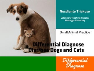

![Small Intestinal Diarrhea
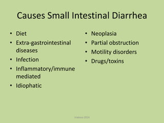
• Diet
– Dietary intolerance [Food hypersensitivity; Food
intolerance; Gluten-sensitive enteropathy]
• Extra-gastrointestinal diseases
– EPI
– Hepatic disease
– Hyperthiroidism
– Hypoadrenocorticism
triakoso 2014](https://image.slidesharecdn.com/differentialdiarrheasmallanimal-140202231524-phpapp01/85/Differential-Diarrhea-Small-Animal-Medicine-10-320.jpg)
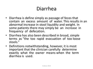
![Small Intestinal Diarrhea
• Infection
– Bacterial [Campyovacter spp; Clostridium spp;
Salmonella spp; E coli; Staphylococcus, SIBO]
– Fungal
– Helminth [Hookworm; Roundworm; Tapeworm;
Whipworm]
– Protozoal [Cryptosporidiosis; Giardia spp]
– Rickettsial
– Viral [Corona virus; Parvovirus; Feline
panleukopenia]
triakoso 2014](https://image.slidesharecdn.com/differentialdiarrheasmallanimal-140202231524-phpapp01/85/Differential-Diarrhea-Small-Animal-Medicine-11-320.jpg)
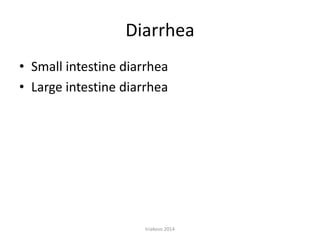
![Small Intestinal Diarrhea
• Inflammatory/immune mediated
–
–
–
–
Basenji enteropathy
Duodenal ulceration
Hemorrhagic gastroenteritis
Inflammatory bowel disease [Eosinophilic;
Granulomatous; Lymphoplasmacytic]
– Protein-losing enteropathy and nephropathy of the
Soft-Coated Weaten Terrier
• Idiophatic
– Lymphangiectasia
triakoso 2014](https://image.slidesharecdn.com/differentialdiarrheasmallanimal-140202231524-phpapp01/85/Differential-Diarrhea-Small-Animal-Medicine-12-320.jpg)
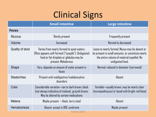
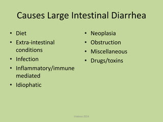
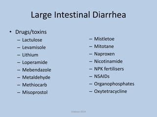
![Large Intestinal Diarrhea
• Diet*
– Dietary hypersensitivity
– Dietary indiscretion
• Extra-intestinal conditions
–
–
–
–
–
Metastatic neoplasia
Neurological disease leading to ulcerative colitis
Pancreatitis
Toxaemia
Uraemia
• Infection
– Bacterial*, e.g. [Campylobacter spp; Clostridium difficile;
Clostridium perfringens; E. coli; Salmonella spp; Yersinia
enterocolitica]
triakoso 2014](https://image.slidesharecdn.com/differentialdiarrheasmallanimal-140202231524-phpapp01/85/Differential-Diarrhea-Small-Animal-Medicine-13-320.jpg)
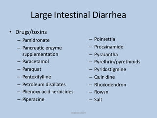
![Large Intestinal Diarrhea
• Infection
– Viral* [Coronavirus; Feline immunodeficiency virus
(C); Feline infectious peritonitis (C); Feline leukaemia
virus (C); Parvovirus]
– Fungal, e.g. [ Histoplasmosis; Protothecosis]
– Parasitic*, e.g. [Amoebiasis; Ancylostoma spp;
Balantidium coli; Cryptosporidiosis; Giardia spp;
Heterobilharzia americana; Roundworm; Tapeworm;
Tritrichomonas foetus(C); Uncinaria spp; Whipworm]
– Protozoal, e.g. [Toxoplasmosis]
triakoso 2014](https://image.slidesharecdn.com/differentialdiarrheasmallanimal-140202231524-phpapp01/85/Differential-Diarrhea-Small-Animal-Medicine-14-320.jpg)
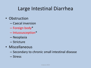
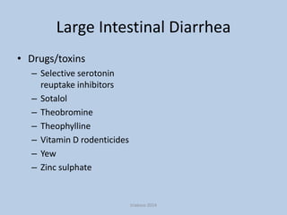
![Large Intestinal Diarrhea
• Immune-mediated/inflammatory disease
– Histiocytic ulcerative colitis of Boxers (D)
– Inflammatory bowel disease*
• Idiopathic conditions
– Fibre-responsive large-bowel diarrhoea
– Irritable bowel syndrome
• Neoplasia*
– Benign, e.g. [Adenomatous polyps; Leiomyoma]
– Malignant, e.g. [Adenocarcinoma; Lymphoma]
triakoso 2014](https://image.slidesharecdn.com/differentialdiarrheasmallanimal-140202231524-phpapp01/85/Differential-Diarrhea-Small-Animal-Medicine-15-320.jpg)


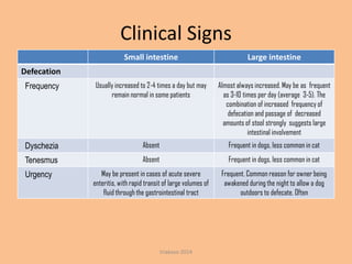
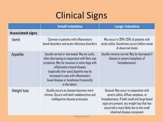
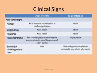
This document discusses the differential diagnosis of diarrhea in small and large intestines. It defines diarrhea and outlines key clinical signs that can indicate the location and underlying cause. For small intestine diarrhea, potential causes include dietary issues, infections, inflammatory/immune diseases, and idiopathic conditions. Large intestine diarrhea may result from similar causes like diet, infections, inflammation, and cancer, as well as obstructions, drugs/toxins, and stress. The document provides extensive lists of specific disorders, pathogens, medications, and toxins that commonly contribute to diarrhea in each intestinal region.


![CTEV [ clubfoot] DR ARUN LAL ,DR MOHAMED ASHRAF travancore medical college k...](https://cdn.slidesharecdn.com/ss_thumbnails/ctevclubfootdrarunlaldrmohamedashraftravancoremedicalcollegekollamkeralaindia-260208063247-18fc466c-thumbnail.jpg?width=640&height=640&fit=bounds)


![PERI-PROSTHETIC FRACTURE NAIL-PLATE CONSTRUCT [NPC].pptx](https://cdn.slidesharecdn.com/ss_thumbnails/drarunkumardrmohamedashrafperiprostheticfrasturenail-plateconstructnpc-260209164459-7e9d15a1-thumbnail.jpg?width=640&height=640&fit=bounds)
