
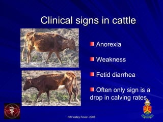
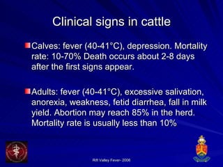
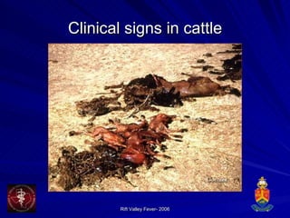
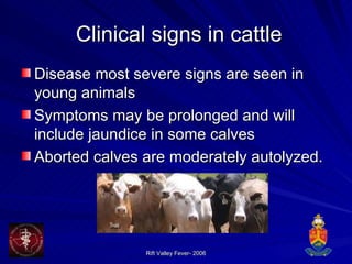
Downloaded 106 times

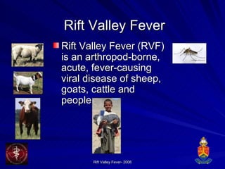
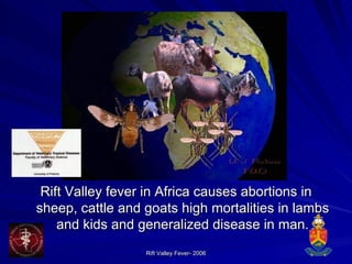
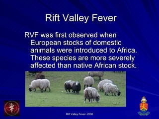
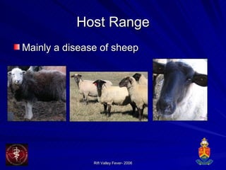
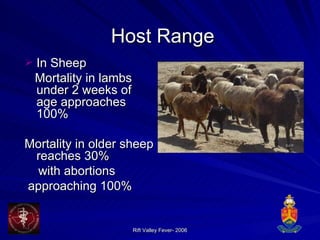
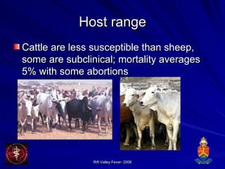


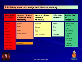

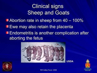
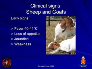
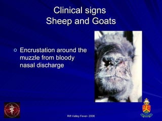
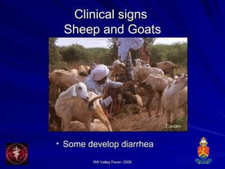
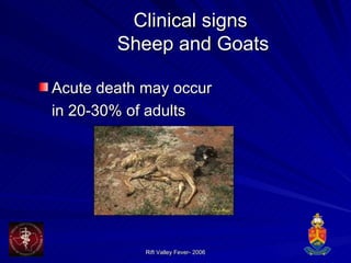
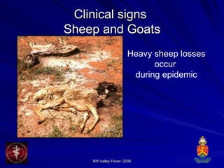
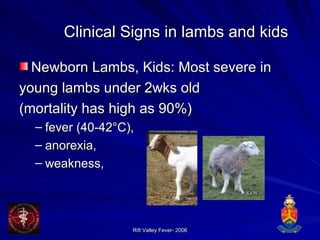
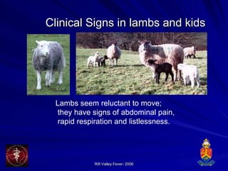
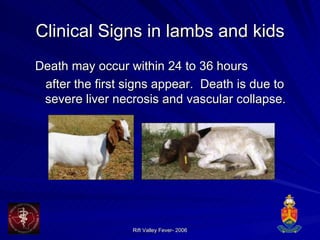
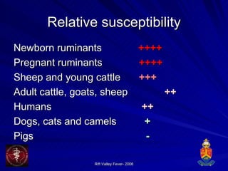
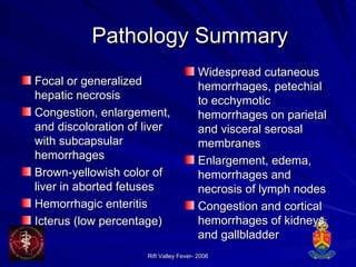
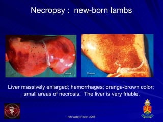
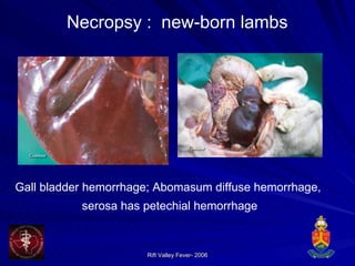
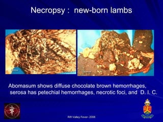
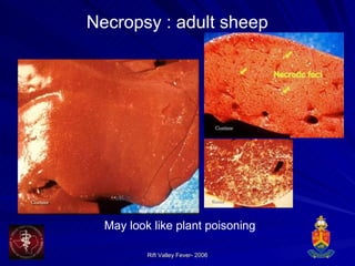

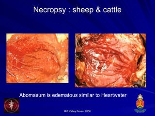
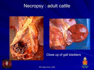
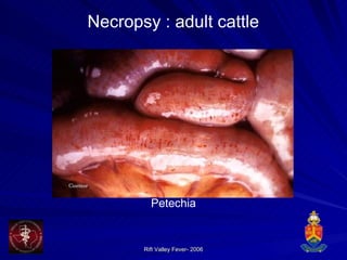
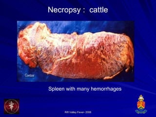

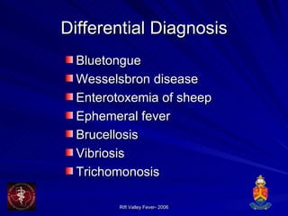
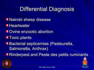
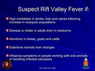


Rift Valley Fever is a viral disease that primarily affects sheep, goats, cattle, and humans. It is transmitted by mosquitoes and causes high rates of abortion and mortality in young animals. Clinical signs in sheep and goats include fever, jaundice, abortion, and death. Lambs and kids often die within 1-3 days of showing signs. Necropsy findings include massive hepatic necrosis and hemorrhages throughout the body. Differential diagnosis includes diseases causing similar symptoms such as bluetongue or hepatitis.


![Epidemiology of malaria [autosaved]](https://cdn.slidesharecdn.com/ss_thumbnails/epidemiologyofmalariaautosaved-201006062122-thumbnail.jpg?width=640&height=640&fit=bounds)


