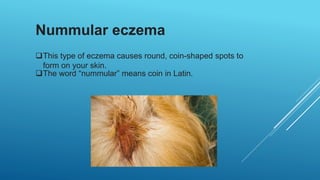
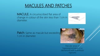
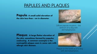
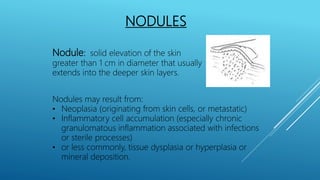
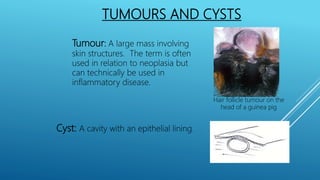
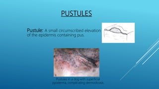
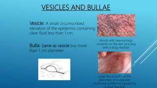
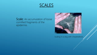
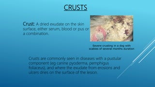
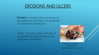
CLASSIFICATION OF
DERMATITIS AND
TYPES OF LESIONS
Introduction
• ECZEMA IS A GROUP OF SKIN DISEASES
CHARACTERIZED BY IRRITATION AND
INFLAMMATION OF THE SKIN.
• ECZEMA IS A GREEK WORD MEANING ‘TO BOIL’
• SYNONYMOUS WITH DERMATITIS
• INCREDIBLY COMMON
• ECZEMA MAY BEINDUCEDBYA WIDERANGE OF EXTERNALAND INTERNAL
FACTORSACTING SINGLYORINCOMBINATION. TWO MAINCLASSIFICATION
ARE:
• ENDOGENOUS ECZEMA: BASEDON AN INHERENTPOTENTIALITYTOHAVE
ECZEMA. NO CONTACT WITHA STIMULUSNEEDED.RECURRENT,SELFABATING
• EXOGENOUS ECZEMA : CONTACT WITHOUTSIDESTIMULANTNEEDED.
CONTACT IRRITANTAND CONTACT ALLERGIC TYPES
Classification
MAJOR TYPES OF DERMATITIS
•Atopic dermatitis
•Contact dermatitis
•Dyshidrotic eczema
•Neurodermatitis
•Nummular eczema
•Stasis dermatitis
 Atopic dermatitis (AD), also known as atopic eczema.
 It is a type of inflammation of the skin (dermatitis).
 Caused by immune hypersensitivity.
 Results in itchy, red, swollen, and cracked skin.
 Clear fluid may come from the affected areas, which often
thickens over time.
Atopic dermatitis
Contact dermatitis
• Red, irritated skin that’s caused by a reaction
to substances you touch.
• It comes in two types:
 Allergic contact dermatitis is an immune
system reaction to an irritant like latex or
metal.
 Irritant contact dermatitis starts when a
chemical or other substance irritates your
skin.
Dyshidrotic eczema
Dyshidrotic eczema causes small blisters to
form on your hands and feet.
Neurodermatitis
• Neurodermatitis is a skin condition of
canines which is caused by the dog
excessively and obsessively licking spots
on their own body.
• Although allergies, insect bites, and
arthritis may initiate the obsessive licking
that is characteristic of this disorder.
Nummular eczema
This type of eczema causes round, coin-shaped spots to
form on your skin.
The word “nummular” means coin in Latin.
Stasis dermatitis
 Stasis dermatitis happens when fluid leaks out of
weakened veins into your skin.
 This fluid causes swelling, redness, itching, and
pain.
PRIMARY LESIONS
 Macules & Patches
 Papules & Plaques
 Nodules
 Tumours & Cysts
 Pustules
 Vesicles & Bullae
 Wheals
 Scales
 Comedones
 Follicular Casts
 Crusts
• Erosions & Ulcers
• Epidermal Collarettes
• Scar
• Excoriation & Fissure
• Lichenification
• Pigmentation
SECONDARY
LESIONS
MACULES AND PATCHES
MACULE: A circumscribed flat area of
change in colour of the skin less than 1 cm in
diameter.
Patch: Same as macule but exceeding
1 cm in diameter.
Macular areas of
hyperpigmentation in a
dachshund with acanthosis
nigricans
PAPULES AND PLAQUES
Papule: A small solid elevation of
the skin less than 1 cm in diameter.
Plaque: A large flatter elevation of
the skin, sometimes formed by papules
coalescing. A common example is the
eosinophilc plaque seen in some cats with
allergic skin disease.
Diffuse papular eruption - the
ventrum of a dog with
dermatophytosis
Plaque on the forelimb of a dog
with dermatophytosis
NODULES
Nodule: solid elevation of the skin
greater than 1 cm in diameter that usually
extends into the deeper skin layers.
Nodules may result from:
• Neoplasia (originating from skin cells, or metastatic)
• Inflammatory cell accumulation (especially chronic
granulomatous inflammation associated with infections
or sterile processes)
• or less commonly, tissue dysplasia or hyperplasia or
mineral deposition.
TUMOURS AND CYSTS
Tumour: A large mass involving
skin structures. The term is often
used in relation to neoplasia but
can technically be used in
inflammatory disease.
Cyst: A cavity with an epithelial lining.
Hair follicle tumour on the
head of a guinea pig
PUSTULES
Pustule: A small circumscribed elevation
of the epidermis containing pus.
Pustules in a dog with superficial
pyoderma, complicating dermodicosis
VESICLES AND BULLAE
Vesicle: A small circumscribed
elevation of the epidermis containing
clear fluid less than 1 cm.
Vesicle with haemorhagic
contents on the skin of a dog
with a drug reaction
Bulla: Same as vesicle but more
than 1 cm diameter.
Large flaccid bulla on the
abdomen of a dog with
erythema multiforme caused by
a drug reaction
WHEALS
Wheal: A circumscribed, raised
lesion consisting of dermal oedema.
Wheal in a horse with acute urticaria
SCALES
Scale: An accumulation of loose
cornified fragments of the
epidermis.
Scaling in a dog with cheyletiellosis
COMEDONES
Comedo: A dilated hair follicle
devoid of hair and plugged with
keratinous debris (a 'blackhead').
Comedones and alopecia in a Boxer with
generalised demodicosis
FOLLICULAR CASTS
Follicular cast: An accumulation of
keratinous debris around the hair shaft
seen protruding from the follicular
ostium or present when hairs are
plucked and examined.
Scaling and follicular casting in a dog with
sedaceous adenitis
CRUSTS
Crust: A dried exudate on the skin
surface, either serum, blood or pus or
a combination.
Severe crusting in a dog with
scabies of several months duration
Crusts are commonly seen in diseases with a pustular
component (eg canine pyoderma, pemphigus
foliaceus), and where the exudate from erosions and
ulcers dries on the surface of the lesion.
EROSIONS AND ULCERS
Erosion: A break in the continuity of
the epidermis that does not penetrate
the basement membrane.
Ulcer: A break in the continuity of
the epidermis that penetrates the
basement membrane.
Footpad ulceration in a
retriever with
epitheliotropic lymphoma
EROSIONS AND ULCERS
Erosions and ulcers can be seen in many
processes. Vesicular and bullous lesions usually
evolve into erosions and ulcers. Ulcers can also
be seen severe infections and in association
with neoplasia (eg squamous cell carcinoma).
EPIDERMAL COLLARETTES
Epidermal collarette: A
circular lesion with a circular
rim of scale and or a peeling
edge.
Epidermal collarete lesion in a dog
with a sterile pustular disease
These are 'footprints' of a vesicular or pustular
lesion. They are therefore a common lesion type in
canine pyoderma.
SCAR
Scar: An area of fibrous tissue replacing
damaged skin.
EXCORIATION AND FISSURE
Excoriation: Erosion or ulceration
caused by scratching, biting or
rubbing.
Severe excoriations in a dog with scabies
Excoriation is usually seen as a consequence of moderate or
severe pruritus.
EXCORIATION AND FISSURE
Fissure: Linear split through the
epidermis into the underlying
dermis.
Severe footpad hyperkeratosis
with fissures (arrow) in a dog with
necrolytic migratory erythema
Fissures are usually seen as a consequence of a loss of skin
elasticity. In dogs with the so-called hepatocutaneous
syndrome, footpad hyperkeratosis and fissuring often
leads to lameness.
LICHENIFICATION
Lichenification: An accentuation of
the skin markings giving an elephant
skin like appearance
Prominent lichenification in a dog
with chronic atopy
and Malassezia dermatitis
Lichenification is associated with chronic inflammation and
trauma, and is usually a feature of severe and chronic canine
allergic disease or Malasseziainfection, for example. Some
dog breeds seem to develop lichenification more readily than
others; WHWT and GSD often present with this lesion type.
PIGMENTATION DISTURBANCES
Hyperpigmentation: Skin
pigmentation increased beyond what
is normal for that area.
Hypopigmentatio: Skin
pigmentation decreased beyond what
is normal for that area.
Leukotrichia: Loss of hair
pigment.
Extensive hyperpigmentation in a
dog with chronic Malassezia
dermatitis
Dermatitis in animals types and lesions

Dermatitis in animals types and lesions

  • 1.
  • 2.
    Introduction • ECZEMA ISA GROUP OF SKIN DISEASES CHARACTERIZED BY IRRITATION AND INFLAMMATION OF THE SKIN. • ECZEMA IS A GREEK WORD MEANING ‘TO BOIL’ • SYNONYMOUS WITH DERMATITIS • INCREDIBLY COMMON
  • 3.
    • ECZEMA MAYBEINDUCEDBYA WIDERANGE OF EXTERNALAND INTERNAL FACTORSACTING SINGLYORINCOMBINATION. TWO MAINCLASSIFICATION ARE: • ENDOGENOUS ECZEMA: BASEDON AN INHERENTPOTENTIALITYTOHAVE ECZEMA. NO CONTACT WITHA STIMULUSNEEDED.RECURRENT,SELFABATING • EXOGENOUS ECZEMA : CONTACT WITHOUTSIDESTIMULANTNEEDED. CONTACT IRRITANTAND CONTACT ALLERGIC TYPES Classification
  • 4.
    MAJOR TYPES OFDERMATITIS •Atopic dermatitis •Contact dermatitis •Dyshidrotic eczema •Neurodermatitis •Nummular eczema •Stasis dermatitis
  • 5.
     Atopic dermatitis(AD), also known as atopic eczema.  It is a type of inflammation of the skin (dermatitis).  Caused by immune hypersensitivity.  Results in itchy, red, swollen, and cracked skin.  Clear fluid may come from the affected areas, which often thickens over time. Atopic dermatitis
  • 6.
    Contact dermatitis • Red,irritated skin that’s caused by a reaction to substances you touch. • It comes in two types:  Allergic contact dermatitis is an immune system reaction to an irritant like latex or metal.  Irritant contact dermatitis starts when a chemical or other substance irritates your skin.
  • 7.
    Dyshidrotic eczema Dyshidrotic eczemacauses small blisters to form on your hands and feet.
  • 8.
    Neurodermatitis • Neurodermatitis isa skin condition of canines which is caused by the dog excessively and obsessively licking spots on their own body. • Although allergies, insect bites, and arthritis may initiate the obsessive licking that is characteristic of this disorder.
  • 9.
    Nummular eczema This typeof eczema causes round, coin-shaped spots to form on your skin. The word “nummular” means coin in Latin.
  • 10.
    Stasis dermatitis  Stasisdermatitis happens when fluid leaks out of weakened veins into your skin.  This fluid causes swelling, redness, itching, and pain.
  • 11.
    PRIMARY LESIONS  Macules& Patches  Papules & Plaques  Nodules  Tumours & Cysts  Pustules  Vesicles & Bullae  Wheals  Scales  Comedones  Follicular Casts  Crusts • Erosions & Ulcers • Epidermal Collarettes • Scar • Excoriation & Fissure • Lichenification • Pigmentation SECONDARY LESIONS
  • 12.
    MACULES AND PATCHES MACULE:A circumscribed flat area of change in colour of the skin less than 1 cm in diameter. Patch: Same as macule but exceeding 1 cm in diameter. Macular areas of hyperpigmentation in a dachshund with acanthosis nigricans
  • 13.
    PAPULES AND PLAQUES Papule:A small solid elevation of the skin less than 1 cm in diameter. Plaque: A large flatter elevation of the skin, sometimes formed by papules coalescing. A common example is the eosinophilc plaque seen in some cats with allergic skin disease. Diffuse papular eruption - the ventrum of a dog with dermatophytosis Plaque on the forelimb of a dog with dermatophytosis
  • 14.
    NODULES Nodule: solid elevationof the skin greater than 1 cm in diameter that usually extends into the deeper skin layers. Nodules may result from: • Neoplasia (originating from skin cells, or metastatic) • Inflammatory cell accumulation (especially chronic granulomatous inflammation associated with infections or sterile processes) • or less commonly, tissue dysplasia or hyperplasia or mineral deposition.
  • 15.
    TUMOURS AND CYSTS Tumour:A large mass involving skin structures. The term is often used in relation to neoplasia but can technically be used in inflammatory disease. Cyst: A cavity with an epithelial lining. Hair follicle tumour on the head of a guinea pig
  • 16.
    PUSTULES Pustule: A smallcircumscribed elevation of the epidermis containing pus. Pustules in a dog with superficial pyoderma, complicating dermodicosis
  • 17.
    VESICLES AND BULLAE Vesicle:A small circumscribed elevation of the epidermis containing clear fluid less than 1 cm. Vesicle with haemorhagic contents on the skin of a dog with a drug reaction Bulla: Same as vesicle but more than 1 cm diameter. Large flaccid bulla on the abdomen of a dog with erythema multiforme caused by a drug reaction
  • 18.
    WHEALS Wheal: A circumscribed,raised lesion consisting of dermal oedema. Wheal in a horse with acute urticaria
  • 19.
    SCALES Scale: An accumulationof loose cornified fragments of the epidermis. Scaling in a dog with cheyletiellosis
  • 20.
    COMEDONES Comedo: A dilatedhair follicle devoid of hair and plugged with keratinous debris (a 'blackhead'). Comedones and alopecia in a Boxer with generalised demodicosis
  • 21.
    FOLLICULAR CASTS Follicular cast:An accumulation of keratinous debris around the hair shaft seen protruding from the follicular ostium or present when hairs are plucked and examined. Scaling and follicular casting in a dog with sedaceous adenitis
  • 22.
    CRUSTS Crust: A driedexudate on the skin surface, either serum, blood or pus or a combination. Severe crusting in a dog with scabies of several months duration Crusts are commonly seen in diseases with a pustular component (eg canine pyoderma, pemphigus foliaceus), and where the exudate from erosions and ulcers dries on the surface of the lesion.
  • 23.
    EROSIONS AND ULCERS Erosion:A break in the continuity of the epidermis that does not penetrate the basement membrane. Ulcer: A break in the continuity of the epidermis that penetrates the basement membrane. Footpad ulceration in a retriever with epitheliotropic lymphoma
  • 24.
    EROSIONS AND ULCERS Erosionsand ulcers can be seen in many processes. Vesicular and bullous lesions usually evolve into erosions and ulcers. Ulcers can also be seen severe infections and in association with neoplasia (eg squamous cell carcinoma).
  • 25.
    EPIDERMAL COLLARETTES Epidermal collarette:A circular lesion with a circular rim of scale and or a peeling edge. Epidermal collarete lesion in a dog with a sterile pustular disease These are 'footprints' of a vesicular or pustular lesion. They are therefore a common lesion type in canine pyoderma.
  • 26.
    SCAR Scar: An areaof fibrous tissue replacing damaged skin.
  • 27.
    EXCORIATION AND FISSURE Excoriation:Erosion or ulceration caused by scratching, biting or rubbing. Severe excoriations in a dog with scabies Excoriation is usually seen as a consequence of moderate or severe pruritus.
  • 28.
    EXCORIATION AND FISSURE Fissure:Linear split through the epidermis into the underlying dermis. Severe footpad hyperkeratosis with fissures (arrow) in a dog with necrolytic migratory erythema Fissures are usually seen as a consequence of a loss of skin elasticity. In dogs with the so-called hepatocutaneous syndrome, footpad hyperkeratosis and fissuring often leads to lameness.
  • 29.
    LICHENIFICATION Lichenification: An accentuationof the skin markings giving an elephant skin like appearance Prominent lichenification in a dog with chronic atopy and Malassezia dermatitis Lichenification is associated with chronic inflammation and trauma, and is usually a feature of severe and chronic canine allergic disease or Malasseziainfection, for example. Some dog breeds seem to develop lichenification more readily than others; WHWT and GSD often present with this lesion type.
  • 30.
    PIGMENTATION DISTURBANCES Hyperpigmentation: Skin pigmentationincreased beyond what is normal for that area. Hypopigmentatio: Skin pigmentation decreased beyond what is normal for that area. Leukotrichia: Loss of hair pigment. Extensive hyperpigmentation in a dog with chronic Malassezia dermatitis