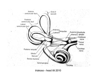
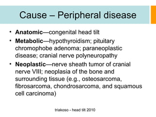
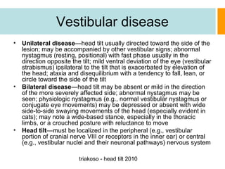
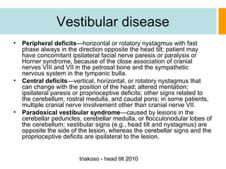
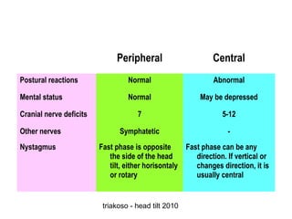
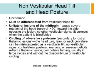
This document discusses head tilt, which is a tilting of the head away from its normal orientation associated with disorders of the vestibular system. It describes the pathophysiology of the vestibular system and how head tilt relates to lesions affecting this system. Potential causes of head tilt are discussed, including peripheral diseases such as otitis and central diseases such as tumors. Signs, diagnostic testing, treatment, and medications associated with different underlying causes of head tilt are outlined.
The document provides information on differential diagnosis of seizures in small animals. It defines seizures and differentiates them from syncope. Seizures are classified as focal or generalized. Potential causes of seizures are categorized as structural epileptic, reactive epileptic, primary epileptic, intracranial, or extracranial. Numerous specific diseases, infections, injuries, metabolic issues, toxins and drugs that can cause seizures are detailed under each category.
This document provides information about dizziness and balance disorders. It defines key terms like dizziness, vertigo, and oscilopsia. It then lists and describes common causes of dizziness including vestibular disorders, central disorders, medical disorders, drugs, and psychological factors. Specific vestibular disorders discussed include benign paroxysmal positional vertigo (BPPV), Meniere's disease, and vestibular neuronitis. Examination techniques are outlined including tests of nystagmus, vestibulospinal function, and the semicircular canals. Investigations and management approaches are also summarized, including pharmacotherapy, physical therapy, repositioning procedures, and surgery.
Vertigo is a symptom of impaired spatial orientation that causes feelings of giddiness, dizziness, and positional insecurity. It can be caused by problems in the inner ear (peripheral), neck (cervical), eyes (ocular), or brain (central). Evaluating a patient with vertigo involves taking a medical history and conducting examinations of the ears, neurological system, and mental status, as well as balance tests, imaging studies, and audiological tests to determine the cause and guide treatment. Vertigo has many potential underlying causes including infections, vascular issues, neurological disorders, tumors, and psychological factors.
Vertigo is caused by disorders that affect the central or peripheral vestibular systems. Peripheral vertigo tends to be intermittent and associated with nystagmus, while central vertigo may occur with or without nystagmus and can include additional neurological signs. Common causes of peripheral vertigo include benign paroxysmal positional vertigo, Meniere's disease, vestibular neuritis, and labyrinthitis. Central causes include strokes, tumors, and migraines. A thorough history and physical exam is needed to localize the lesion and establish the likely diagnosis.
1. Dizziness can be caused by issues with balance, vision, or blood flow and can manifest as vertigo, presyncope, or disequilibrium.
2. Vertigo is a false sense of spinning or rotational movement, while presyncope involves lightheadedness and impending fainting. Disequilibrium refers to impaired balance.
3. Diagnosis involves determining if the cause is peripheral (related to balance organs) or central (in the brain) based on symptoms and presence of nystagmus.
4. Peripheral disorders like vestibular neuronitis typically cause sudden vertigo and nausea with eye movements, while central issues like stroke can have milder symptoms or progress over time.
This document presents a clinical approach to diagnosing vertigo based on identifying key syndromes. It discusses 4 main syndromes: 1) acute vestibulopathy which includes vestibular neuritis likely caused by reactivation of herpes simplex virus, 2) recurrent vestibulopathy including migraine and Meniere's disease, 3) motion-induced vertigo including benign positional vertigo treated with maneuvers like Epley and Brandt-Daroff exercises, and 4) disequilibrium from central or peripheral causes. Differentiating central from peripheral causes is important, and a HINTS exam can help identify stroke.
This document provides an overview of evaluating patients presenting with dizziness or vertigo. It discusses classifying vertigo based on duration, whether it is central or peripheral in origin, and non-vestibular causes. A thorough history and physical exam including neurologic and vestibular testing is outlined. Key diagnostic tests like Dix-Hallpike, caloric testing, and VEMP are described. Common causes of vertigo like BPPV, vestibular neuronitis, and Meniere's disease are highlighted based on presenting symptoms, exam findings, and test results.
Ataxia refers to poor coordination of movement and balance. It is a symptom of neurological dysfunction rather than a specific disease. The summary discusses the causes, presentation, and classification of ataxia:
1. Ataxia can be caused by disorders of the cerebellum, sensory pathways, vestibular system, or cortical regions. It affects coordination of gait, limbs, speech, and eye movements.
2. Common signs include titubation, nystagmus, hypotonia, intentional tremors, dysarthria, and dysmetria. Causes include genetic, vascular, infectious, autoimmune, metabolic, and neoplastic etiologies.
3. Ataxias are
This document discusses the evaluation of vertigo. It defines vertigo and classifies it according to duration. It describes the characteristics of peripheral vertigo such as associated hearing loss, episodic nature, and association with head movement. The document outlines tests used to investigate peripheral vertigo, including the caloric test and Fitzgerald-Hallpike test, and how to differentiate between peripheral vertigo and central vertigo based on features like nystagmus characteristics and neurological deficits.
This document provides an overview of central vestibular disorders. It discusses how the vestibular system senses head motion and distributes signals to control eye movements, posture, and balance. Central vestibular disorders can cause pathological sensations of self-motion and conflicts between visual and vestibular inputs. Common causes include vascular issues like strokes, inflammation, tumors, inherited conditions, and migraines. Central vestigular disorders are challenging to diagnose but it is important to differentiate them from peripheral disorders due to their potential medical urgency and risk of long-term neurological effects.
The document discusses various causes of central vertigo including:
1. Wallenberg syndrome which is caused by occlusion of the posterior inferior cerebellar artery and presents with nausea, vomiting, nystagmus, ataxia and other neurological signs.
2. Brainstem or cerebellar infarcts which present with abrupt vertigo and accompanying neurological symptoms depending on location and size of infarct.
3. Cerebellar hemorrhage which is a neurosurgical emergency that can cause sudden onset headache, vertigo and vomiting.
4. Other causes discussed include multiple sclerosis, central nervous system tumors, acoustic neuromas, neurodegenerative disorders, epilepsy, familial atax
Here are the answers to the quiz questions:
1. Nystagmus is away from the lesion side in peripheral vertigo.
2. Fitzgerald-Hallpike Test
3. Canalith repositioning maneuvers like Epley maneuver or Semont maneuver.
4. Vestibular neuronitis
5. Aminoglycoside antibiotics, quinine, aspirin, etc. can be vestibulotoxic.
Vertigo is a sensation of rotational or linear movement that is not actually occurring. It is caused by disturbances in the vestibular system of the inner ear. Benign paroxysmal positional vertigo (BPPV) and labyrinthitis are two common causes of peripheral vertigo. BPPV involves detached calcium crystals in the inner ear that cause vertigo with certain head movements and is treated with repositioning maneuvers. Labyrinthitis is an inner ear infection that causes both vertigo and hearing loss. It is usually viral in origin and causes sudden onset vertigo, nausea, and unilateral hearing loss.
This document outlines the approach to evaluating a patient presenting with dizziness. It discusses the overview, epidemiology, major etiologies including vertigo, disequilibrium, syncope, and nonspecific dizziness. For each etiology, the summary includes defining the condition, taking a relevant history, performing a physical exam including special maneuvers, generating a differential diagnosis, ordering appropriate investigations, management, identifying red flags, providing health education, follow up, and the role of family medicine. The approach emphasizes taking a thorough history to determine the type and characteristics of dizziness and using physical exam findings to distinguish peripheral from central causes of vertigo.
Vertigo or positional giddyness is a very common condition. Ayurveda has a better treatment option for Vertigo. This is how we treat our vertigo patients at Ukkiandas Ayurveda.
This document presents information on giddiness/dizziness, including definitions, types, causes, investigations, and management. It discusses the difference between true vertigo and non-vertiginous giddiness. Common causes of giddiness include cardiac, orthostatic, metabolic, and hematological conditions. Initial workup of a patient with significant giddiness should include vital sign checks, glucose testing, ECG, and lab tests. Unsteady patients require monitoring and treatment in an intermediate care area.
Vertigo is caused by abnormalities in the vestibular system and can have many underlying causes. It is useful to classify vertigo into four main groups: positional vertigo, vertigo as an isolated symptom, vertigo with deafness and tinnitus, and vertigo with neurological signs. Common causes of positional vertigo include benign paroxysmal positional vertigo and disequilibrium of aging. Vertigo as an isolated symptom can be due to conditions like vestibular neuronitis. Vertigo accompanied by deafness and tinnitus is often caused by Meniere's disease or labyrinthitis. Neurological causes of vertigo include tumors, multiple sclerosis, and vertebrobasilar insufficiency.
A concise presentation about BPPV and Ménière's disease and other causes of vertigo, the difference between central and peripheral vertigo, symptoms and etiology and approach to physical examination and treatment.
This document discusses the assessment and management of patients presenting with vertigo. It outlines various central and peripheral causes of vertigo including vestibular migraine, brainstem ischemia, and BPPV. It also presents a case study of a 32 year old woman presenting with acute onset vertigo following viral symptoms who is assessed as likely having vestibular neuritis. The document recommends using the TiTrATE approach to classify vertigo presentations as episodic or constant, triggered or spontaneous to guide further testing and management. Physical exam findings of nystagmus patterns are also discussed to help differentiate central from peripheral causes of vertigo.
The document discusses dizziness, including its types (vertigo, lightheadedness, disequilibrium, syncope), symptoms (headache, vomiting, difficulty walking, vision/speech/hearing changes, fainting, numbness), causes (inner ear infection, migraine, stress, low blood sugar, postural hypotension, dehydration), risk factors (age, past episodes), complications (falling, accidents, untreated issues), and approaches to diagnosis (taking history of current and past issues and medications, performing examination, ordering investigations).
Vertigo is an illusion of movement that can be rotatory or postural. The clinical evaluation of patients with vertigo involves determining if they have true vertigo or other similar symptoms, identifying the type of vertigo, assessing precipitating and accompanying factors, and performing a neuro-otological examination. Common causes of vertigo include BPPV, vestibular migraine, Meniere's disease, and vestibular neuronitis. Management depends on correct diagnosis and may involve medications, repositioning maneuvers like the Epley maneuver for BPPV, or surgery.
A 40-year-old man presented with worsening episodes of rotational vertigo for the last 2 weeks. He reports a history of similar episodes starting 8 years ago, initially occurring every 2-3 months and lasting 1-2 hours, resolving with sleep. Recently the episodes increased in frequency to weekly, then daily, and were no longer resolving with sleep. Examination findings were normal. He was referred to neurology for suspected vestibular migraine based on his history and lack of improvement with previous treatments. Vestibular migraine is one of the most common causes of vertigo and can present with episodic vertigo, positional vertigo, and constant imbalance. Diagnosis is based on history in the absence of abnormal examination findings
Ataxia and Vertigo can be caused by disorders of the peripheral or central nervous system. Peripheral causes include benign paroxysmal positional vertigo (BPPV), Meniere's disease, and vestibular neuronitis. BPPV is treated with particle repositioning maneuvers like the Epley maneuver. Meniere's disease causes episodes of vertigo, hearing loss, and tinnitus. Vestibular neuronitis causes sudden, intense vertigo that lasts for days. Central causes of ataxia and vertigo include lesions in the cerebellum or brainstem. Physical exams can help distinguish peripheral from central etiologies.
Vertigo is a problem commonly encountered in daily clinical practice.So an uniform approach to a patient with Vertigo is essential to identify the underlying aetiology of Vertigo.
The document discusses various causes of vertigo including peripheral causes like benign positional vertigo and Meniere's disease, as well as central causes like stroke and migraine. It outlines the diagnostic approach including taking a history of symptoms, performing a physical exam with tests like the Dix-Hallpike maneuver, and considering audiometric testing, imaging, or other workup based on findings. The goal is to distinguish between peripheral and central causes of vertigo based on characteristics of the nystagmus, hearing loss, imbalance, and other associated neurological symptoms.
Understanding & Managing Vertigo : Dr Vijay SardanaVijay Sardana
The document discusses vertigo, including its prevalence, causes, mechanisms, types, clinical evaluation, and treatment. Vertigo is a common symptom that can be caused by disturbances in the peripheral or central vestibular system. Treatment involves identifying the specific cause and providing symptomatic relief through vestibular suppression or rehabilitation to aid compensation. Medications like antihistamines and betahistine that affect the vestibular system can help manage vertigo symptoms.
The document discusses various syndromes associated with craniosynostosis including Apert syndrome, Crouzon syndrome, Saethre-Chotzen syndrome, Pfeiffer syndrome, and Muenke syndrome. It describes the characteristic features of each syndrome and treatments for cranial vault expansion using distraction osteogenesis as well as bilateral orbital advancement.
This document discusses vertigo, including its causes and management. It defines vertigo as a subjective sense of imbalance or spinning. The vestibular system, which includes the inner ear and brainstem regions, provides input about movement and spatial orientation. Disruptions in the vestibular system or its central connections can cause vertigo. Common causes include benign paroxysmal positional vertigo (BPPV), Ménière's disease, vestibular neuronitis, and acoustic neuromas. Treatment depends on the cause but may include repositioning maneuvers, medications, or surgery. A thorough history and physical exam are important for diagnosis.
The document discusses head injuries and nursing management. It defines a head injury as any trauma to the scalp, skull, or brain. It describes the causes, anatomy, types of injuries including skull fractures and concussions, clinical manifestations, complications, diagnostic tests, and management including medications, surgery, and nursing care. Nursing management focuses on maintaining cerebral perfusion and airway clearance, thermoregulation, preventing infection, and reducing anxiety. Head injury is commonly caused by motor vehicle accidents and falls and requires careful monitoring.
Head injuries can range from minor scalp lacerations to severe traumatic brain injuries. The document defines different types of head injuries including closed and open injuries, skull fractures, and brain injuries such as concussions, contusions, and intracranial hemorrhages. Treatment depends on the severity but may include managing increased intracranial pressure, antibiotics, anti-seizure medications, surgery, and supportive care including monitoring neurological status, maintaining hydration and oxygenation, and preventing complications.
This document discusses various congenital abnormalities including neural tube defects. It begins by defining congenital abnormalities as defects present at birth or in early life. It then discusses specific defects like anencephaly, microcephaly, and megalencephaly in more detail, covering their causes, symptoms, diagnosis, and management. It also covers other central nervous system abnormalities like septal-optic dysplasia, diastematomyelia, polymicrogyria, encephalocele, hydrocephalus, and several types of spina bifida. The document provides an overview of major congenital abnormalities and their characteristics.
Cerebral palsy is a disorder of movement and posture caused by an injury to the developing brain. It has a variety of presentations ranging from mild motor impairment to severe involvement of the entire body. Risk factors include preterm birth, infections, genetic factors, and complications during delivery. The main types are spastic, athetoid, ataxic, and hypotonic cerebral palsy. Treatment is multidisciplinary and focuses on rehabilitation, physical therapy, medications, and surgery to improve symptoms and quality of life. Hydrocephalus is an excess of cerebrospinal fluid in the brain which can occur as a complication of cerebral palsy.
This document discusses headache syndromes and provides details on evaluating and diagnosing different types of headaches. It covers primary headaches like migraines and tension headaches. It also discusses secondary headache disorders and dangerous causes of sudden onset headaches like subarachnoid hemorrhage. Key factors for diagnosis are discussed like headache location, character, duration and associated symptoms. Diagnostic criteria for specific conditions like migraines are also provided.
This document provides information on the differential diagnosis of vertigo from central nervous system causes. It discusses the pathophysiology, clinical presentation, evaluation, and treatment of various central causes of vertigo including migraine, vertebrobasilar insufficiency, cerebellar and brainstem infarction, cerebello-pontine angle tumors, and multiple sclerosis. The clinical history and physical exam aim to localize the lesion, while imaging, vestibular testing, and occasionally lumbar puncture aid diagnosis. Treatment involves managing the underlying condition, controlling risk factors, and using anti-vertigo medications.
The document discusses several craniofacial anomalies including craniosynostosis. Craniosynostosis occurs when one or more of the fibrous sutures in the skull fuse prematurely, restricting skull growth. It can be primary, due to a defect in ossification, or secondary, due to inadequate brain growth. Primary craniosynostosis affects a single suture and causes specific head shapes like scaphocephaly or brachycephaly, while secondary craniosynostosis involves multiple sutures fusing. Treatment involves surgery to reshape the skull if increased intracranial pressure develops by age 2-4 months.
This document discusses different types of vertigo and how to distinguish between peripheral and central causes. [1] Peripheral vertigo refers to issues in the inner ear or vestibular nerve, is more common, and clues include signs of ear involvement and nystagmus that beats in one direction. [2] Central vertigo is rarer and involves the brain, with clues being vascular risk factors and inability to stand. [3] Benign positional vertigo is a common cause after ear damage, producing vertigo when changing position, and is treated with maneuvers like the Epley maneuver to move debris.
This document provides guidance on evaluating and treating patients presenting with vertigo. It outlines key factors to determine such as whether the patient is experiencing true vertigo, orthostatic hypotension, or vague unsteadiness. Examination should include testing for nystagmus and evaluating the ears, cranial nerves, and cerebellar function. Based on findings, the etiology can be determined as central (involving the brainstem) or peripheral (involving the vestibular organs or eighth nerve). Treatment differs based on severity and includes anti-vertigo medications or hospitalization. Provocative maneuvers should only be used if not symptomatic, and anti-vertigo drugs avoided in elderly with dysequilibrium.
This document discusses disorders of head and teeth growth. It provides details on measuring head circumference and normal growth rates. Microcephaly is defined as a head circumference more than 3 standard deviations below the mean. Causes of primary microcephaly include familial, genetic diseases, structural brain abnormalities, and craniosynostosis. Secondary microcephaly results from insults affecting brain growth and has causes such as maternal infections/diseases, perinatal brain injuries, and postnatal illnesses. Macrocephaly is a head circumference over 2 standard deviations above the mean and can be caused by abnormalities of the cranial vault, brain, CSF, or space occupying lesions. Craniosynostosis is premature fusion of cranial sut
Clinical APPROACH TO a patient with ATAXIA.pptxvishalsingh1756
1) Ataxia refers to a lack of coordination that can be caused by lesions in different areas of the brain or spine. A thorough history and neurological exam are important for determining the cause.
2) Acute ataxia occurring within 72 hours may be caused by trauma, toxins, infections like labyrinthitis, or intracranial lesions. Subacute ataxia suggests a progressive intracranial condition, while chronic ataxia is rare in children.
3) Key aspects of the exam include tests of gait, coordination, cerebellar signs, and involvement of other neurological functions. Imaging of the brain, especially MRI, is usually needed except in clear cases of toxin ingestion
The document discusses definitions of seizures and epilepsy, providing that a seizure is abnormal neuronal activity in the brain and epilepsy is recurrent unprovoked seizures. Epilepsy syndromes describe unique conditions defined by signs and symptoms. Epilepsies are classified based on electroclinical criteria into idiopathic, symptomatic, and cryptogenic types and can be focal, generalized, or undetermined. Causes of focal and generalized seizures are outlined. The management of epilepsy involves a thorough history, physical exam, and differential diagnosis to determine seizure type and etiology.
Definition
Classification
Causes of tinnitus
Treatment of tinnitus
Definition
Classification
Causes of tinnitus
Treatment of tinnitus
Definition of vertigo
It’s Causes
Specific Question for History
Differential diagnosis
Investigation
Management Plan
Pathology of equilibrium - DR ADITYA GOELAditya Goel
The document provides information on the pathology of equilibrium and diseases related to it. It discusses the anatomy and physiology of the vestibular system including the otolith organs and semicircular canals. It then describes specific diseases like benign paroxysmal positional vertigo (BPPV), Meniere's disease, and superior semicircular canal dehiscence. BPPV is the most common cause of vertigo and involves displacement of otoconia within the inner ear. Diagnosis is made using tests like Dix-Hallpike and treatment involves repositioning maneuvers. Superior semicircular canal dehiscence involves a hole in the bone over the canal and causes both vestibular and
Dokumen tersebut merupakan pedoman kesiagaan darurat veteriner Indonesia untuk penyakit mulut dan kuku edisi ketiga tahun 2014. Pedoman ini merupakan revisi besar dari edisi sebelumnya dengan mempertimbangkan peraturan terbaru dan simulasi yang dilakukan di berbagai pulau. Pedoman ini digunakan sebagai acuan nasional dalam mencegah dan menangani kemungkinan masuknya penyakit mulut dan kuku ke Indonesia.
This document does not provide any apparent essential information in 3 sentences or less. It consists of a series of blank lines without any text content.
[Ringkasan]
Dokumen tersebut membahas tentang zoonosis dan beberapa penyakit zoonosis pada hewan dan manusia, seperti rabies, avian influenza, swine flu, Japanese encephalitis, cowpox, anthrax, dan leptospirosis. Penyakit-penyakit tersebut ditularkan melalui kontak langsung atau vektor seperti nyamuk, dan menyebabkan gejala seperti demam, batuk, dan paralisis pada hewan maupun manusia. Pencegahannya melalui
Dokumen tersebut membahas beberapa penyakit hewan yang strategis di Indonesia, termasuk anthrax, surra, paratuberkulosis, dan MCF. Informasi kunci mencakup gejala, diagnosis, terapi, dan upaya pencegahan penyakit-penyakit tersebut.
Dokumen tersebut membahas tentang pembuatan urea molases block (UMB) yang bertujuan memberikan suplemen pakan untuk ternak. Terdapat beberapa formula campuran yang menggunakan bahan seperti molases, urea, dedak, tepung kedelai sebagai sumber karbohidrat, nitrogen dan mineral. Ada tiga cara pembuatan UMB yaitu cara dingin, hangat dan panas, di mana cara panas yang menghasilkan UMB berkualitas terbaik karena padat dan keras.
Differential Dyspnea-Tachypnea - Small Animal MedicineNusdianto Triakoso
This document provides a differential diagnosis for dyspnea and tachypnea in dogs and cats. It lists potential physiological, upper airway, lower airway, restrictive, systemic, and toxic causes. Physiological causes include exercise, fear, heat, and pain. Upper airway disorders involve the cervical trachea, pharynx, larynx, and nasal passages. Lower airway issues affect the thoracic trachea, bronchi, and pulmonary parenchyma. Restrictive disorders relate to the diaphragm, masses, tumors, and chest wall abnormalities. Systemic factors involve neurologic diseases, metabolic derangements, and shock. Various drugs and toxins can also induce respiratory signs.
Peraturan ini mengatur pedoman pelayanan jasa medik veteriner yang meliputi ketentuan perizinan, persyaratan pelayanan, hak dan kewajiban tenaga kesehatan hewan, serta pembinaan dan pengawasan pelaksanaan pelayanan. Tujuannya untuk memberikan arahan bagi terlaksananya sistem kesehatan hewan nasional melalui pelayanan jasa medik veteriner sesuai standar.
This document discusses the differential diagnosis of diarrhea in small and large intestines. It defines diarrhea and outlines key clinical signs that can indicate the location and underlying cause. For small intestine diarrhea, potential causes include dietary issues, infections, inflammatory/immune diseases, and idiopathic conditions. Large intestine diarrhea may result from similar causes like diet, infections, inflammation, and cancer, as well as obstructions, drugs/toxins, and stress. The document provides extensive lists of specific disorders, pathogens, medications, and toxins that commonly contribute to diarrhea in each intestinal region.
The document is from the Veterinary Teaching Hospital at Airlangga University and discusses differential diagnosis of vomiting in small animals. It lists potential causes of acute and chronic vomiting including gastrointestinal, endocrine, metabolic/systemic, drug/toxin-related, and miscellaneous conditions. Causes are grouped by etiology and marked with asterisks if commonly associated with vomiting. Mechanisms of vomiting and the three phase process are also outlined.
Menjelaskan peran penting kucing dalam penyebaran Toksoplasma, termasuk kaitan dengan hewan-hewan lain dalam penyakit zoonosis, mengenali gejala klinis, pengobatan dan pencegahan.
Presentasi ini pernah diberikan pada kegiatan Program Kreatifitas Mahasiswa, khususnya mahasiswa Kedokteran Hewan Universitas Airlangga pada bulan Maret 2012 di Surabaya
Meningkatnya prevalensi obesitas dan faktor faktor penyebabnya pada anjingNusdianto Triakoso
Memaparkan hasil riset terjadinya peningkatan kejadian obesitas pada anjing di Surabaya beserta faktor faktor penyebabnya. Pernah diberikan pada Konferensi Ilmiah Veteriner Nasional di Jogjakarta tahun 2012.
penjelasan tentang syndroma ambruk dan bloat pada ruminansia, disampaikan pada pembekalan pengabdian pada masyarakat mahasiswa kedokteran hewan universitas arlangga di nganjuk tahun 2008
Dokumen tersebut memberikan ringkasan singkat tentang tips dan trik dasar dalam menguasai aplikasi PowerPoint dalam waktu singkat, meliputi cara membuat, menyimpan, membuka file presentasi, menambahkan slide, mengatur gambar, teks, animasi, dan menjalankan slide show.
Aspek klinis dan penyebaran pada pengendalian penyakit ternak - triakosoNusdianto Triakoso
Dokumen tersebut membahas tentang aspek klinis dan penyebaran penyakit ternak. Terdapat penjelasan mengenai diagnosis penyakit, penyebabnya baik yang bersifat infeksius maupun non-infeksius, jalur penularannya, serta dampak ekonomi yang ditimbulkan. Pengetahuan akan hal tersebut penting untuk menegakkan diagnosis dengan benar dan memutus mata rantai penyebaran penyakit.
Dokumen ini memberikan 5 formula pembuatan urea molasses block (UMB) sebagai sumber protein dan mineral untuk ternak. Bahan utama yang digunakan antara lain molases, dedak, onggok, tepung kedelai, tepung tulang, kapur, urea, garam dan mineral. Adonan dicampur merata, dipanaskan, lalu dituang ke cetakan untuk mengeras. UMB dapat diberikan pada ternak atau disimpan.
Dokumen tersebut membahas beberapa penyakit non infeksius pada ternak, seperti asidosis rumen, indigesti, milk fever, downer cow syndrome, grass tetany, asetonemia, dan pregnancy toxemia. Penyakit-penyakit tersebut disebabkan oleh berbagai faktor seperti pemberian pakan yang tidak seimbang, produksi susu tinggi, perubahan lingkungan, dan kebuntingan lanjut.
Insanony: Watch Instagram Stories Secretly - A Complete GuideTrending Blogers
Welcome to the world of social media, where Instagram reigns supreme! Today, we're going to explore a fascinating tool called Insanony that lets you watch Instagram Stories secretly. If you've ever wanted to view someone's story without them knowing, this blog is for you. We'll delve into everything you need to know about Insanony with Trending Blogers!
Amid the constant barrage of distractions and dwindling motivation, self-discipline emerges as the unwavering beacon that guides individuals toward triumph. This vital quality serves as the key to unlocking one’s true potential, whether the aspiration is to attain personal goals, ascend the career ladder, or refine everyday habits.
Understanding Self-Discipline
MISS RAIPUR 2024 - WINNER POONAM BHARTI.DK PAGEANT
Poonam Bharti, a guide of ability and diligence, has been chosen as the champ from Raipur for Mrs. India 2024, Pride of India, from the DK Show. Her journey to this prestigious title is a confirmation of her commitment, difficult work, and multifaceted gifts. At fair 23 a long time ago, Poonam has as of now made noteworthy strides in both her proficient and individual lives, encapsulating the soul of present-day Indian ladies who adjust different parts with beauty and competence. This article dives into Poonam Bharti’s foundation, achievements, and qualities that separated her as a meriting champion of this award.
MISS TEEN LUCKNOW 2024 - WINNER ASIYA 2024DK PAGEANT
In the dynamic city of Lucknow, known for its wealthy social legacy and authentic importance, a youthful star has developed, capturing the hearts of numerous with her elegance, insights, and eagerness. Asiya, as of late delegated as the champ from Lucknow for Miss Youngster India 2024 by the DK Pageant, stands as a confirmation of the monstrous ability and potential dwelling inside the youth of India. This exceptional young lady is a signal of excellence and a paragon of devotion and aspiration.
2. Definition
• Tilting of the head away from its normal
orientation with the trunk and limbs;
associated with disorders of the vestibular
system
triakoso - head tilt 2010
3. Pathophysiology
• Vestibular system—coordinates position
and movement of the head with that of the
eyes, trunk, and limbs by detecting linear
acceleration and rotational movements of
the head; includes vestibular nuclei in the
rostral medulla of the brainstem, vestibular
portion of the vestibulocochlear nerve
(cranial nerve VIII), and receptors in the
semicircular canals of the inner ear
triakoso - head tilt 2010
4. Pathophysiology
• Head tilt—most consistent sign of
diseases affecting the vestibular system
and its projections to the cerebellum,
spinal cord, cerebral cortex, reticular
formation, and extraocular eye muscles
via the medial longitudinal fasciculus;
usually directed toward the same side as
the lesion
triakoso - head tilt 2010
5. Risks factor
• Hypothyroidism
• Administration of ototoxic drugs
• Thiamine-deficient diet (e.g., exclusively
fish diet)
• Otitis externa, media, and interna
triakoso - head tilt 2010
8. Signs
• Be sure that abnormal head posture is not
head turning (turning the head and neck to
the side as if to turn in a circle), which is of
thalamocortical origin and is not
associated with other vestibular signs
(e.g., abnormal nystagmus).
triakoso - head tilt 2010
9. Signs
• Head tilt
• Paralisis fasialis, Horner’s syndrome
Falling
• Leaning
• Turning
triakoso - head tilt 2010
18. Vestibular disease
•
Unilateral disease—head tilt usually directed toward the side of the
lesion; may be accompanied by other vestibular signs; abnormal
nystagmus (resting, positional) with fast phase usually in the
direction opposite the tilt; mild ventral deviation of the eye (vestibular
strabismus) ipsilateral to the tilt that is exacerbated by elevation of
the head; ataxia and disequilibrium with a tendency to fall, lean, or
circle toward the side of the tilt
• Bilateral disease—head tilt may be absent or mild in the direction
of the more severely affected side; abnormal nystagmus may be
seen; physiologic nystagmus (e.g., normal vestibular nystagmus or
conjugate eye movements) may be depressed or absent with wide
side-to-side swaying movements of the head (especially evident in
cats); may note a wide-based stance, especially in the thoracic
limbs, or a crouched posture with reluctance to move
• Head tilt—must be localized in the peripheral (e.g., vestibular
portion of cranial nerve VIII or receptors in the inner ear) or central
(e.g., vestibular nuclei and their neuronal pathways) nervous system
triakoso - head tilt 2010
19. Vestibular disease
•
Peripheral deficits—horizontal or rotatory nystagmus with fast
phase always in the direction opposite the head tilt; patient may
have concomitant ipsilateral facial nerve paresis or paralysis or
Horner syndrome, because of the close association of cranial
nerves VIII and VII in the petrosal bone and the sympathetic
nervous system in the tympanic bulla.
• Central deficits—vertical, horizontal, or rotatory nystagmus that
can change with the position of the head; altered mentation;
ipsilateral paresis or proprioceptive deficits; other signs related to
the cerebellum, rostral medulla, and caudal pons; in some patients,
multiple cranial nerve involvement other than cranial nerve VII.
• Paradoxical vestibular syndrome—caused by lesions in the
cerebellar peduncles, cerebellar medulla, or flocculonodular lobes of
the cerebellum; vestibular signs (e.g., head tilt and nystagmus) are
opposite the side of the lesion, whereas the cerebellar signs and the
proprioceptive deficits are ipsilateral to the lesion.
triakoso - head tilt 2010
20. Peripheral
Central
Postural reactions
Normal
Abnormal
Mental status
Normal
May be depressed
7
5-12
Symphatetic
-
Cranial nerve deficits
Other nerves
Nystagmus
Fast phase is opposite
Fast phase can be any
the side of the head
direction. If vertical or
tilt, either horisontaly
changes direction, it is
or rotary
usually central
triakoso - head tilt 2010
21. Non Vestibular Head Tilt
and Head Posture
• Uncommon
• Must be differentiated from vestibular head tilt
• Unilateral lesions of the midbrain—cause severe
rotation of the head (rare) of > 90° toward the side
opposite the lesion; no other vestibular signs; tilt corrects
when the patient is blindfolded
• Circling of adversive syndrome (secondary to rostral
thalamic lesions)—the head turn, lean, or neck curvature
can be misinterpreted as a vestibular tilt; no vestibular
signs; contralateral postural, menace, or sensory deficits
reflect a thalamic lesion; compulsive turning, usually in
large circles and without the disequilibrium of vestibular
circling
triakoso - head tilt 2010
22. CBC/Biochemistry
• Usually normal
• Mild anemia—hypothyroidism
• Leucocytosis with neutrophilia—otitis
media or interna
• Thrombocytopenia—ehrlichiosis
• Hypercholesterolemia—hypothyroidism
• High serum globulin concentration—FIP
triakoso - head tilt 2010
24. Treatment
• Inpatient vs. outpatient—depends on severity of the
signs (especially vestibular ataxia), size, and age of the
patient, and need for supportive care
• Supportive fluids—replacement or maintenance fluids
(depend on clinical state); may be required in the acute
phase when disorientation, nausea, and vomiting
preclude oral intake; especially important in geriatric
patients
• Activity—restrict (e.g., avoid stairs and slippery
surfaces) according to the degree of disequilibrium
• Diet—usually no need for modification unless the cause
is thiamine deficiency (e.g., exclusively fish diet without
vitamin supplementation); oral intake may need to be
restricted with nausea and vomiting
triakoso - head tilt 2010
25. Treatment
• CAUTION: be aware of aspiration secondary to
abnormal body posture in patients with severe
head tilt and vestibular disequilibrium or
brainstem dysfunction.
– Advise client that the prognosis for central vestibular
disorders is usually poorer than that for peripheral
disorders.
– Inform client of the risks associated with biopsy,
surgery, and radiation of a brainstem mass.
– Surgical treatment—may be required to drain bulla
with otitis media or interna, to remove
nasopharyngeal polyps in cats, and to resect tumor, if
accessible
triakoso - head tilt 2010
26. Medications
• Otitis media or interna—broad-spectrum antibiotic
(parenteral or oral) that penetrates bone while awaiting
culture results; trimethoprim-sulfa (15 mg/kg PO q12h or
30 mg/kg PO q12–24h); first-generation cephalosporins,
such as cephalexin (10–30 mg/kg PO q6–8h) and
amoxicillin/clavulanic acid (12.2–25 mg/kg PO q12h for
dogs or 62.5 mg/cat PO q12h); treatment often required
for 4–6 weeks
• Hypothyroidism—T4 replacement (dogs, levothyroxine
22 mg/kg PO q12h) should be introduced gradually in
geriatric patients, especially with cardiac disease;
response varies, partly depending on the duration of
signs (e.g., in some patients, neuropathy is not
reversible)
triakoso - head tilt 2010
27. Medications
• Drug affecting vestibular function—discontinue
offending agent; signs are usually, but not always,
reversible.
• Infectious—specific treatment, if indicated; for bacterial
diseases, antibiotic that penetrates the blood–brain
barrier (e.g., trimerhoprim-sulfa, 15 mg/kg PO q12h); for
protozoal diseases, sulfa or clindamycin (12.5–25 mg/kg
PO q12h); for fungal diseases, itraconazole (dogs, 2.5
mg/kg PO q12h or 5 mg/kg PO q24h; cats, 5 mg/kg PO
q12h); prognosis usually grave for protozoal, fungal, and
viral diseases (e.g., canine distemper and FIP)
triakoso - head tilt 2010
28. Medications
• Granulomatous meningoencephalomyelitis—usually
initially treated with steroids: dexamethasone (dogs, 0.25
mg/kg PO, IM q12h for 3 days; then 0.25 mg/kg PO q24h
for 3 days), followed by prednisone (1 mg/kg PO q24h
for 1–2 weeks; then decrease slowly); depending on
progress, may need stronger immunosuppression—
azathioprine (dogs, 2 mg/kg PO q24h initially; then 0.5–1
mg/kg PO q48h)—or radiation
• Trauma—supportive care (e.g., antiinflammatory drugs,
antibiotics, intravenous fluid administration); specific
fracture repair or hematoma removal is difficult,
considering the location.
triakoso - head tilt 2010
29. Medications
• Canine geriatric and feline idiopathic vestibular
disease—supportive care only
• Cranial polyneuropathy—response to prednisone
usually good if the patient has a primary immune
disorder
• Thiamine deficiency—diet modification and thiamine
replacement
triakoso - head tilt 2010
30. Medications
• CONTRAINDICATIONS
– Drugs potentially toxic to the vestibular
system—aminoglycoside antibiotics;
prolonged high-dose metronidazole
• PRECAUTIONS
– Long-term trimethoprim sulfa administration—
keratoconjunctivitis sicca (dry eye)
– Avoid topical drugs (especially oil based) if
the tympanic membrane is ruptured
triakoso - head tilt 2010