This document summarizes different types of enteritis:
1. Acute enteritis is an acute inflammation of the small intestine most commonly caused by food or drink contaminated with pathogens. Symptoms include abdominal pain, cramping, diarrhea and fever.
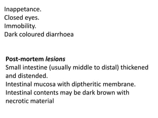
2. Chronic enteritis is the chronic inflammation of the small intestine which may be hemorrhagic, necrotic, or granulomatous. Hemorrhagic enteritis in turkeys is caused by a Type II Adenovirus and causes sudden death and blood in the vent. Necrotic enteritis in poultry is caused by Clostridium perfringens and results in intestinal thickening and dark diarrhea.
3


![ONFH[AVN HIP] -TRIPLE REGIME -A NOVAL SURGICAL CONCEPT .pptx](https://cdn.slidesharecdn.com/ss_thumbnails/onfhavnhip2026koaconcalicutdrgokuldevdrmashraf-260210064517-213ec005-thumbnail.jpg?width=640&height=640&fit=bounds)


