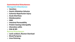
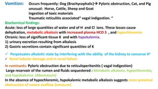
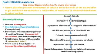
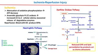
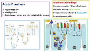
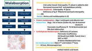
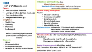
Biochemistry of Gastrointestinal disorders
Dr T V Chaitanya Kumar
Assistant Professor
Dept of Vety Biocemistry
CVSc, Tirupati
Gastrointestinal Disturbances
Monogastric Disturbances
o Vomition
o Gastric Dilatation-Volvulus
o Ischemia-Reperfusion Injury
o Acute Diarrheas
o Malabsorption
o SIBO
o Intestinal Permeability
o Protein loosing enteropathy
o Ulcerative colitis
o EHA & DPJ
Ruminal Disturbances
o Lactic acidosis /Rumen Overload
o Bloat/Tympony
o Urea Poisining
Vomition: Occurs frequently: Dog (Brachycephalic) Pyloric obstruction, Cat, and Pig
unusual: Horse, Cattle, Sheep and Goat
Ingestion of toxic materials
Traumatic reticulitis associated“ vagal indigestion. ”
Biochemical findings:
Acute: loss of large quantities of water and of H and Cl ions. These losses cause
dehydration, metabolic alkalosis with increased plasma HCO 3 , and hypochloremia.
Chronic: loss of significant tissue K and with hypokalemia.
1) urinary excretion resulting from alkalosis
2) Gastric secretions contain significant quantities of K
 Perpetuates alkalotic state by interfering with the ability of the kidney to conserve H+
 Renal tubular damage and in renal failure
In ruminants: Pyloric obstruction due to reticuloperitonitis ( vagal indigestion)
Large reservoir of the rumen and fluids sequestered : Metabolic alkalosis, hypochloremia,
and hypokalemia. (Abomasum)
In the absence of hypochloremic, hypokalemic metabolic alkalosis suggests more proximal
obstruction of rumen outflow (omasum)
Gastric Dilatation-Volvulus
Deep-chested dogs and smaller dogs, the cat, and other species
 Gastric dilatation precedes development of volvulus and is the result of the accumulation
of gas and fluid in the stomach as a result either of mechanical or functional disturbances
in pyloric outflow
Stomach distends
Rotates about the distal esophagus
Displacement and occlusion of the pylorus and duodenum
Necrosis and perforation of the stomach wall
Peritonitis (common causes of death)
Obstruction of the caudal vena cava and portal vein
Hypovolemic shock and Septic shock
Myocardial depressant factor released by ischemic pancreas
Intravascular coagulopathy
Biochemical findings:
 Increased plasma gastrin
 Hyperkalemia and hyperphosphataemia
(Damaged tissue)
 Hypovolemia  decreased renal perfusion
 renal insufficiency -Increased BUN
and creatinine (even after decompression)
 Smooth muscle damage and hepatocytes
 Increased ALT and AST
 Venous stasis Tissue Hypoxia--
Increased CK and Lactic acid in blood
Ischemia-Reperfusion Injury
Released OFR and lipid
peroxidation by-products act
as chemo attractants
Ischemia:
 Attenuation of oxidative phosphorylation-
ATP decrease
 Anaerobic glycolysis I/C acidosis 
increased i/C Ca 2 cellular edema, lysosomal
release of degradative enzymes
Reperfusion: Blood with O2 produce OFRs
Ca2+
MDA
MPO activity
Acute Diarrheas
 Hyper motility
 Maldigestion
 Secretion of water and electrolytes into lumen
Biochemical Findings:
Hemoconcentration Hypovolemic shock
Metabolic acidosis: Hyperkalemia
Decreased renal perfusion  decreased H+
Increased organic acids
Malabsorption
 Protein malabsorption : Alpa 1 antitrypsin and albumin test
 Steatorrhea: Dogs : Fat in faeces (Sudan III), Van de krammer
steatocrit
Pancreatic Insufficiency TLI & Bentiromide test
Bile duct obstruction
 Carbohydrate Malabsorption: Deficiency of Lactases
Pancreatic Insufficiency
H2 Breath test (SIBO, Transit time )
OGT vs IVGT
D-Xylose Malabsorption test
Fat malabsorption test: Olein I131 and triolein I131tests
SIBO: H2 test, Nitrosonaptol test
Cobalamin (Schillings test) and folate absorption tests:
methyl malonic acid levels
Idiopathic intestinal malabsorption in dogs:
Irish setter breed: Enteropathy  wheat is added to diet
Decreased mucosal ALP and peptidases activity
German shepherds: Enteropathy  Sprue
Deficiency of Ig A Bacterial overload enteropathy
Cats: IBS
Horses : Amino acid malabsorption in JD
SIBO
> 105 CFU/ml Bacterial count
Causes:
• Ileocaecal valve resection
• Low Ig A levels in German shepherds
• Poor luminal motility
• Beagles with normal Ig A
Symptoms:
• Weight loss
• Vomiting
• Diarhhea
• Borborygmi
Chronic cases IBD (lymphocytes and
plasmacytes in lamina propria, villus
atrophy)
Diagnosis:
• H2 test
• Nitrosonapthol test
• Unconjugated Bile acids
• Decreased ALP activity of brush border
Intestinal permeability: Dogs
Gluten induced enteropathy
Biochemical Tests: Disaccharides : Cellobiose, lactulose
Disaccharides/ Monosaccharide's ratio
Protein loosing enteropathy:
Causes:
• Ulcerative intestines
• Increased permeability
• Lymphangectasia
• Chronic ileitis in swine
• Parasitic infestation
• JD and PLE in horses
Biochemical tests: Radiolabelled Cr51 Albumin and ceruloplasmin
Faecal alpha 1 proteinase inhibitor test
Decrease in serum albumin levels
Ulcerative colitis: Et: FeLV, tricuriasis, Eosinopilic UC, Histoplasmosis,
Balantidiasis
Hypoalbunemia –loss through denuded Colon
Hyper gamma globulenemia– Inflammation
Equine Hyper ammonemia: Clostridium sordelli
DPJ: Clostridium  Increased GGT, AST, ALP Diff diagnosis SISO
Helicobacter Pylori: Gastric ulcers
Rumen Disturbances
Acute rumen overload/Lactic acidosis
High Roughages
(Grain/Apple)
Streptococcus bovis
D and L Lactate
Bloat
Galacturonic acid increase rumen viscosity
Ribulose diphosphate carboxylase activity
Foam formation and gas trap
Rx: Sodium alkyl sulfonate
Poloxalene, Silicone
Urea poisoning
Urea Ammonia
Ammonia poisining
Rx: Acetic acid
NH3 + H+→ NH4+
(Not absorbed in rumen)
Bacterial Urease
Free Gas bloat
• Defects in
eructation of
gas
• Vagal
indigestion
• Obstruction in
oesophagous
Feed lot bloat
Biochemistry of free radical generation
and oxidative stress
Pickled pigs and malignant hyperthermia
Suucinyl choline : depolarising muscle relaxant
GI DISORDERS.pptx
GI DISORDERS.pptx
GI DISORDERS.pptx
GI DISORDERS.pptx
GI DISORDERS.pptx
GI DISORDERS.pptx
GI DISORDERS.pptx
GI DISORDERS.pptx
GI DISORDERS.pptx
GI DISORDERS.pptx
GI DISORDERS.pptx

GI DISORDERS.pptx

  • 1.
    Biochemistry of Gastrointestinaldisorders Dr T V Chaitanya Kumar Assistant Professor Dept of Vety Biocemistry CVSc, Tirupati
  • 2.
    Gastrointestinal Disturbances Monogastric Disturbances oVomition o Gastric Dilatation-Volvulus o Ischemia-Reperfusion Injury o Acute Diarrheas o Malabsorption o SIBO o Intestinal Permeability o Protein loosing enteropathy o Ulcerative colitis o EHA & DPJ Ruminal Disturbances o Lactic acidosis /Rumen Overload o Bloat/Tympony o Urea Poisining
  • 3.
    Vomition: Occurs frequently:Dog (Brachycephalic) Pyloric obstruction, Cat, and Pig unusual: Horse, Cattle, Sheep and Goat Ingestion of toxic materials Traumatic reticulitis associated“ vagal indigestion. ” Biochemical findings: Acute: loss of large quantities of water and of H and Cl ions. These losses cause dehydration, metabolic alkalosis with increased plasma HCO 3 , and hypochloremia. Chronic: loss of significant tissue K and with hypokalemia. 1) urinary excretion resulting from alkalosis 2) Gastric secretions contain significant quantities of K  Perpetuates alkalotic state by interfering with the ability of the kidney to conserve H+  Renal tubular damage and in renal failure In ruminants: Pyloric obstruction due to reticuloperitonitis ( vagal indigestion) Large reservoir of the rumen and fluids sequestered : Metabolic alkalosis, hypochloremia, and hypokalemia. (Abomasum) In the absence of hypochloremic, hypokalemic metabolic alkalosis suggests more proximal obstruction of rumen outflow (omasum)
  • 4.
    Gastric Dilatation-Volvulus Deep-chested dogsand smaller dogs, the cat, and other species  Gastric dilatation precedes development of volvulus and is the result of the accumulation of gas and fluid in the stomach as a result either of mechanical or functional disturbances in pyloric outflow Stomach distends Rotates about the distal esophagus Displacement and occlusion of the pylorus and duodenum Necrosis and perforation of the stomach wall Peritonitis (common causes of death) Obstruction of the caudal vena cava and portal vein Hypovolemic shock and Septic shock Myocardial depressant factor released by ischemic pancreas Intravascular coagulopathy Biochemical findings:  Increased plasma gastrin  Hyperkalemia and hyperphosphataemia (Damaged tissue)  Hypovolemia  decreased renal perfusion  renal insufficiency -Increased BUN and creatinine (even after decompression)  Smooth muscle damage and hepatocytes  Increased ALT and AST  Venous stasis Tissue Hypoxia-- Increased CK and Lactic acid in blood
  • 5.
    Ischemia-Reperfusion Injury Released OFRand lipid peroxidation by-products act as chemo attractants Ischemia:  Attenuation of oxidative phosphorylation- ATP decrease  Anaerobic glycolysis I/C acidosis  increased i/C Ca 2 cellular edema, lysosomal release of degradative enzymes Reperfusion: Blood with O2 produce OFRs Ca2+ MDA MPO activity
  • 6.
    Acute Diarrheas  Hypermotility  Maldigestion  Secretion of water and electrolytes into lumen Biochemical Findings: Hemoconcentration Hypovolemic shock Metabolic acidosis: Hyperkalemia Decreased renal perfusion  decreased H+ Increased organic acids
  • 7.
    Malabsorption  Protein malabsorption: Alpa 1 antitrypsin and albumin test  Steatorrhea: Dogs : Fat in faeces (Sudan III), Van de krammer steatocrit Pancreatic Insufficiency TLI & Bentiromide test Bile duct obstruction  Carbohydrate Malabsorption: Deficiency of Lactases Pancreatic Insufficiency H2 Breath test (SIBO, Transit time ) OGT vs IVGT D-Xylose Malabsorption test Fat malabsorption test: Olein I131 and triolein I131tests SIBO: H2 test, Nitrosonaptol test Cobalamin (Schillings test) and folate absorption tests: methyl malonic acid levels Idiopathic intestinal malabsorption in dogs: Irish setter breed: Enteropathy  wheat is added to diet Decreased mucosal ALP and peptidases activity German shepherds: Enteropathy  Sprue Deficiency of Ig A Bacterial overload enteropathy Cats: IBS Horses : Amino acid malabsorption in JD
  • 8.
    SIBO > 105 CFU/mlBacterial count Causes: • Ileocaecal valve resection • Low Ig A levels in German shepherds • Poor luminal motility • Beagles with normal Ig A Symptoms: • Weight loss • Vomiting • Diarhhea • Borborygmi Chronic cases IBD (lymphocytes and plasmacytes in lamina propria, villus atrophy) Diagnosis: • H2 test • Nitrosonapthol test • Unconjugated Bile acids • Decreased ALP activity of brush border Intestinal permeability: Dogs Gluten induced enteropathy Biochemical Tests: Disaccharides : Cellobiose, lactulose Disaccharides/ Monosaccharide's ratio Protein loosing enteropathy: Causes: • Ulcerative intestines • Increased permeability • Lymphangectasia • Chronic ileitis in swine • Parasitic infestation • JD and PLE in horses Biochemical tests: Radiolabelled Cr51 Albumin and ceruloplasmin Faecal alpha 1 proteinase inhibitor test Decrease in serum albumin levels Ulcerative colitis: Et: FeLV, tricuriasis, Eosinopilic UC, Histoplasmosis, Balantidiasis Hypoalbunemia –loss through denuded Colon Hyper gamma globulenemia– Inflammation Equine Hyper ammonemia: Clostridium sordelli DPJ: Clostridium  Increased GGT, AST, ALP Diff diagnosis SISO Helicobacter Pylori: Gastric ulcers
  • 9.
    Rumen Disturbances Acute rumenoverload/Lactic acidosis High Roughages (Grain/Apple) Streptococcus bovis D and L Lactate
  • 10.
    Bloat Galacturonic acid increaserumen viscosity Ribulose diphosphate carboxylase activity Foam formation and gas trap Rx: Sodium alkyl sulfonate Poloxalene, Silicone Urea poisoning Urea Ammonia Ammonia poisining Rx: Acetic acid NH3 + H+→ NH4+ (Not absorbed in rumen) Bacterial Urease Free Gas bloat • Defects in eructation of gas • Vagal indigestion • Obstruction in oesophagous Feed lot bloat
  • 11.
    Biochemistry of freeradical generation and oxidative stress
  • 20.
    Pickled pigs andmalignant hyperthermia
  • 22.
    Suucinyl choline :depolarising muscle relaxant