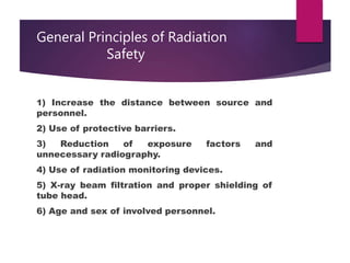
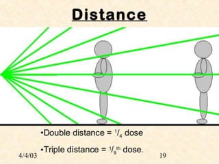
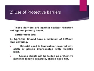
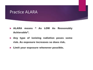
Radiation Protection
Methods
Radiation protection
 Radiation protection, sometimes known as
radiological protection, is defined by the
International Atomic Energy Agency (IAEA) as
"The protection of people from harmful
effects of exposure to ionizing radiation, and
the means for achieving this". The IAEA also
states "The accepted understanding of the
term radiation protection is restricted to
protection of people
Why it is necessary ?
 Exposure above than threshold level produces
following biological effects:
1. Somatic effect
2. Genetic effect
3. Teratogenic effect
 Radiation may…….
1. Deposit energy
2. Damage DNA
3. Create ionization in the body leading to free
radicals formation leads to biological damage.
Susceptibility to radiation
 The LD 50/30 ( the dose that kill 50%
population in 30 days) is influenced by the :
I. Source of radiation
II. Dose rate
 The LD 50/30 of x rays for humans is 250-450
Rad.
 Rad*… is a unit of absorbed dose for any
ionizing radiation.
Radiosensitivity of living cells
 Low sensitivity : Mature RBCs , Ganglion
cells, Muscle cells, Mature connective tissue
 Intermediate sensitivity: Gastric mucosa,
mucus membrane, esophageal epithelium,
urinary bladder epithelium.
 High sensitivity: Primitive blood cells ,
spermatogonia, Lymphocytes.
General Principles of Radiation
Safety
1) Increase the distance between source and
personnel.
2) Use of protective barriers.
3) Reduction of exposure factors and
unnecessary radiography.
4) Use of radiation monitoring devices.
5) X-ray beam filtration and proper shielding of
tube head.
6) Age and sex of involved personnel.
1) Increase in Distance between
Source and Personnel.
 Distance is an important factor in reducing
exposure.
 Doubling the distance from source will reduce
the radiation exposure by four.
 Use tongs, forceps and trays to increase the
distance between yourself and radiation
source.
 Always know the radiation intensity, where
you perform most of your work and if it is high
then move to lower dose areas during work
delays.
Continue…….
 No individual other than operator and
essentially needed person should be there.
 Animal should not be restrained manually use
either chemical or physical method of
restraining.
 Operator should be on shielding booth or
behind the screen or at least 6 feet away from
source when exposure is made.
 No part of body should be exposed to primary
beam.
2) Use of Protective Barriers
These barriers are against scatter radiation
not against primary beam.
Barrier used are;
a) Aprons: Should have a minimum of 0.25mm
lead covering.
 Material used is lead rubber covered with
cloth or plastic impregnated with metallic
lead.
 Aprons should not be folded as protective
material tend to separate, should keep flat.
Use of Protective Barriers
b) Gloves and Goggles: The lead equivalent of
gloves should not be less than 0.33mm.
 Gloves should be checked by radiography
for cracks which can easily be missed on visual
inspection.
 Lead goggles should be used during fluoroscopic
examination
c) X ray room and equipment: Room should be
away from public places
 Equipment should be checked for possible
leakage.
Continue……
 Room should also be well coated with lead
along with free from any leakage.
 The wall of room should be at least 22cm
thick and should be of concrete into which
iron may be introduced.
 Warning signs must be placed near X ray
room regarding potential hazards.
3) Reduction of Exposure factors and
unnecessary Radiography
 Correct exposure factors must be used in
first attempt. Repeated exposures definitely
enhance exposure level.
 Many time owners demand radiography
when it is not justified, unnecessary exposure
should be avoided.
4) Use of Radiation Monitoring
Devices
 Radiation monitoring devices like “film
badges” should be worn all the time by
individuals involved in radiographic work.
 Ideally one film badge should be at belt
level to monitor whole body exposure while
other at neckline to estimate exposure to skin
of head neck and eyed.
 Radiation monitoring badges available in
rings, clips and wrist badges.
5) X-ray beam Filtration and
Shielding of tube head
a) Filtration: Aluminum filters of at least 2.5mm
thickness should be used to absorb the soft X
rays to reduce the amount of scatter radiation.
By this exposure dose can be reduced upto three
to four times.
b) X-ray tube head: It should be well shielded
from all sides except the exit window.
6) Age and Sex of involved
Personnel
 Persons under 18 years of age should not
be involved in radiography because of the
sensitivity of the growing tissues.
 Pregnant females should not be involved
because of extreme sensitivity of embryos at
certain stages of development.
Practice ALARA
 ALARA means “ As LOW As Reasonably
Achievable”.
 Any type of ionizing radiation poses some
risk. As exposure increases so does risk.
 Limit your exposure whenever possible.
References
 An Introduction to Radiation Protection in Medicine by
Jamie V. Trapp, Tomas Kron
 An Introduction to Radiation Protection by Alan Martin,
Sam Harbison.
 Introduction to Radiation Protection by J Sabol
Radiation protection

Radiation protection

  • 1.
  • 2.
    Radiation protection  Radiationprotection, sometimes known as radiological protection, is defined by the International Atomic Energy Agency (IAEA) as "The protection of people from harmful effects of exposure to ionizing radiation, and the means for achieving this". The IAEA also states "The accepted understanding of the term radiation protection is restricted to protection of people
  • 3.
    Why it isnecessary ?  Exposure above than threshold level produces following biological effects: 1. Somatic effect 2. Genetic effect 3. Teratogenic effect  Radiation may……. 1. Deposit energy 2. Damage DNA 3. Create ionization in the body leading to free radicals formation leads to biological damage.
  • 4.
    Susceptibility to radiation The LD 50/30 ( the dose that kill 50% population in 30 days) is influenced by the : I. Source of radiation II. Dose rate  The LD 50/30 of x rays for humans is 250-450 Rad.  Rad*… is a unit of absorbed dose for any ionizing radiation.
  • 5.
    Radiosensitivity of livingcells  Low sensitivity : Mature RBCs , Ganglion cells, Muscle cells, Mature connective tissue  Intermediate sensitivity: Gastric mucosa, mucus membrane, esophageal epithelium, urinary bladder epithelium.  High sensitivity: Primitive blood cells , spermatogonia, Lymphocytes.
  • 6.
    General Principles ofRadiation Safety 1) Increase the distance between source and personnel. 2) Use of protective barriers. 3) Reduction of exposure factors and unnecessary radiography. 4) Use of radiation monitoring devices. 5) X-ray beam filtration and proper shielding of tube head. 6) Age and sex of involved personnel.
  • 7.
    1) Increase inDistance between Source and Personnel.  Distance is an important factor in reducing exposure.  Doubling the distance from source will reduce the radiation exposure by four.  Use tongs, forceps and trays to increase the distance between yourself and radiation source.  Always know the radiation intensity, where you perform most of your work and if it is high then move to lower dose areas during work delays.
  • 8.
    Continue…….  No individualother than operator and essentially needed person should be there.  Animal should not be restrained manually use either chemical or physical method of restraining.  Operator should be on shielding booth or behind the screen or at least 6 feet away from source when exposure is made.  No part of body should be exposed to primary beam.
  • 11.
    2) Use ofProtective Barriers These barriers are against scatter radiation not against primary beam. Barrier used are; a) Aprons: Should have a minimum of 0.25mm lead covering.  Material used is lead rubber covered with cloth or plastic impregnated with metallic lead.  Aprons should not be folded as protective material tend to separate, should keep flat.
  • 13.
    Use of ProtectiveBarriers b) Gloves and Goggles: The lead equivalent of gloves should not be less than 0.33mm.  Gloves should be checked by radiography for cracks which can easily be missed on visual inspection.  Lead goggles should be used during fluoroscopic examination c) X ray room and equipment: Room should be away from public places  Equipment should be checked for possible leakage.
  • 14.
    Continue……  Room shouldalso be well coated with lead along with free from any leakage.  The wall of room should be at least 22cm thick and should be of concrete into which iron may be introduced.  Warning signs must be placed near X ray room regarding potential hazards.
  • 18.
    3) Reduction ofExposure factors and unnecessary Radiography  Correct exposure factors must be used in first attempt. Repeated exposures definitely enhance exposure level.  Many time owners demand radiography when it is not justified, unnecessary exposure should be avoided.
  • 19.
    4) Use ofRadiation Monitoring Devices  Radiation monitoring devices like “film badges” should be worn all the time by individuals involved in radiographic work.  Ideally one film badge should be at belt level to monitor whole body exposure while other at neckline to estimate exposure to skin of head neck and eyed.  Radiation monitoring badges available in rings, clips and wrist badges.
  • 21.
    5) X-ray beamFiltration and Shielding of tube head a) Filtration: Aluminum filters of at least 2.5mm thickness should be used to absorb the soft X rays to reduce the amount of scatter radiation. By this exposure dose can be reduced upto three to four times. b) X-ray tube head: It should be well shielded from all sides except the exit window.
  • 22.
    6) Age andSex of involved Personnel  Persons under 18 years of age should not be involved in radiography because of the sensitivity of the growing tissues.  Pregnant females should not be involved because of extreme sensitivity of embryos at certain stages of development.
  • 23.
    Practice ALARA  ALARAmeans “ As LOW As Reasonably Achievable”.  Any type of ionizing radiation poses some risk. As exposure increases so does risk.  Limit your exposure whenever possible.
  • 24.
    References  An Introductionto Radiation Protection in Medicine by Jamie V. Trapp, Tomas Kron  An Introduction to Radiation Protection by Alan Martin, Sam Harbison.  Introduction to Radiation Protection by J Sabol