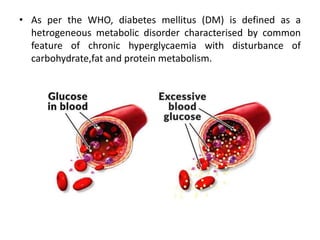
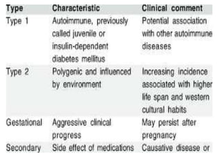
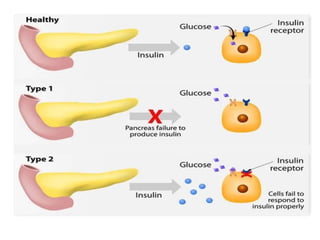
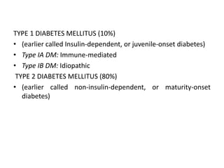
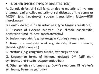
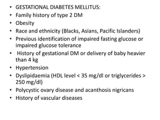
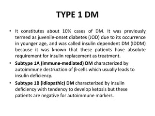
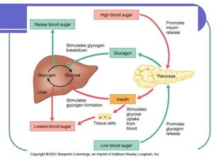
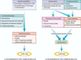
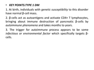
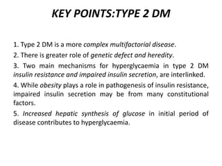
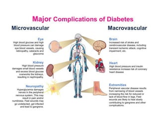
Diabetic Mellitus
• As per the WHO, diabetes mellitus (DM) is defined as a
hetrogeneous metabolic disorder characterised by common
feature of chronic hyperglycaemia with disturbance of
carbohydrate,fat and protein metabolism.
TYPE 1 DIABETES MELLITUS (10%)
• (earlier called Insulin-dependent, or juvenile-onset diabetes)
• Type IA DM: Immune-mediated
• Type IB DM: Idiopathic
TYPE 2 DIABETES MELLITUS (80%)
• (earlier called non-insulin-dependent, or maturity-onset
diabetes)
• III. OTHER SPECIFIC TYPES OF DIABETES (10%)
A. Genetic defect of β-cell function due to mutations in various
enzymes (earlier called maturity-onset diabetes of the young or
MODY) (e.g. hepatocyte nuclear transcription factor—HNF,
glucokinase)
B. Genetic defect in insulin action (e.g. type A insulin resistance)
C. Diseases of exocrine pancreas (e.g. chronic pancreatitis,
pancreatic tumours, post-pancreatectomy)
D. Endocrinopathies (e.g. acromegaly, Cushing's syndrome)
E. Drug- or chemical-induced (e.g. steroids, thyroid hormone,
thiazides, β-blockers etc)
F. Infections (e.g. congenital rubella, cytomegalovirus)
G. Uncommon forms of immune-mediated DM (stiff man
syndrome, anti-insulin receptor antibodies)
H. Other genetic syndromes (e.g. Down's syndrome, Klinefelter's
syndrome, Turner's syndrome)
• GESTATIONAL DIABETES MELLITUS:
• Family history of type 2 DM
• Obesity
• Race and ethnicity (Blacks, Asians, Pacific Islanders)
• Previous identification of impaired fasting glucose or
impaired glucose tolerance
• History of gestational DM or delivery of baby heavier
than 4 kg
• Hypertension
• Dyslipidaemia (HDL level < 35 mg/dl or triglycerides >
250 mg/dl)
• Polycystic ovary disease and acanthosis nigricans
• History of vascular diseases
TYPE 1 DM
• It constitutes about 10% cases of DM. It was previously
termed as juvenile-onset diabetes (JOD) due to its occurrence
in younger age, and was called insulin dependent DM (IDDM)
because it was known that these patients have absolute
requirement for insulin replacement as treatment.
• Subtype 1A (immune-mediated) DM characterized by
autoimmune destruction of β-cells which usually leads to
insulin deficiency.
• Subtype 1B (idiopathic) DM characterized by insulin
deficiency with tendency to develop ketosis but these
patients are negative for autoimmune markers.
TYPE 2 DM.
• This type comprises about 80% cases of DM. It was previously
called maturity-onset diabetes, or non-insulin dependent
diabetes mellitus (NIDDM) of obese and no obese type.
• Although type 2 DM predominantly affects older individuals, it
is now known that it also occurs in obese adolescent children;
OTHER SPECIFIC ETIOLOGIC TYPES OF
DM.
• One important subtype in this group is maturity-onset
diabetes of the young (MODY) which has autosomal dominant
inheritance, early onset of hyperglycaemia and impaired
insulin secretion.
GESTATIONAL DM.
• About 4% pregnant women develop DM due to metabolic
changes during pregnancy.
• Although they revert back to normal glycaemia after delivery,
these women are prone to develop DM later in their life.
Pathogenesis
• Depending upon etiology of DM
hyperglycaemia may result from the following:
• Reduced insulin secretion
• Decreased glucose use by the body
• Increased glucose production.
Pathogenesis of two main types of DM and its
complications is distinct.
NORMAL INSULIN METABOLISM
• Synthesis.
• i) It is initially formed as pre-proinsulin which is single-chain
• ii) Subsequent proteolysis removes the amino terminal
• iii) A (21 amino acids) and B (30 amino acids) chains of insulin,
linked together by connecting segment called C-peptide
• For therapeutic purposes, human insulin is now produced by
recombinant DNA technology.
• Release
• Hypoglycaemia (glucose level below 70 mg/dl) stimulates
transport into β-cells of a glucose transporter, GLUT2. include
nutrients in the meal, ketones, amino acids etc.
• An islet transcription factor, glucokinase, causes
glucosephosphorylation.
• glucose to glucose-6-phosphate by glycolysis generates ATP.
• It causes inhibition of ATP-sensitive K+ channel on the cell
membrane and opening up of calcium channel with resultant
influx of calcium, which stimulates insulin release.
• Action.
• Insulin receptor has intrinsic tyrosine kinase activity.
• activates post-receptor intracellular signalling pathway
molecules, insulin receptor substrates (IRS) 1 and 2 proteins,
which initiate sequence of phosphorylation and DE
phosphorylation reactions.
• These reactions on the target cells are responsible for the
main mitogenic and anabolic actions of insulin—glycogen
synthesis, glucose transport, protein synthesis, lipogenesis.
PATHOGENESIS
• KEY POINTS:TYPE 1 DM
1. At birth, individuals with genetic susceptibility to this disorder
have normal β-cell mass.
2. β-cells act as autoantigens and activate CD4+ T lymphocytes,
bringing about immune destruction of pancreatic β-cells by
autoimmune phenomena and takes months to years.
3. The trigger for autoimmune process appears to be some
infectious or environmental factor which specifically targets β-
cells.
KEY POINTS:TYPE 2 DM
1. Type 2 DM is a more complex multifactorial disease.
2. There is greater role of genetic defect and heredity.
3. Two main mechanisms for hyperglycaemia in type 2 DM
insulin resistance and impaired insulin secretion, are interlinked.
4. While obesity plays a role in pathogenesis of insulin resistance,
impaired insulin secretion may be from many constitutional
factors.
5. Increased hepatic synthesis of glucose in initial period of
disease contributes to hyperglycaemia.
Clinical Features
• Patients of type 1 DM usually manifest at early age, generally
below the age of 35.
ii) The onset of symptoms is often abrupt.
iii) At presentation, these patients have polyuria, polydipsia and
polyphagia.
iv) The patients are not obese but have generally progressive loss
of weight.
v) These patients are prone to develop metabolic complications
such as ketoacidosis and hypoglycaemic episodes.
• Type 2 DM:
i) This form of diabetes generally manifests in middle life or
beyond, usually above the age of 40.
ii) The onset of symptoms in type 2 DM is slow.
iii) Generally, the patient is asymptomatic when the diagnosis is
made on the basis of glucosuria or hyperglycaemia during
physical examination, or may present with polyuria and
polydipsia.
iv) The patients are frequently obese and have unexplained
weakness and loss of weight.
v) Metabolic complications such as ketoacidosis are infrequent.
I. Acute metabolic complications:
These include diabetic ketoacidosis,
hyperosmolar nonketotic coma, and
hypoglycaemia.
II. Late systemic complications: These are
atherosclerosis, diabetic microangiopathy,
diabetic nephropathy, diabetic neuropathy,
diabetic retinopathy and infections.
Diagnosis of Diabetes
confirmed by finding glucosuria and a random plasma glucose
concentration above 200 mg/dl.
• The severity of clinical symptoms of polyuria and polydipsia is
directly related .
• In asymptomatic cases, when there is persistently elevated
fasting plasma glucose level, diagnosis again poses no
difficulty.
• The problem arises in asymptomatic patients who have
normal fasting glucose level in the plasma but are suspected
to have diabetes on other grounds and are thus subjected to
oral glucose tolerance test (GTT).
• If abnormal GTT values are found, these subjects are said to
have ‘chemical diabetes’
I. URINE TESTING.
Urine is tested for the presence of glucose and ketones.
1. Glucosuria.
• Benedict’s qualitative test detects any reducing substance in
the urine and is not specific for glucose. dipstick method
based on enzyme-coated paper strip which turns purple when
dipped in urine containing glucose.
II. SINGLE BLOOD SUGAR ESTIMATION.
III. SCREENING BY FASTING GLUCOSE TEST.
IV. ORAL GLUCOSE TOLERANCE TEST.
Oral GTT is performed principally for patients with borderline
fasting plasma glucose value (i.e. between 100-140 mg/dl).
V. OTHER TESTS.
1. Glycosylated haemoglobin (HbA1C).
2. Glycated albumin.
3. Extended GTT
4.Insulin assay.
5.Islet autoantibodies
6.Screening for diabetes-associated
complications.
Diabetic mellitus pathophysiology

Diabetic mellitus pathophysiology

  • 1.
  • 2.
    • As perthe WHO, diabetes mellitus (DM) is defined as a hetrogeneous metabolic disorder characterised by common feature of chronic hyperglycaemia with disturbance of carbohydrate,fat and protein metabolism.
  • 6.
    TYPE 1 DIABETESMELLITUS (10%) • (earlier called Insulin-dependent, or juvenile-onset diabetes) • Type IA DM: Immune-mediated • Type IB DM: Idiopathic TYPE 2 DIABETES MELLITUS (80%) • (earlier called non-insulin-dependent, or maturity-onset diabetes)
  • 7.
    • III. OTHERSPECIFIC TYPES OF DIABETES (10%) A. Genetic defect of β-cell function due to mutations in various enzymes (earlier called maturity-onset diabetes of the young or MODY) (e.g. hepatocyte nuclear transcription factor—HNF, glucokinase) B. Genetic defect in insulin action (e.g. type A insulin resistance) C. Diseases of exocrine pancreas (e.g. chronic pancreatitis, pancreatic tumours, post-pancreatectomy) D. Endocrinopathies (e.g. acromegaly, Cushing's syndrome) E. Drug- or chemical-induced (e.g. steroids, thyroid hormone, thiazides, β-blockers etc) F. Infections (e.g. congenital rubella, cytomegalovirus) G. Uncommon forms of immune-mediated DM (stiff man syndrome, anti-insulin receptor antibodies) H. Other genetic syndromes (e.g. Down's syndrome, Klinefelter's syndrome, Turner's syndrome)
  • 8.
    • GESTATIONAL DIABETESMELLITUS: • Family history of type 2 DM • Obesity • Race and ethnicity (Blacks, Asians, Pacific Islanders) • Previous identification of impaired fasting glucose or impaired glucose tolerance • History of gestational DM or delivery of baby heavier than 4 kg • Hypertension • Dyslipidaemia (HDL level < 35 mg/dl or triglycerides > 250 mg/dl) • Polycystic ovary disease and acanthosis nigricans • History of vascular diseases
  • 9.
    TYPE 1 DM •It constitutes about 10% cases of DM. It was previously termed as juvenile-onset diabetes (JOD) due to its occurrence in younger age, and was called insulin dependent DM (IDDM) because it was known that these patients have absolute requirement for insulin replacement as treatment. • Subtype 1A (immune-mediated) DM characterized by autoimmune destruction of β-cells which usually leads to insulin deficiency. • Subtype 1B (idiopathic) DM characterized by insulin deficiency with tendency to develop ketosis but these patients are negative for autoimmune markers.
  • 10.
    TYPE 2 DM. •This type comprises about 80% cases of DM. It was previously called maturity-onset diabetes, or non-insulin dependent diabetes mellitus (NIDDM) of obese and no obese type. • Although type 2 DM predominantly affects older individuals, it is now known that it also occurs in obese adolescent children;
  • 11.
    OTHER SPECIFIC ETIOLOGICTYPES OF DM. • One important subtype in this group is maturity-onset diabetes of the young (MODY) which has autosomal dominant inheritance, early onset of hyperglycaemia and impaired insulin secretion.
  • 12.
    GESTATIONAL DM. • About4% pregnant women develop DM due to metabolic changes during pregnancy. • Although they revert back to normal glycaemia after delivery, these women are prone to develop DM later in their life.
  • 13.
    Pathogenesis • Depending uponetiology of DM hyperglycaemia may result from the following: • Reduced insulin secretion • Decreased glucose use by the body • Increased glucose production. Pathogenesis of two main types of DM and its complications is distinct.
  • 14.
    NORMAL INSULIN METABOLISM •Synthesis. • i) It is initially formed as pre-proinsulin which is single-chain • ii) Subsequent proteolysis removes the amino terminal • iii) A (21 amino acids) and B (30 amino acids) chains of insulin, linked together by connecting segment called C-peptide • For therapeutic purposes, human insulin is now produced by recombinant DNA technology.
  • 16.
    • Release • Hypoglycaemia(glucose level below 70 mg/dl) stimulates transport into β-cells of a glucose transporter, GLUT2. include nutrients in the meal, ketones, amino acids etc. • An islet transcription factor, glucokinase, causes glucosephosphorylation. • glucose to glucose-6-phosphate by glycolysis generates ATP. • It causes inhibition of ATP-sensitive K+ channel on the cell membrane and opening up of calcium channel with resultant influx of calcium, which stimulates insulin release.
  • 17.
    • Action. • Insulinreceptor has intrinsic tyrosine kinase activity. • activates post-receptor intracellular signalling pathway molecules, insulin receptor substrates (IRS) 1 and 2 proteins, which initiate sequence of phosphorylation and DE phosphorylation reactions. • These reactions on the target cells are responsible for the main mitogenic and anabolic actions of insulin—glycogen synthesis, glucose transport, protein synthesis, lipogenesis.
  • 19.
  • 21.
    • KEY POINTS:TYPE1 DM 1. At birth, individuals with genetic susceptibility to this disorder have normal β-cell mass. 2. β-cells act as autoantigens and activate CD4+ T lymphocytes, bringing about immune destruction of pancreatic β-cells by autoimmune phenomena and takes months to years. 3. The trigger for autoimmune process appears to be some infectious or environmental factor which specifically targets β- cells.
  • 22.
    KEY POINTS:TYPE 2DM 1. Type 2 DM is a more complex multifactorial disease. 2. There is greater role of genetic defect and heredity. 3. Two main mechanisms for hyperglycaemia in type 2 DM insulin resistance and impaired insulin secretion, are interlinked. 4. While obesity plays a role in pathogenesis of insulin resistance, impaired insulin secretion may be from many constitutional factors. 5. Increased hepatic synthesis of glucose in initial period of disease contributes to hyperglycaemia.
  • 24.
    Clinical Features • Patientsof type 1 DM usually manifest at early age, generally below the age of 35. ii) The onset of symptoms is often abrupt. iii) At presentation, these patients have polyuria, polydipsia and polyphagia. iv) The patients are not obese but have generally progressive loss of weight. v) These patients are prone to develop metabolic complications such as ketoacidosis and hypoglycaemic episodes.
  • 25.
    • Type 2DM: i) This form of diabetes generally manifests in middle life or beyond, usually above the age of 40. ii) The onset of symptoms in type 2 DM is slow. iii) Generally, the patient is asymptomatic when the diagnosis is made on the basis of glucosuria or hyperglycaemia during physical examination, or may present with polyuria and polydipsia. iv) The patients are frequently obese and have unexplained weakness and loss of weight. v) Metabolic complications such as ketoacidosis are infrequent.
  • 27.
    I. Acute metaboliccomplications: These include diabetic ketoacidosis, hyperosmolar nonketotic coma, and hypoglycaemia. II. Late systemic complications: These are atherosclerosis, diabetic microangiopathy, diabetic nephropathy, diabetic neuropathy, diabetic retinopathy and infections.
  • 29.
    Diagnosis of Diabetes confirmedby finding glucosuria and a random plasma glucose concentration above 200 mg/dl. • The severity of clinical symptoms of polyuria and polydipsia is directly related . • In asymptomatic cases, when there is persistently elevated fasting plasma glucose level, diagnosis again poses no difficulty. • The problem arises in asymptomatic patients who have normal fasting glucose level in the plasma but are suspected to have diabetes on other grounds and are thus subjected to oral glucose tolerance test (GTT). • If abnormal GTT values are found, these subjects are said to have ‘chemical diabetes’
  • 30.
    I. URINE TESTING. Urineis tested for the presence of glucose and ketones. 1. Glucosuria. • Benedict’s qualitative test detects any reducing substance in the urine and is not specific for glucose. dipstick method based on enzyme-coated paper strip which turns purple when dipped in urine containing glucose. II. SINGLE BLOOD SUGAR ESTIMATION. III. SCREENING BY FASTING GLUCOSE TEST. IV. ORAL GLUCOSE TOLERANCE TEST. Oral GTT is performed principally for patients with borderline fasting plasma glucose value (i.e. between 100-140 mg/dl).
  • 31.
    V. OTHER TESTS. 1.Glycosylated haemoglobin (HbA1C). 2. Glycated albumin. 3. Extended GTT 4.Insulin assay. 5.Islet autoantibodies 6.Screening for diabetes-associated complications.