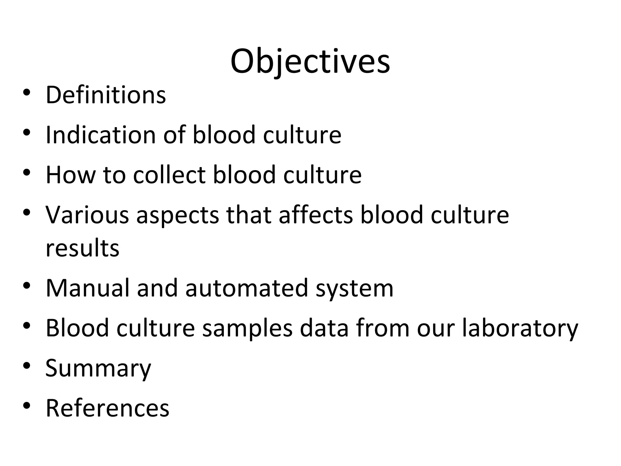
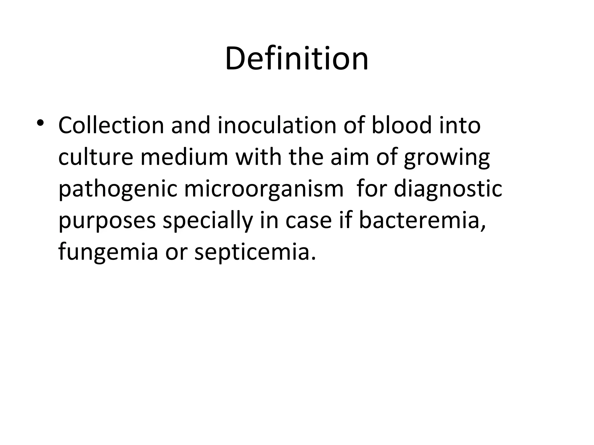
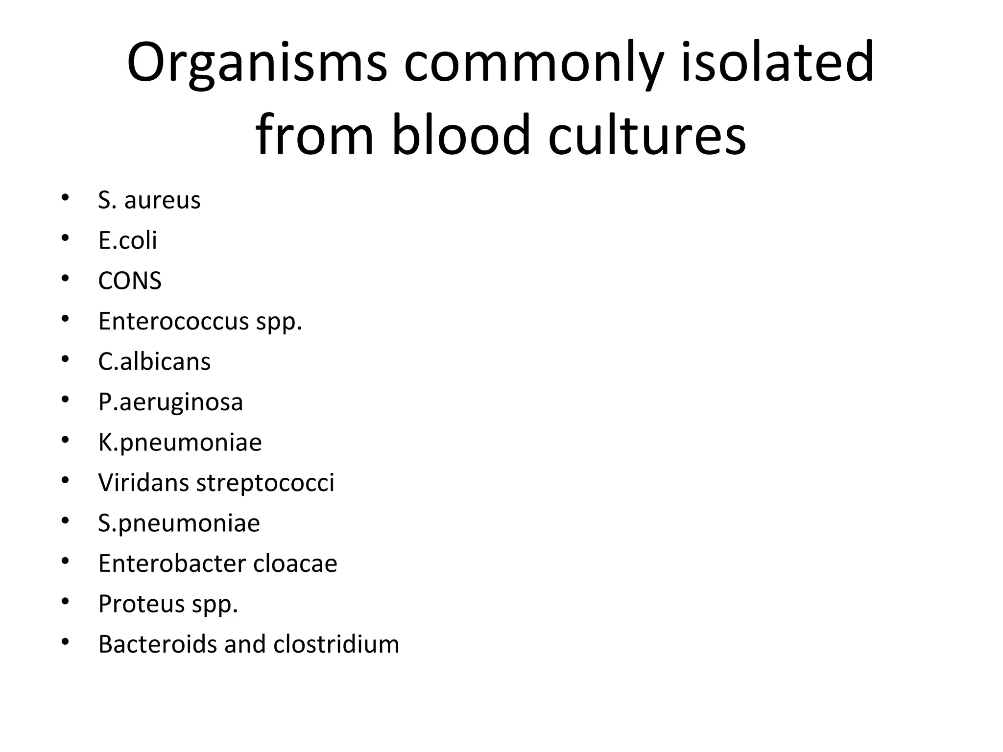
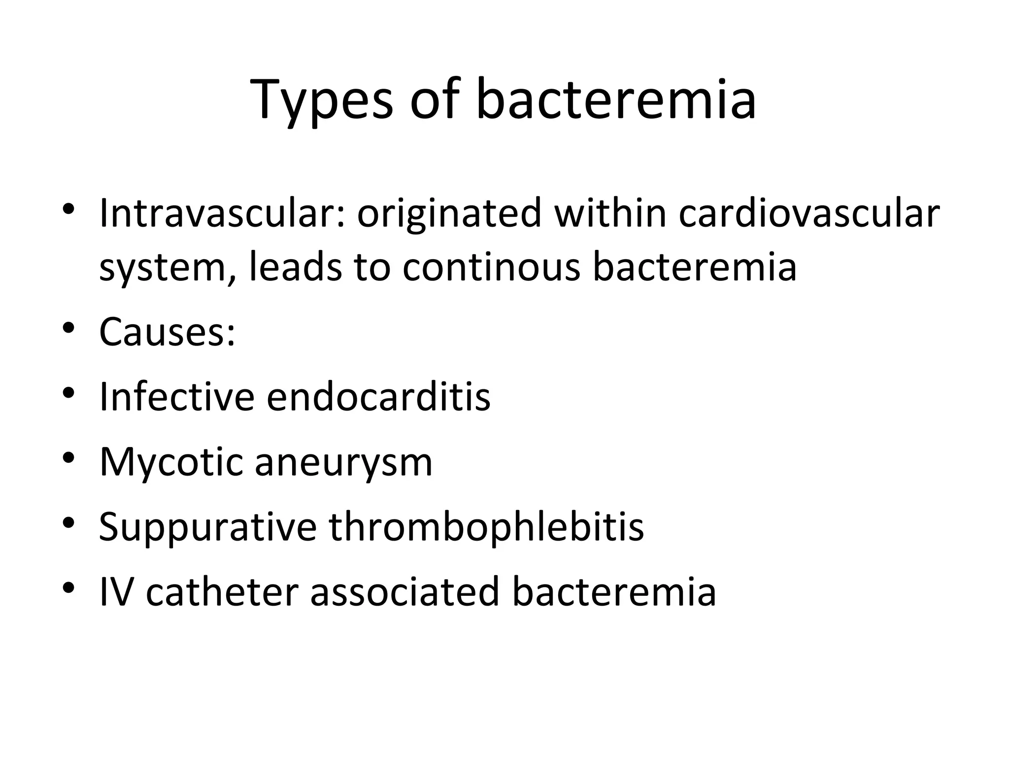
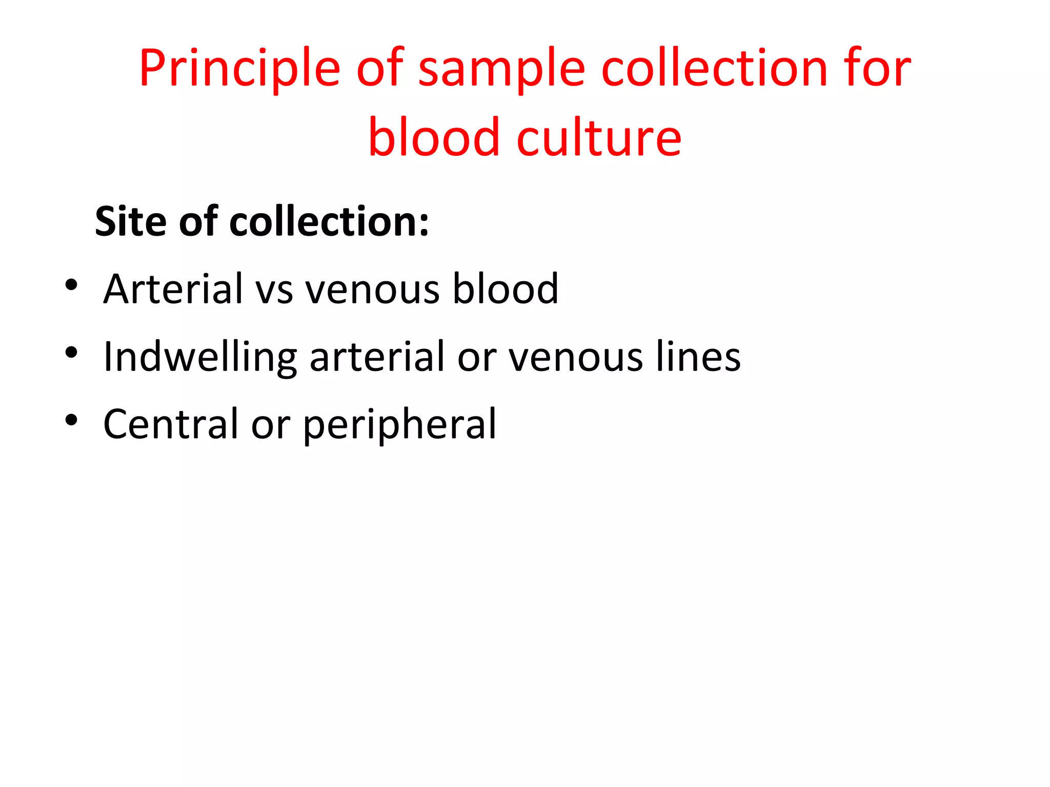
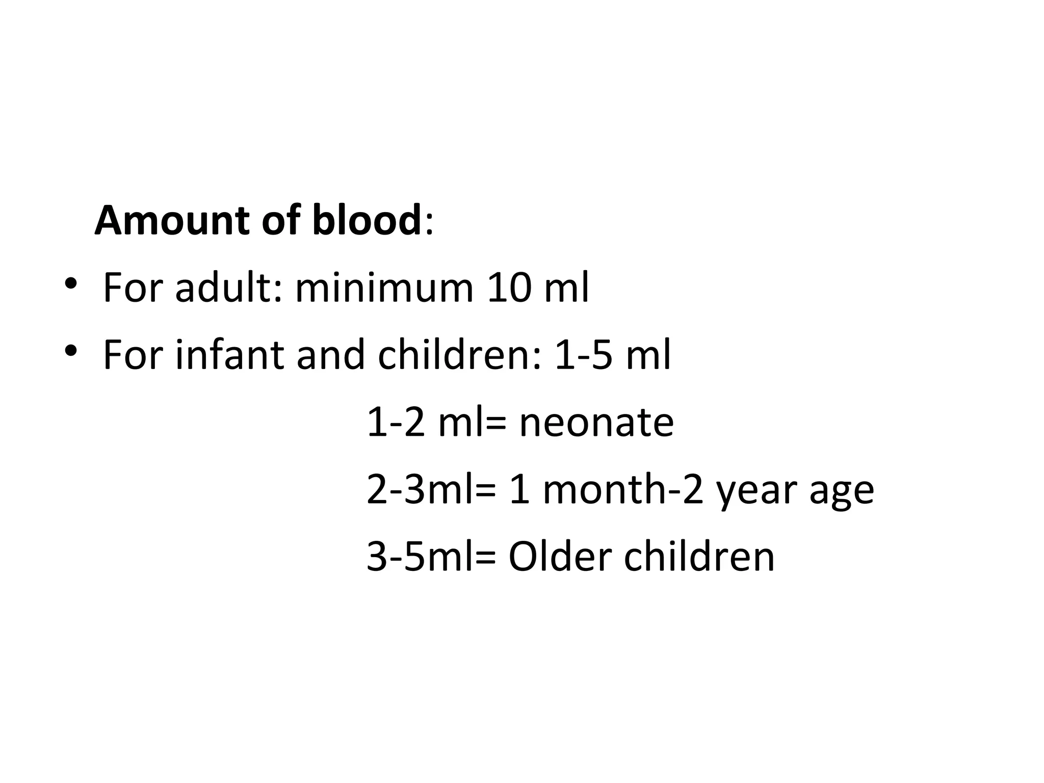
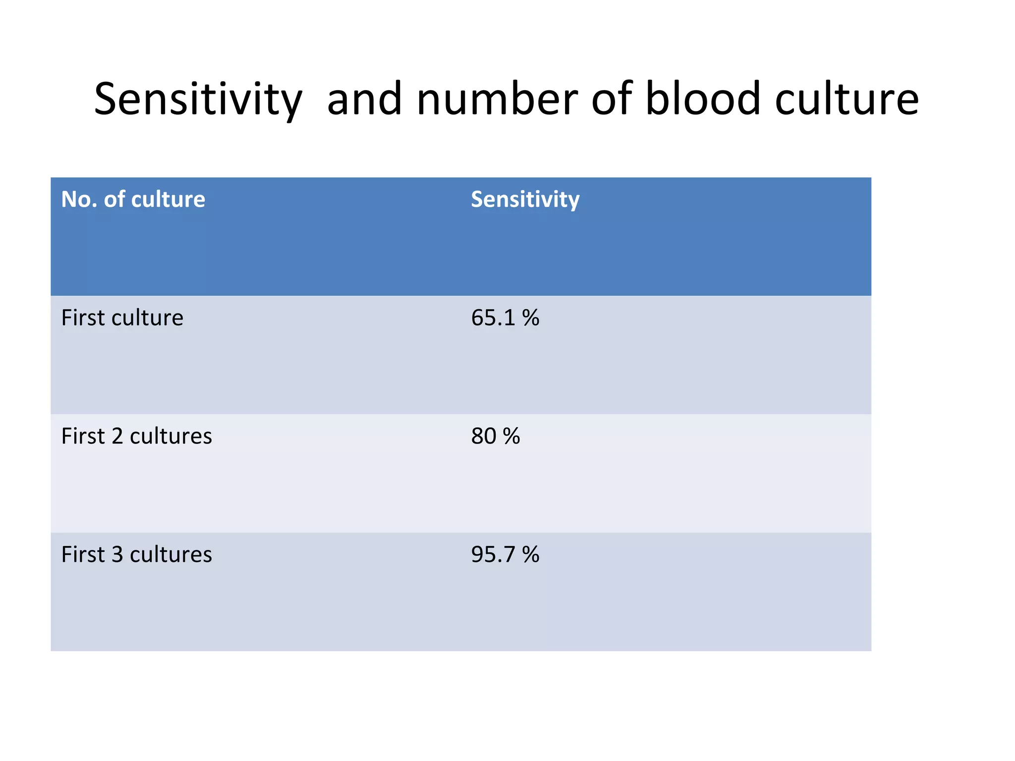
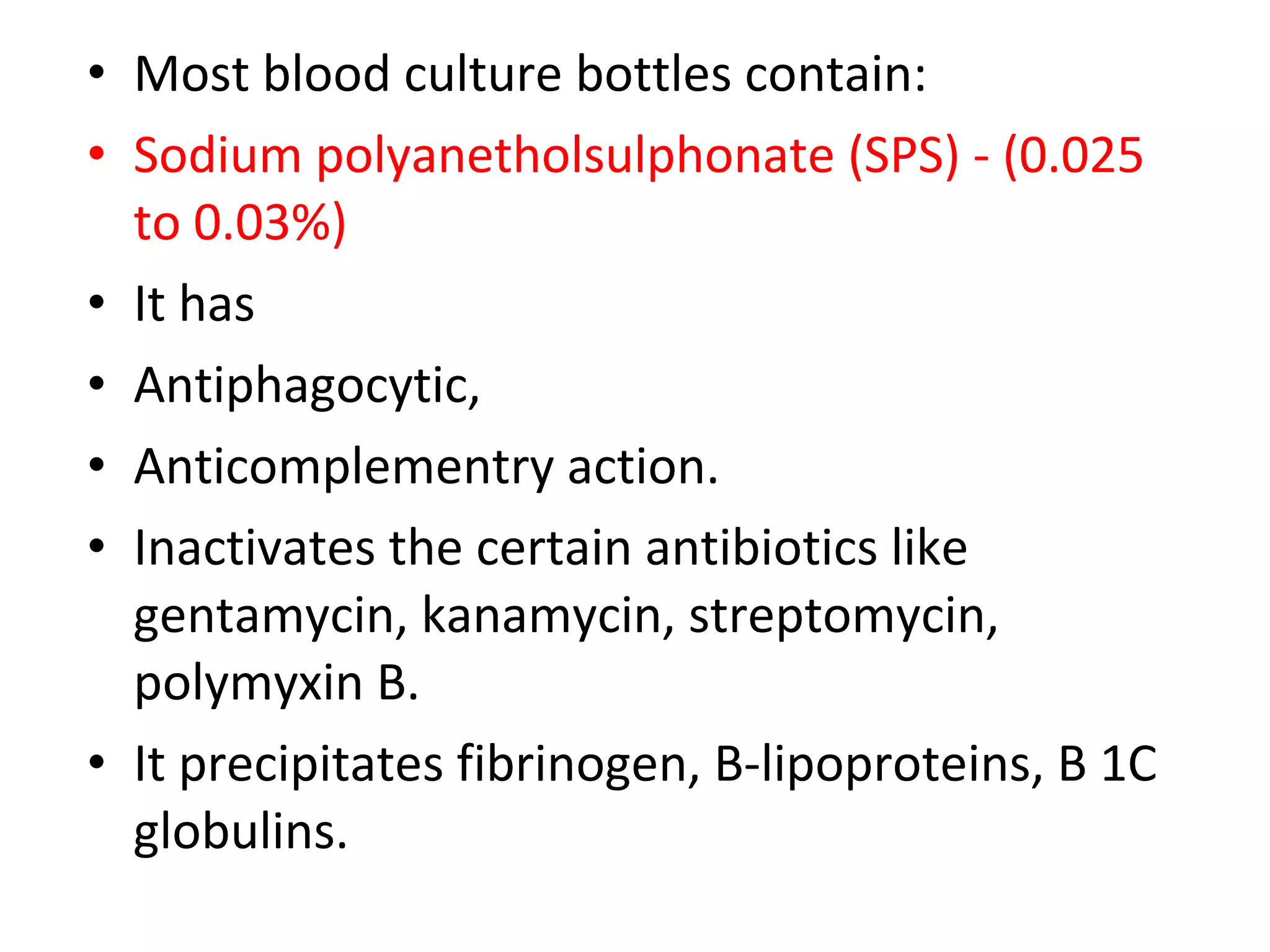
Blood culture
Dr.Yogita Mistry
GMC,Surat
Objectives
• Definitions
• Indication of blood culture
• How to collect blood culture
• Various aspects that affects blood culture
results
• Manual and automated system
• Blood culture samples data from our laboratory
• Summary
• References
Definition
• Collection and inoculation of blood into
culture medium with the aim of growing
pathogenic microorganism for diagnostic
purposes specially in case if bacteremia,
fungemia or septicemia.
• Bacteraemia: The presence of viable bacteria in the
bloodstream.
• It may be transient (e.g. following dental procedures,
vigorous chewing,instrumentation of genitourinary
tract),
• intermittent (e.g. undrained abscesses),
• or continuous (e.g. endovascular infection).
• Septicemia : Is a condition when microorganisms
(mainly bacteria) circulate and multiply to form toxic
products in the patient's blood.
Bacteremia Septicemia
Simple presence of bacteria in the blood It is the presence and active
multiplication of bacteria in blood
Not as dangerous as septicemia Potentially life threatening infection
Less amount of bacteria are present in
blood
Large amount of bacteria present in
blood
May result through a wound infection,
surgical procedure, injection.
Can arise from infections throughout the
body, including infections in lungs,
abdomen , urinary tract
Toxins are not produced Toxins are produced
No symptoms/mild fever Fever, chills, fast respiration, increase
heart rate,
Can resolve without treatment Quickly leads to spesis if untreated
Rapidly removed from blood stream by
immune system
Antibiotics are needed.
Organisms commonly isolated
from blood cultures
• S. aureus
• E.coli
• CONS
• Enterococcus spp.
• C.albicans
• P.aeruginosa
• K.pneumoniae
• Viridans streptococci
• S.pneumoniae
• Enterobacter cloacae
• Proteus spp.
• Bacteroids and clostridium
Organism found with
immunocompromised patients
• C. jeikeinum
• Bartonella henselae
• Shigella flexneri
• H. capsulatum
• C. neoformans
Organisms commonly associated with vlood stream infection from
extravascular sites of infections
Organism Extravascular site of infcetion
S.aureus Wound, soft tissue, meninges
S.payogen wound, soft tissue
S. pneumoniae Respiratory, meninges
S.typhi Small intestine, regional lymph nodes of
intestine, reticuloendothelial system
P. aeruginosa Wound, soft tissue, CNS
N. Meningitidis meninges
Listeria monocytogens meninges
Legionella Respiratory
H. influenza Meninges, epiglotitis, periorbital region,
respiratory
Enterobacteriaceae CNS, genitourinary region
CONS Wound, soft tissue
Clostridium spp. Wound, soft tissue
Candida albicans Genitourinary tract
Types of bacteremia
• Intravascular: originated within cardiovascular
system, leads to continous bacteremia
• Causes:
• Infective endocarditis
• Mycotic aneurysm
• Suppurative thrombophlebitis
• IV catheter associated bacteremia
• Infective endocarditis:
Infection of endocardium mostly by bacteria due to damage
to cardiac endothelium by congenital abnormality or direct
trauma or by transient bacteremia
Deposition of platelets , fibrin over damaged valve
Enhance the colonization of bacteria with protective layer of
fibrin and platelets
Favorable for multiplication of bacteria and forms a
vegetation
Seeding of bacteria at a slow but constant rate occurs
• Bacteria causing IE:
Natural heart valves: Viridans steptococci
(S.sanguis, S.mutan, S.intermedius.
S.mitis,S.bovis)
Prosthetic valve : Coagulase negative
staphylococci, viridans streptococci
In IV drug abusers: S.aureus
Organisms of infective endocarditis
• Aggregatibacter aprophilus
• Nutritionally deficient streptococci
(Abiotrophia spp. and Granulicatella spp.)
• Hemophilus spp.
• Unusual gram negative bacilli ( Actinobacillus,
Cardiobacterium, Eikenella, Kingella)
• Yeast
IV catheter associated bacteremia
• occurs by 2 routes:
• Extraluminal spread: By hospital staff/
patients microflora
• Intraluminal spread: By contaminated
infusion/ hospital staff
• In both the route S.epidermidis is most common due to its
ability to produce biofilm or slime , which helps in adherence
to catheter surface
• Other causes :
• S.aureus
• Enterobacteriaceae
• P.aeruginosa
• Candida spp.
• Other gram negative rods
• Extra vascular: Bacteria enter the blood stream
through lymphatic from other sites
• Causes:
• Respiratory disease-20%
• Genitourinary disease-10%
• Surgical wound infection-15%
• Billiary infection-5%
• Uncertain sites-25%
• CNS infection-5%
Indication of blood culture
• Where the possibility of septicemia or bacteremia is suggested by
the presence of fever, shock or other signs and symptoms occurring
in association with a known or suspected local infection such as
sepsis in a surgical wound, Puerperal sepsis, Pneumonia, Bacterial
Meningitis, Endocarditis.Osteomyelitis Peritonitis , Arthritis Enteric
fever ,Brucellosis, leptospirosis
• Pyrexia of unknown origin(temperatures of >38.3°C (>101°F) on
several occasions with fever of >3 weeks and failure to reach a
diagnosis despite 1 week of inpatient investigation. )
• Unexplained leucocytosis or leucopenia
• Suspected fungemia specially in Immunocompromised
patients, HIV patients
Purpose of inoculation of blood into culture
medium:
• Since density of bacteria in blood is usually
very low, it will helps in increase their number
to detectable level.
• Dilutes any antibacterial substance/ inhibitory
substances present in blood
Principle of sample collection for
blood culture
Site of collection:
• Arterial vs venous blood
• Indwelling arterial or venous lines
• Central or peripheral
• The peripheral vein sample should be collected first.
• Sets taken from either CVAD , peripheral or both sites must be obtained within 15 minutes of
each other and the volumes of blood obtained from both sites must match to ensure accuracy.
e.g. if only 10mL is obtained from the peripheral vein , obtain 10mL from the CVAD
• When taking blood from both the CVAD and from a peripheral vein, ensure that the site of each
sample is clearly labelled on the culture bottles and the request form.
• Collecting peripheral blood cultures:
• Blood cultures should be taken from a suitable, previously unused venepuncture site.
• Blood cultures should not routinely be taken from existing central catheters or peripheral venous
cannulae where there is no indication of sepsis.
• Collecting Central Venous Access Device (CVAD) blood cultures:
• If the CVAD is the suspected source of sepsis then taking blood cultures from the CVAD is
appropriate.
• Blood cultures should be taken from a CVAD in combination with a separate peripheral IV sample
when investigating potential central venous catheter-related septicaemia.
• EXCEPTIONS
• Obtaining CVAD blood cultures as the single source
• When peripheral IV access is clinically impossible and the only available source is the
CVAD, a blood culture specimen may be taken from the CVAD. The source of the
blood culture and the reasons for using a CVAD must be clearly documented on the
blood request form and in the patient's clinical notes.
• Blood Cultures should NOT be taken from the following sites
• Veins which are immediately proximal to an existing peripheral IV cannula.
• A femoral vein due to difficulty in skin disinfection of the site. This area poses a high
risk of contamination.
Amount of blood:
• For adult: minimum 10 ml
• For infant and children: 1-5 ml
1-2 ml= neonate
2-3ml= 1 month-2 year age
3-5ml= Older children
• However …….
• 20 ml of blood obtain in sequence is more effective and
sensitive (98%) specially in intermittent bacteremia.
• Patients who have received antibiotics should give 3 separate
collections of blood. Also one or two of which on 2nd
day also.
Sensitivity and number of blood culture
No. of culture Sensitivity
First culture 65.1 %
First 2 cultures 80 %
First 3 cultures 95.7 %
Timing of blood culture:
• Before starting antimicrobial therapy
• At the time of fever peak
• Minimum 30-60 minute interval between 2
samples except in critically ill septic patient.
• In continous bacteremia-timing of blood
culture is not important, but in intermittent
bacteremia 2 or 3 culture should be spaced an
hour apart.
• Blood to broth ratio: 1:5 only, should not be <1:5 or > 1:10
• Agitation during incubation
• Atmosphere of incubation: aerobic and anaerobic
Different broth:
• Glucose broth: useful in endocarditis
• Bile broth: in enteric fever
• Trypticase soy broth (inhibits Neisseria and
S.pneumoniae)
• Brain heart infusion broth: multipurpose broth
• Thioglycolate broth: for anaerobes
• Columbia or brucella broth
• Mycobacteria: Middlebrook 7H9 with 0.05% SPS, BHI with 0.5 %
polysorbate 80
• Fungus broth
• Additives in broth:
• Anticoagulant- bacteria are trapped in blood clot
• Antimicrobial- if patient is already in antibiotics
• Anticomplementry agents- to inactivates complement
action
• Antiphagocytic
• Most blood culture bottles contain:
• Sodium polyanetholsulphonate (SPS) - (0.025
to 0.03%)
• It has
• Antiphagocytic,
• Anticomplementry action.
• Inactivates the certain antibiotics like
gentamycin, kanamycin, streptomycin,
polymyxin B.
• It precipitates fibrinogen, B-lipoproteins, B 1C
globulins.
• However, SPS can also be toxic to certain
fastidious organisms like
• Neisseria,
• Mycoplasma,
• Peptostreptococcus,
• Streptobacillus moniliformis (Gelatin-1.2%)
• Other additives:
• Penicillinase- to inactivate penicillin
• Resin/charcol- inactivates most antibiotics by
adsorbing them to surface of resin
• Osmosic stabiliser like sucrose, mannitol,
sorbose- for cell wall deficient bacteria, but
RBC are lysed.
Length of incubation:
• Not more than 7 days
• 5 days is sufficient
• >5 day-contaminants
• 7 days is useful for:
• Fungemia
• Bacteremia due to fastidious organisms like HACEK group,
brucella, legionella
• For patients suspected of endocarditis who has been treated
with antimicrobial before obtaining blood culture
• Mycobacterial culture > 4 weeks
Proper asepsis:
• More important ways to decrease
contamination of blood culture bottles
include:
• Use of tincture of iodine as a disinfectant,
chlorhexidine also useful
• Avoidance of drawing blood through existing
intravenous lines and
• Disinfecting the membrane of the blood
culture bottle.
• Proper sample collection method:
• Instructions:
• Follow routine procedure for hand washing, gloving, equipment
preparation and organization ,patient approach, patient
identification and selection of venipuncture site.
• Explain to the patient.
• Ask the patient about allergies to iodine.
• Apply the tourniquet, select the site.
• Be careful that the ends of the tourniquet do not fall onto the
puncture site, thereby contaminating it, if the tourniquet does
accidentally touch the prepared puncture site, the site must be
recleaned.
• Apply alcohol/acetone pad at the puncture site for 30 seconds
( till it dry)
• Apply the iodine swab, apply to puncture site, move the iodine
in concentric circles outward. Keep it for 60 seconds ( till it
dry).
• Again clean the site with alcohol/acetone and allow it to dry.
• Perform the venipuncture, following routine venipuncture
procedures. Do not repalpate.
• If the blood culture is one of a series of samples to be drawn
from a patient, the blood culture must be collected first.
• Withdraw needle from vein and insert into the top of the
blood culture container.
• Other than syringe and needle, by closed system, consisting of
vacuum bottle and double needle collection tube can be done.
• Do not change the needle.
• Do not hold the container in your hand, this may result in a
needle exposure.
• Do not push the blood. Mix the content.
• Keep at room temperature.
• Label the blood specimen collected, following standard
labeling procedures. Be sure to include the site used and the
number of the specimen in the series ordered.
• Changing needles between venepuncture and
inoculation into culture bottles is
controversial. Discontinuing this practice
owing to lack of evidence that it decreases
contamination and the increased risk of sharp
injuries has been recommended.
• However, a meta-analysis challenges this
recommendation, suggesting a slight overall
benefit of switching needles.
Type of blood culture bottle
• Most blood culture systems are now
automated, with some form of continuous
monitoring to detect growth. The bottles
contain broth supplemented with additives.
Different bottles are designed either to be
used in different patient groups, or to isolate
different classes of pathogens.
• Standard aerobic bottles are suitable for the recovery of most
common bacterial pathogens, including aerobes ,facultative
anaerobes and for candidemia.
• These are not as suitable for supporting the growth of strict
anaerobes, and therefore traditional advice has been to use a
paired set of aerobic and anaerobic bottles when taking blood
cultures.
• Although routine use of anaerobic bottles is not warranted,
particularly in resource-limited settings
• Mycobacterial blood culture bottles contain broth
suitable for the isolation of mycobacteria and are
valuable for the detection of disseminated
mycobacterial infections, both M. tuberculosis and
non-tuberculous mycobacteria.
• Blood culture methods:
• Traditional/ conventional system
• Commercially developed system
• Automated system
Traditional blood culture system:
• Blood culture system:
• Broth are dispensed in special flat blood
culture bottle of 100-120 ml capacity.
• Fitted with a screw cap with a central hole
giving access to a rubber or neoprene washer
seal.
• An adequate space above broth ensures that
blood is not injected under undue pressure
and some air is still available for strict
aerobes.
• Smaller bottles are used for neonates and
young children, from whom appropriate small
volumes of blood are taken.
• After inoculation , bottles are incubated
aerobically.
• Examination of bottles for bacterial growth:
• Macroscopically:
• Generalized turbidity
• Hemolysis
• Gas production
• Discreate colonies on the surface of the sedimented red cells
• However, recoverable bacterial growth may
occur before turbidity is evident.
• So it is important to make subculture from
bottles as a routine.
• For subculture:
• Care should be taken to avoid contamination
• The procedure should be done quickly so that
bottles do not cool appreciably
• Subcuture should be done at least once during
the first day after 5-6 hours and at interval
thereafter which should be at least twice
during first 2-3 days.
• Gram stain:
• Should be made and examined at the
subculture stage.
• Any positive finding should be reported at
once to clinician as the morphological type of
organism may guide the physician to start
antibiotic.
• Quantitative counts of bacteria in blood:
• Inoculate 1 ml amounts of blood into several tubes of
melted agar and make pore plates either directly
from patients.
• Another method is treat the patient sample with lytic
agent. Then lysed sample is centrifuged and
harvested organism cultured directly on a suitable
solid medium to allow identification and to give a
semi-quantitative indication of its presence in blood.
( When monitoring colonization associated with a
prosthesis or catheter.)
• Mannual systems are flexible, not require
expensive instruments but are labour
intensive, chances of contamination are more.
Commercially available blood
culture systems:
• Biphasic septicheck system
• Opticult blood culture system
• Oxoid signal broth displacement system
• These are labour saving variations of
conventional mannual system
• Biphasic septicheck and opticult
blood culture system
• Castaneda method:
• An agar coated paddles / slants are
attached to the broth containing bottles.
• Allows the subculture to be done
without opening the bottle by tilting the
bottle and allowing the broth to flow
over agar at interval of times.
• Agar surface is examined for colonies.
• Oxoid signal broth
displacement system:
• After blood is inoculated into bottle, a
clear plastic cylindrical signal device is
attached to the top of bottle .
• A long needle from the lower end of
device is extended below the surface of
blood broth mixture, creating a closed
system.
• Gases produced as a by product of
microbial growth causes increase in
pressure in head space and it forces some
of blood broth mixture into cylinder----
>“Signaling” positive culture.
• Isolator:
• Unique manual method
• Useful particularly for fastidious or slow growing
organisms.
• It is also a method of choice for filamentous molds,
dimorphic fungi, Malassezia furfur and Legionella.
• It causes reduction in mean recovery time for yeast
from 4.9 days to 2.12 days, for histoplasma
capsulatum from 24.14 days to 8 days in comparison
to conventional methods.
• An overall 36.6 % increase in the recovery rate of
fungi. .
• Method:
• A special tube containing saponin, a chemical that lyses both
the RBC and white blood cells is used.
• Approximately 7.5 to 10 ml of blood is added in tube,
thoroughly mixed by inverting the tubes several times, so
complete lysis can occur.
• The tube is then placed into an angle cetrifuge for 3000 rpm for
15 minutes to concentrate any microorganism.
• Then a sediment is aspirated and subcultured in appropriate
medium.
• Advantage:
• It can be use as a quantitative method for blood
culture.
• The CFU/ml can be calculated from the volume of
blood processed and number of colonies present on
the agar surface.
• Disadvantage:
• Lysis solution is toxic to microorganism.
• Specimen must be processed within 8 hours of
collection.
• Labourous for intial processing.
• Contamination rate are high.
Automated systems:
• Available from 1970- radiometric method
• From mid 1980- Non-radiometric type.
• Three most commonly used systems are:
• BACTEC 9240/9120/9050
• BAC T /Alert
• TREK ESP culture system
• These system alerts that the culture is positive, after which
the relevant should be gram stained and sub-cultured.
• If the organism are not visualized , a blind subculture should
be performed and bottle returned in to the instrument for
continuous monitoring system.
BAC T/Alert system
• First continuously monitoring
system
• Bottles : having capacity to
receive 10 ml of blood.
• Principle:
• As the organism will grow, a CO2 is liberated, which is sensed by a
CO2-sensitive chemicals , which is bounded at the bottom of each
bottle , which is also separated from blood broth mixture by a
unidirectional CO2 permeable membrane.
• In the presence of CO2 , the color of sensor turns from green to
yellow.
• Each bottle is placed bottom down into a receiving well,
directed by a barcode on the bottle, which is integrated in the
computer to match patient information.
• The wells containing rack that gently rocks back and forth
when door is closed.
• At 10 minutes interval, a light beam from emitting diodes is
projected through an excitation filter to reflect off the CO2
sensitive sensor in the bottom of each bottle.
• The reflecting light is directed through an emission
filter to a photosensitive detection that in turn is
connected to a computer compiler.
• As soon as the accumulation of CO2 is sufficient in
the bottle to alert the sensor , an audible or visible
‘alert’ is generated, and the position of bottle is
immediately flagged by computer, which is in the
form of graph .
• Bactec:
• Fully automated computerized
system
• Same as BAC T/Alert, except that it
uses fluorescent, rather than spectral
light to detect changes in the
concentration of CO2.
• .
• Bottle should be loaded in detection portion
of instrument, where needle perforate the
bottle diaphragm and gas contents in head
space are screened once/twice a day.
• Instrument:
• BACTEC 9240/9120/9050(fluroscence series).
• Computer
• Barcode scanner-in front of door
• Diiferent media:
• BACTEC PLUS Aerobic/F culture vial
• BACTEC PLUS Anaerobic/F culture vial
• BACTEC PEDS PLUS/F culture vial
• BACTEC MYCO/F LYTIC
The TREK ESP culture system
• It differs from other two
methods by
• The production of CO2 is
measured
manometrically
• Both gas consumption
and production are
monitored
• Changes in the
concentration of H2 and
O2 in addition to CO2 are
also noted.
• Vital (bioMerieux)
• This measures exitor-induced flurescence but
is sensitive to changes of CO2 and also to pH
and EH.
• Difco Sentinel:
• Two electrodes are inserted into bottle which
read the potential differences that is due to
bacterial metabolism causing alteration in the
EH of the medium.
Quality control of blood culture
bottle
• For contamination -plane media incubate and
subculture
• For performance efficacy:
• Inoculate 1.0 ml a 0.5 ml MCFarland standard of either E.coli
ATCC or S.aureus ATCC prepared from fresh 18-24 hour
culture.
• It should be detected positive within 72 hours.
• It varifies that media is not subjected to adverse storage or
shipping condition prior to recipient in laboratory.
• For Mycological or Mycobacterial blood
culture system
• C. Glabrata ATCC 15545 = <3 days
• C.neoformance ATCC 15545= <3 days
• M. intracellulare ATCC 13950= 8-16 days
• For instrument maintenance:
• Cabinet / rack temperature=35±1.5˚C
• Rack indicator for barcode scanner, audible
alarm, signal detection
Advantages of continuous
monitoring systems:
• Decrease the laboratory work
• Decrease the pseudobacteremia due to
decrease in sampling and handling
• Increase in the speed of detection and rate of
recovery
• Disadvantage:
• Require an instrument
• Limited selection of medium
• Expensive
• Adjustment of staff during off time is required
• So each lab must decide the hours during
which the instrument will be attended.
Other use of blood culture
• Sterile body fluids cultures like
• CSF
• Pleural fluid
• Ascetic fluid
• Pericardial fluid
• Synovial fluid
• Most common contaminants:
• Coagulase negative s.aureus
• Corynebacterium spp.
• Proprionibacterium acnes
• Bacillus species
• Bacteremia due to unusual organisms due to
• Presence of one or more predisposing metabolic, physiologic, or
immunologic factors
• Cancer chemotherapy
• Intravenous catheterization
• liver disease
• hematologic malignancies
• Source: Environmental/endogenous
• Can occur even after a years of invasive procedure, instrumentation.
• Organisms: Aeromonas hydrophilia, Bacillus spp., Capnocytophaga spp.,
Corynebacterium jeikeium, Listeria monocytogen, Rodococcus equi, S.
bovis.
• The recovery of any unusual bacterium from blood should warrant a
careful consideration of the possibility of underlying, especially cancer.
Microbiologists should advise clinicians of the unusual nature of the
identified organism and provide the counsel that certain neoplastic
processes, often accompanied by neutropenia, render the human host
susceptible to invasion by almost any bacterium.
Fungemia in non-HIV-infected patients: a five-year
review
Siriluck Anunnatsir , Ploenchan Chetchotisakd, Piroon Mootsikap.Division of Infectious Diseases and Tropical
Medicine. January 2009Volume 13, Issue 1, Pages 90–96
• 147 episodes of fungemia due to Candida spp and Trichosporon spp in adult patients
admitted to a university hospital in Northeast Thailand between 1999 and 2003.
• Results:
• The overall incidence of fungemia was 14.1 per 10 000 hospital admissions. 
• Candida was the most common isolate (138 episodes, 93.9%) with non-albicans
Candida accounting for 68.7%.
• The major non-albicans Candida isolates were
• Candida parapsilosis and Candida tropicalis.
• Fungemia caused by Trichosporon accounted for 6.1% of the cases, but their clinical
features could not be distinguished from fungemia due to Candida.
• The overall in-hospital mortality rate was 56.1%.
Summary
• Blood culture is a method for detection of
bacteremia and septicemia.
• Manual and automated systems are available.
• Care should be taken to rule out
contaminants.
References:
• A method for early detection of antibiotic resistance in positive blood cultures:
experience from an oncology centre in eastern India.G Goel, D Das, S Mukherjee, S
Bose, K Das, R Mahato, *S Bhattacharya.Indian Journal of Medical Microbiology,
(2015) 33(Supplement 1): S513-58.
• Utility of Extended Blood Culture Incubation for Isolation of Haemophilus,
Actinobacillus, Cardiobacterium, Eikenella, and Kingella Organisms: a
Retrospective multicenter Evaluation. Hasan S. Bhally, Melvin P. Weinstein, Kim
Joho, Teresa Wakefield. Journal of clinical microbiology, jan. 2006, p. 257–259 vol.
44.
• Mackie & McCarteny practical medical microbiology 14th
edition, page: 98-101.
• Konemman’s color atlas and textbook of Diagnostic Microbiology, 6th
edition,Page: 100-104.
• Monica cheesebrough-Laboratory practice in tropical countries, 2nd
edition,part:2,
page: 67-71.
• Thank you

Blood culture

  • 1.
  • 2.
    Objectives • Definitions • Indicationof blood culture • How to collect blood culture • Various aspects that affects blood culture results • Manual and automated system • Blood culture samples data from our laboratory • Summary • References
  • 3.
    Definition • Collection andinoculation of blood into culture medium with the aim of growing pathogenic microorganism for diagnostic purposes specially in case if bacteremia, fungemia or septicemia.
  • 4.
    • Bacteraemia: Thepresence of viable bacteria in the bloodstream. • It may be transient (e.g. following dental procedures, vigorous chewing,instrumentation of genitourinary tract), • intermittent (e.g. undrained abscesses), • or continuous (e.g. endovascular infection). • Septicemia : Is a condition when microorganisms (mainly bacteria) circulate and multiply to form toxic products in the patient's blood.
  • 5.
    Bacteremia Septicemia Simple presenceof bacteria in the blood It is the presence and active multiplication of bacteria in blood Not as dangerous as septicemia Potentially life threatening infection Less amount of bacteria are present in blood Large amount of bacteria present in blood May result through a wound infection, surgical procedure, injection. Can arise from infections throughout the body, including infections in lungs, abdomen , urinary tract Toxins are not produced Toxins are produced No symptoms/mild fever Fever, chills, fast respiration, increase heart rate, Can resolve without treatment Quickly leads to spesis if untreated Rapidly removed from blood stream by immune system Antibiotics are needed.
  • 6.
    Organisms commonly isolated fromblood cultures • S. aureus • E.coli • CONS • Enterococcus spp. • C.albicans • P.aeruginosa • K.pneumoniae • Viridans streptococci • S.pneumoniae • Enterobacter cloacae • Proteus spp. • Bacteroids and clostridium
  • 7.
    Organism found with immunocompromisedpatients • C. jeikeinum • Bartonella henselae • Shigella flexneri • H. capsulatum • C. neoformans
  • 8.
    Organisms commonly associatedwith vlood stream infection from extravascular sites of infections Organism Extravascular site of infcetion S.aureus Wound, soft tissue, meninges S.payogen wound, soft tissue S. pneumoniae Respiratory, meninges S.typhi Small intestine, regional lymph nodes of intestine, reticuloendothelial system P. aeruginosa Wound, soft tissue, CNS N. Meningitidis meninges Listeria monocytogens meninges Legionella Respiratory H. influenza Meninges, epiglotitis, periorbital region, respiratory Enterobacteriaceae CNS, genitourinary region CONS Wound, soft tissue Clostridium spp. Wound, soft tissue Candida albicans Genitourinary tract
  • 9.
    Types of bacteremia •Intravascular: originated within cardiovascular system, leads to continous bacteremia • Causes: • Infective endocarditis • Mycotic aneurysm • Suppurative thrombophlebitis • IV catheter associated bacteremia
  • 10.
    • Infective endocarditis: Infectionof endocardium mostly by bacteria due to damage to cardiac endothelium by congenital abnormality or direct trauma or by transient bacteremia Deposition of platelets , fibrin over damaged valve Enhance the colonization of bacteria with protective layer of fibrin and platelets Favorable for multiplication of bacteria and forms a vegetation Seeding of bacteria at a slow but constant rate occurs
  • 11.
    • Bacteria causingIE: Natural heart valves: Viridans steptococci (S.sanguis, S.mutan, S.intermedius. S.mitis,S.bovis) Prosthetic valve : Coagulase negative staphylococci, viridans streptococci In IV drug abusers: S.aureus
  • 12.
    Organisms of infectiveendocarditis • Aggregatibacter aprophilus • Nutritionally deficient streptococci (Abiotrophia spp. and Granulicatella spp.) • Hemophilus spp. • Unusual gram negative bacilli ( Actinobacillus, Cardiobacterium, Eikenella, Kingella) • Yeast
  • 13.
    IV catheter associatedbacteremia • occurs by 2 routes: • Extraluminal spread: By hospital staff/ patients microflora • Intraluminal spread: By contaminated infusion/ hospital staff
  • 14.
    • In boththe route S.epidermidis is most common due to its ability to produce biofilm or slime , which helps in adherence to catheter surface • Other causes : • S.aureus • Enterobacteriaceae • P.aeruginosa • Candida spp. • Other gram negative rods
  • 15.
    • Extra vascular:Bacteria enter the blood stream through lymphatic from other sites • Causes: • Respiratory disease-20% • Genitourinary disease-10% • Surgical wound infection-15% • Billiary infection-5% • Uncertain sites-25% • CNS infection-5%
  • 16.
    Indication of bloodculture • Where the possibility of septicemia or bacteremia is suggested by the presence of fever, shock or other signs and symptoms occurring in association with a known or suspected local infection such as sepsis in a surgical wound, Puerperal sepsis, Pneumonia, Bacterial Meningitis, Endocarditis.Osteomyelitis Peritonitis , Arthritis Enteric fever ,Brucellosis, leptospirosis • Pyrexia of unknown origin(temperatures of >38.3°C (>101°F) on several occasions with fever of >3 weeks and failure to reach a diagnosis despite 1 week of inpatient investigation. ) • Unexplained leucocytosis or leucopenia • Suspected fungemia specially in Immunocompromised patients, HIV patients
  • 17.
    Purpose of inoculationof blood into culture medium: • Since density of bacteria in blood is usually very low, it will helps in increase their number to detectable level. • Dilutes any antibacterial substance/ inhibitory substances present in blood
  • 18.
    Principle of samplecollection for blood culture Site of collection: • Arterial vs venous blood • Indwelling arterial or venous lines • Central or peripheral
  • 19.
    • The peripheralvein sample should be collected first. • Sets taken from either CVAD , peripheral or both sites must be obtained within 15 minutes of each other and the volumes of blood obtained from both sites must match to ensure accuracy. e.g. if only 10mL is obtained from the peripheral vein , obtain 10mL from the CVAD • When taking blood from both the CVAD and from a peripheral vein, ensure that the site of each sample is clearly labelled on the culture bottles and the request form. • Collecting peripheral blood cultures: • Blood cultures should be taken from a suitable, previously unused venepuncture site. • Blood cultures should not routinely be taken from existing central catheters or peripheral venous cannulae where there is no indication of sepsis. • Collecting Central Venous Access Device (CVAD) blood cultures: • If the CVAD is the suspected source of sepsis then taking blood cultures from the CVAD is appropriate. • Blood cultures should be taken from a CVAD in combination with a separate peripheral IV sample when investigating potential central venous catheter-related septicaemia.
  • 20.
    • EXCEPTIONS • ObtainingCVAD blood cultures as the single source • When peripheral IV access is clinically impossible and the only available source is the CVAD, a blood culture specimen may be taken from the CVAD. The source of the blood culture and the reasons for using a CVAD must be clearly documented on the blood request form and in the patient's clinical notes. • Blood Cultures should NOT be taken from the following sites • Veins which are immediately proximal to an existing peripheral IV cannula. • A femoral vein due to difficulty in skin disinfection of the site. This area poses a high risk of contamination.
  • 21.
    Amount of blood: •For adult: minimum 10 ml • For infant and children: 1-5 ml 1-2 ml= neonate 2-3ml= 1 month-2 year age 3-5ml= Older children
  • 22.
    • However ……. •20 ml of blood obtain in sequence is more effective and sensitive (98%) specially in intermittent bacteremia. • Patients who have received antibiotics should give 3 separate collections of blood. Also one or two of which on 2nd day also.
  • 23.
    Sensitivity and numberof blood culture No. of culture Sensitivity First culture 65.1 % First 2 cultures 80 % First 3 cultures 95.7 %
  • 24.
    Timing of bloodculture: • Before starting antimicrobial therapy • At the time of fever peak • Minimum 30-60 minute interval between 2 samples except in critically ill septic patient. • In continous bacteremia-timing of blood culture is not important, but in intermittent bacteremia 2 or 3 culture should be spaced an hour apart.
  • 25.
    • Blood tobroth ratio: 1:5 only, should not be <1:5 or > 1:10 • Agitation during incubation • Atmosphere of incubation: aerobic and anaerobic
  • 26.
    Different broth: • Glucosebroth: useful in endocarditis • Bile broth: in enteric fever • Trypticase soy broth (inhibits Neisseria and S.pneumoniae) • Brain heart infusion broth: multipurpose broth • Thioglycolate broth: for anaerobes • Columbia or brucella broth • Mycobacteria: Middlebrook 7H9 with 0.05% SPS, BHI with 0.5 % polysorbate 80 • Fungus broth
  • 27.
    • Additives inbroth: • Anticoagulant- bacteria are trapped in blood clot • Antimicrobial- if patient is already in antibiotics • Anticomplementry agents- to inactivates complement action • Antiphagocytic
  • 28.
    • Most bloodculture bottles contain: • Sodium polyanetholsulphonate (SPS) - (0.025 to 0.03%) • It has • Antiphagocytic, • Anticomplementry action. • Inactivates the certain antibiotics like gentamycin, kanamycin, streptomycin, polymyxin B. • It precipitates fibrinogen, B-lipoproteins, B 1C globulins.
  • 29.
    • However, SPScan also be toxic to certain fastidious organisms like • Neisseria, • Mycoplasma, • Peptostreptococcus, • Streptobacillus moniliformis (Gelatin-1.2%)
  • 30.
    • Other additives: •Penicillinase- to inactivate penicillin • Resin/charcol- inactivates most antibiotics by adsorbing them to surface of resin • Osmosic stabiliser like sucrose, mannitol, sorbose- for cell wall deficient bacteria, but RBC are lysed.
  • 31.
    Length of incubation: •Not more than 7 days • 5 days is sufficient • >5 day-contaminants • 7 days is useful for: • Fungemia • Bacteremia due to fastidious organisms like HACEK group, brucella, legionella • For patients suspected of endocarditis who has been treated with antimicrobial before obtaining blood culture • Mycobacterial culture > 4 weeks
  • 32.
    Proper asepsis: • Moreimportant ways to decrease contamination of blood culture bottles include: • Use of tincture of iodine as a disinfectant, chlorhexidine also useful • Avoidance of drawing blood through existing intravenous lines and • Disinfecting the membrane of the blood culture bottle.
  • 33.
    • Proper samplecollection method: • Instructions: • Follow routine procedure for hand washing, gloving, equipment preparation and organization ,patient approach, patient identification and selection of venipuncture site. • Explain to the patient. • Ask the patient about allergies to iodine.
  • 34.
    • Apply thetourniquet, select the site. • Be careful that the ends of the tourniquet do not fall onto the puncture site, thereby contaminating it, if the tourniquet does accidentally touch the prepared puncture site, the site must be recleaned. • Apply alcohol/acetone pad at the puncture site for 30 seconds ( till it dry) • Apply the iodine swab, apply to puncture site, move the iodine in concentric circles outward. Keep it for 60 seconds ( till it dry). • Again clean the site with alcohol/acetone and allow it to dry.
  • 35.
    • Perform thevenipuncture, following routine venipuncture procedures. Do not repalpate. • If the blood culture is one of a series of samples to be drawn from a patient, the blood culture must be collected first. • Withdraw needle from vein and insert into the top of the blood culture container. • Other than syringe and needle, by closed system, consisting of vacuum bottle and double needle collection tube can be done. • Do not change the needle. • Do not hold the container in your hand, this may result in a needle exposure. • Do not push the blood. Mix the content. • Keep at room temperature. • Label the blood specimen collected, following standard labeling procedures. Be sure to include the site used and the number of the specimen in the series ordered.
  • 36.
    • Changing needlesbetween venepuncture and inoculation into culture bottles is controversial. Discontinuing this practice owing to lack of evidence that it decreases contamination and the increased risk of sharp injuries has been recommended. • However, a meta-analysis challenges this recommendation, suggesting a slight overall benefit of switching needles.
  • 37.
    Type of bloodculture bottle • Most blood culture systems are now automated, with some form of continuous monitoring to detect growth. The bottles contain broth supplemented with additives. Different bottles are designed either to be used in different patient groups, or to isolate different classes of pathogens.
  • 38.
    • Standard aerobicbottles are suitable for the recovery of most common bacterial pathogens, including aerobes ,facultative anaerobes and for candidemia. • These are not as suitable for supporting the growth of strict anaerobes, and therefore traditional advice has been to use a paired set of aerobic and anaerobic bottles when taking blood cultures. • Although routine use of anaerobic bottles is not warranted, particularly in resource-limited settings
  • 39.
    • Mycobacterial bloodculture bottles contain broth suitable for the isolation of mycobacteria and are valuable for the detection of disseminated mycobacterial infections, both M. tuberculosis and non-tuberculous mycobacteria.
  • 40.
    • Blood culturemethods: • Traditional/ conventional system • Commercially developed system • Automated system
  • 41.
    Traditional blood culturesystem: • Blood culture system: • Broth are dispensed in special flat blood culture bottle of 100-120 ml capacity. • Fitted with a screw cap with a central hole giving access to a rubber or neoprene washer seal. • An adequate space above broth ensures that blood is not injected under undue pressure and some air is still available for strict aerobes.
  • 42.
    • Smaller bottlesare used for neonates and young children, from whom appropriate small volumes of blood are taken. • After inoculation , bottles are incubated aerobically.
  • 43.
    • Examination ofbottles for bacterial growth: • Macroscopically: • Generalized turbidity • Hemolysis • Gas production • Discreate colonies on the surface of the sedimented red cells • However, recoverable bacterial growth may occur before turbidity is evident. • So it is important to make subculture from bottles as a routine.
  • 44.
    • For subculture: •Care should be taken to avoid contamination • The procedure should be done quickly so that bottles do not cool appreciably • Subcuture should be done at least once during the first day after 5-6 hours and at interval thereafter which should be at least twice during first 2-3 days.
  • 45.
    • Gram stain: •Should be made and examined at the subculture stage. • Any positive finding should be reported at once to clinician as the morphological type of organism may guide the physician to start antibiotic.
  • 46.
    • Quantitative countsof bacteria in blood: • Inoculate 1 ml amounts of blood into several tubes of melted agar and make pore plates either directly from patients. • Another method is treat the patient sample with lytic agent. Then lysed sample is centrifuged and harvested organism cultured directly on a suitable solid medium to allow identification and to give a semi-quantitative indication of its presence in blood. ( When monitoring colonization associated with a prosthesis or catheter.)
  • 47.
    • Mannual systemsare flexible, not require expensive instruments but are labour intensive, chances of contamination are more.
  • 48.
    Commercially available blood culturesystems: • Biphasic septicheck system • Opticult blood culture system • Oxoid signal broth displacement system • These are labour saving variations of conventional mannual system
  • 49.
    • Biphasic septicheckand opticult blood culture system • Castaneda method: • An agar coated paddles / slants are attached to the broth containing bottles. • Allows the subculture to be done without opening the bottle by tilting the bottle and allowing the broth to flow over agar at interval of times. • Agar surface is examined for colonies.
  • 50.
    • Oxoid signalbroth displacement system: • After blood is inoculated into bottle, a clear plastic cylindrical signal device is attached to the top of bottle . • A long needle from the lower end of device is extended below the surface of blood broth mixture, creating a closed system. • Gases produced as a by product of microbial growth causes increase in pressure in head space and it forces some of blood broth mixture into cylinder---- >“Signaling” positive culture.
  • 51.
    • Isolator: • Uniquemanual method • Useful particularly for fastidious or slow growing organisms. • It is also a method of choice for filamentous molds, dimorphic fungi, Malassezia furfur and Legionella. • It causes reduction in mean recovery time for yeast from 4.9 days to 2.12 days, for histoplasma capsulatum from 24.14 days to 8 days in comparison to conventional methods. • An overall 36.6 % increase in the recovery rate of fungi. .
  • 52.
    • Method: • Aspecial tube containing saponin, a chemical that lyses both the RBC and white blood cells is used. • Approximately 7.5 to 10 ml of blood is added in tube, thoroughly mixed by inverting the tubes several times, so complete lysis can occur. • The tube is then placed into an angle cetrifuge for 3000 rpm for 15 minutes to concentrate any microorganism. • Then a sediment is aspirated and subcultured in appropriate medium.
  • 53.
    • Advantage: • Itcan be use as a quantitative method for blood culture. • The CFU/ml can be calculated from the volume of blood processed and number of colonies present on the agar surface. • Disadvantage: • Lysis solution is toxic to microorganism. • Specimen must be processed within 8 hours of collection. • Labourous for intial processing. • Contamination rate are high.
  • 54.
    Automated systems: • Availablefrom 1970- radiometric method • From mid 1980- Non-radiometric type.
  • 55.
    • Three mostcommonly used systems are: • BACTEC 9240/9120/9050 • BAC T /Alert • TREK ESP culture system • These system alerts that the culture is positive, after which the relevant should be gram stained and sub-cultured. • If the organism are not visualized , a blind subculture should be performed and bottle returned in to the instrument for continuous monitoring system.
  • 56.
    BAC T/Alert system •First continuously monitoring system • Bottles : having capacity to receive 10 ml of blood.
  • 57.
    • Principle: • Asthe organism will grow, a CO2 is liberated, which is sensed by a CO2-sensitive chemicals , which is bounded at the bottom of each bottle , which is also separated from blood broth mixture by a unidirectional CO2 permeable membrane. • In the presence of CO2 , the color of sensor turns from green to yellow.
  • 59.
    • Each bottleis placed bottom down into a receiving well, directed by a barcode on the bottle, which is integrated in the computer to match patient information. • The wells containing rack that gently rocks back and forth when door is closed. • At 10 minutes interval, a light beam from emitting diodes is projected through an excitation filter to reflect off the CO2 sensitive sensor in the bottom of each bottle.
  • 60.
    • The reflectinglight is directed through an emission filter to a photosensitive detection that in turn is connected to a computer compiler. • As soon as the accumulation of CO2 is sufficient in the bottle to alert the sensor , an audible or visible ‘alert’ is generated, and the position of bottle is immediately flagged by computer, which is in the form of graph .
  • 61.
    • Bactec: • Fullyautomated computerized system • Same as BAC T/Alert, except that it uses fluorescent, rather than spectral light to detect changes in the concentration of CO2. • .
  • 62.
    • Bottle shouldbe loaded in detection portion of instrument, where needle perforate the bottle diaphragm and gas contents in head space are screened once/twice a day.
  • 63.
    • Instrument: • BACTEC9240/9120/9050(fluroscence series). • Computer • Barcode scanner-in front of door
  • 64.
    • Diiferent media: •BACTEC PLUS Aerobic/F culture vial • BACTEC PLUS Anaerobic/F culture vial • BACTEC PEDS PLUS/F culture vial • BACTEC MYCO/F LYTIC
  • 65.
    The TREK ESPculture system • It differs from other two methods by • The production of CO2 is measured manometrically • Both gas consumption and production are monitored • Changes in the concentration of H2 and O2 in addition to CO2 are also noted.
  • 66.
    • Vital (bioMerieux) •This measures exitor-induced flurescence but is sensitive to changes of CO2 and also to pH and EH.
  • 67.
    • Difco Sentinel: •Two electrodes are inserted into bottle which read the potential differences that is due to bacterial metabolism causing alteration in the EH of the medium.
  • 68.
    Quality control ofblood culture bottle • For contamination -plane media incubate and subculture • For performance efficacy: • Inoculate 1.0 ml a 0.5 ml MCFarland standard of either E.coli ATCC or S.aureus ATCC prepared from fresh 18-24 hour culture. • It should be detected positive within 72 hours. • It varifies that media is not subjected to adverse storage or shipping condition prior to recipient in laboratory.
  • 69.
    • For Mycologicalor Mycobacterial blood culture system • C. Glabrata ATCC 15545 = <3 days • C.neoformance ATCC 15545= <3 days • M. intracellulare ATCC 13950= 8-16 days
  • 70.
    • For instrumentmaintenance: • Cabinet / rack temperature=35±1.5˚C • Rack indicator for barcode scanner, audible alarm, signal detection
  • 71.
    Advantages of continuous monitoringsystems: • Decrease the laboratory work • Decrease the pseudobacteremia due to decrease in sampling and handling • Increase in the speed of detection and rate of recovery
  • 72.
    • Disadvantage: • Requirean instrument • Limited selection of medium • Expensive • Adjustment of staff during off time is required • So each lab must decide the hours during which the instrument will be attended.
  • 73.
    Other use ofblood culture • Sterile body fluids cultures like • CSF • Pleural fluid • Ascetic fluid • Pericardial fluid • Synovial fluid
  • 74.
    • Most commoncontaminants: • Coagulase negative s.aureus • Corynebacterium spp. • Proprionibacterium acnes • Bacillus species
  • 75.
    • Bacteremia dueto unusual organisms due to • Presence of one or more predisposing metabolic, physiologic, or immunologic factors • Cancer chemotherapy • Intravenous catheterization • liver disease • hematologic malignancies • Source: Environmental/endogenous • Can occur even after a years of invasive procedure, instrumentation. • Organisms: Aeromonas hydrophilia, Bacillus spp., Capnocytophaga spp., Corynebacterium jeikeium, Listeria monocytogen, Rodococcus equi, S. bovis.
  • 76.
    • The recoveryof any unusual bacterium from blood should warrant a careful consideration of the possibility of underlying, especially cancer. Microbiologists should advise clinicians of the unusual nature of the identified organism and provide the counsel that certain neoplastic processes, often accompanied by neutropenia, render the human host susceptible to invasion by almost any bacterium.
  • 77.
    Fungemia in non-HIV-infectedpatients: a five-year review Siriluck Anunnatsir , Ploenchan Chetchotisakd, Piroon Mootsikap.Division of Infectious Diseases and Tropical Medicine. January 2009Volume 13, Issue 1, Pages 90–96 • 147 episodes of fungemia due to Candida spp and Trichosporon spp in adult patients admitted to a university hospital in Northeast Thailand between 1999 and 2003. • Results: • The overall incidence of fungemia was 14.1 per 10 000 hospital admissions.  • Candida was the most common isolate (138 episodes, 93.9%) with non-albicans Candida accounting for 68.7%. • The major non-albicans Candida isolates were • Candida parapsilosis and Candida tropicalis. • Fungemia caused by Trichosporon accounted for 6.1% of the cases, but their clinical features could not be distinguished from fungemia due to Candida. • The overall in-hospital mortality rate was 56.1%.
  • 78.
    Summary • Blood cultureis a method for detection of bacteremia and septicemia. • Manual and automated systems are available. • Care should be taken to rule out contaminants.
  • 79.
    References: • A methodfor early detection of antibiotic resistance in positive blood cultures: experience from an oncology centre in eastern India.G Goel, D Das, S Mukherjee, S Bose, K Das, R Mahato, *S Bhattacharya.Indian Journal of Medical Microbiology, (2015) 33(Supplement 1): S513-58. • Utility of Extended Blood Culture Incubation for Isolation of Haemophilus, Actinobacillus, Cardiobacterium, Eikenella, and Kingella Organisms: a Retrospective multicenter Evaluation. Hasan S. Bhally, Melvin P. Weinstein, Kim Joho, Teresa Wakefield. Journal of clinical microbiology, jan. 2006, p. 257–259 vol. 44. • Mackie & McCarteny practical medical microbiology 14th edition, page: 98-101. • Konemman’s color atlas and textbook of Diagnostic Microbiology, 6th edition,Page: 100-104. • Monica cheesebrough-Laboratory practice in tropical countries, 2nd edition,part:2, page: 67-71.
  • 80.