Download to read offline


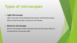
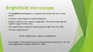
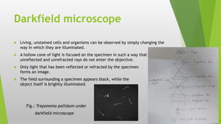

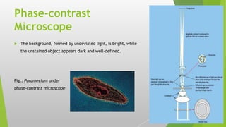


The document provides an overview of microscopy, detailing its history, components, types, and usage. It outlines the development of various microscopes, including light microscopes (such as brightfield, darkfield, phase-contrast, and fluorescence) and electron microscopes (transmission and scanning). Key care instructions for microscopes are also highlighted, ensuring proper maintenance and optimal functionality.