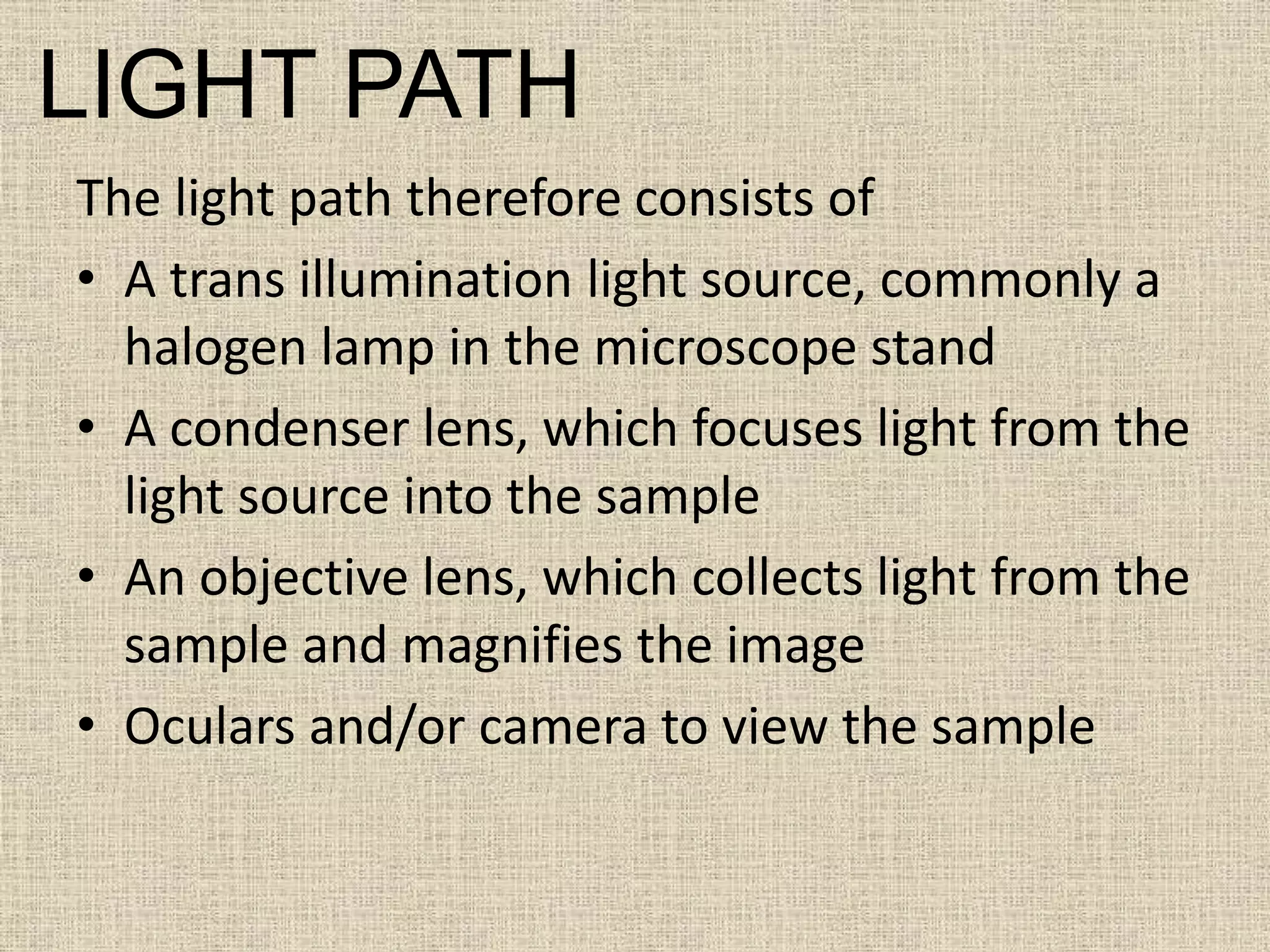
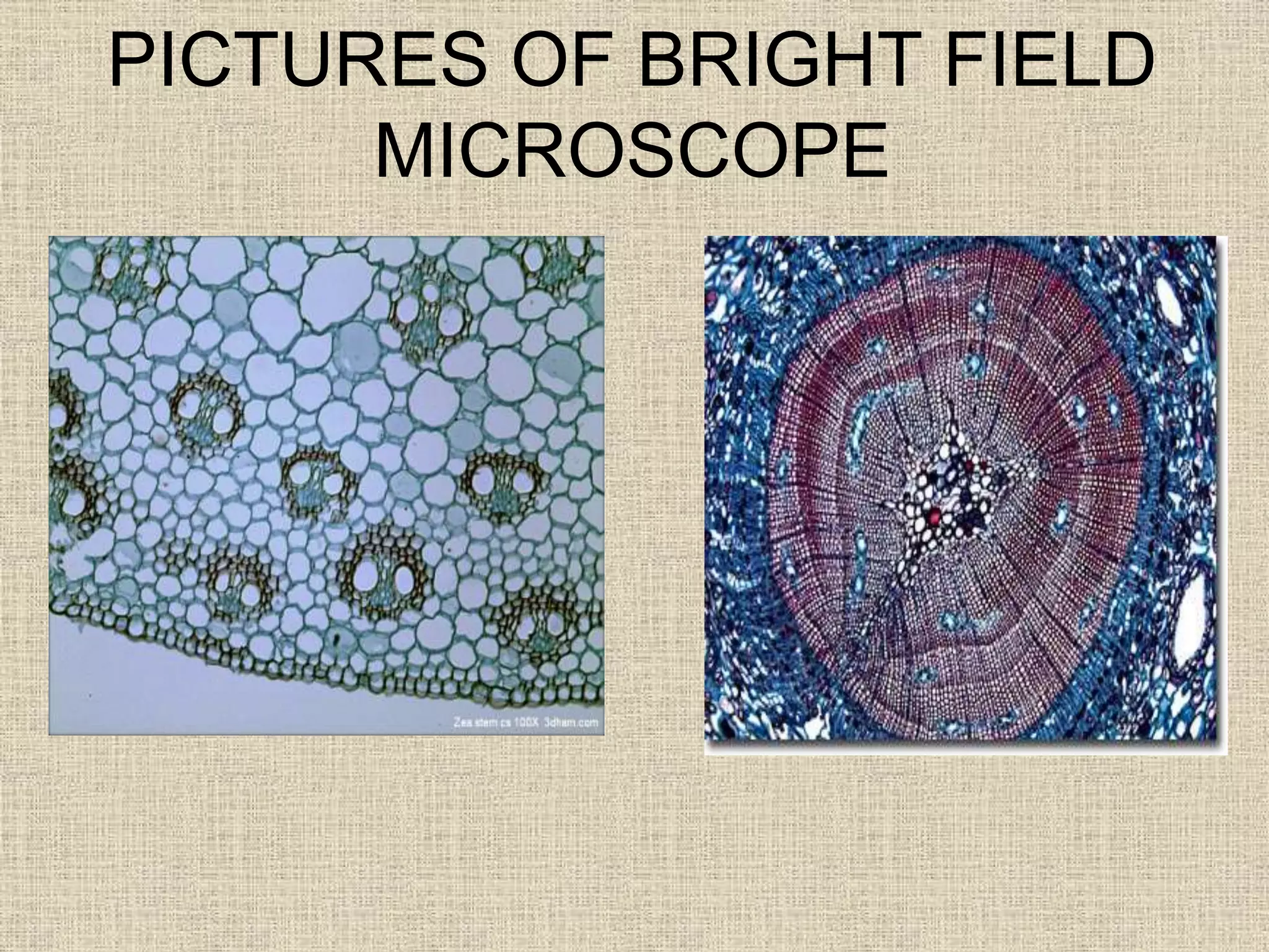
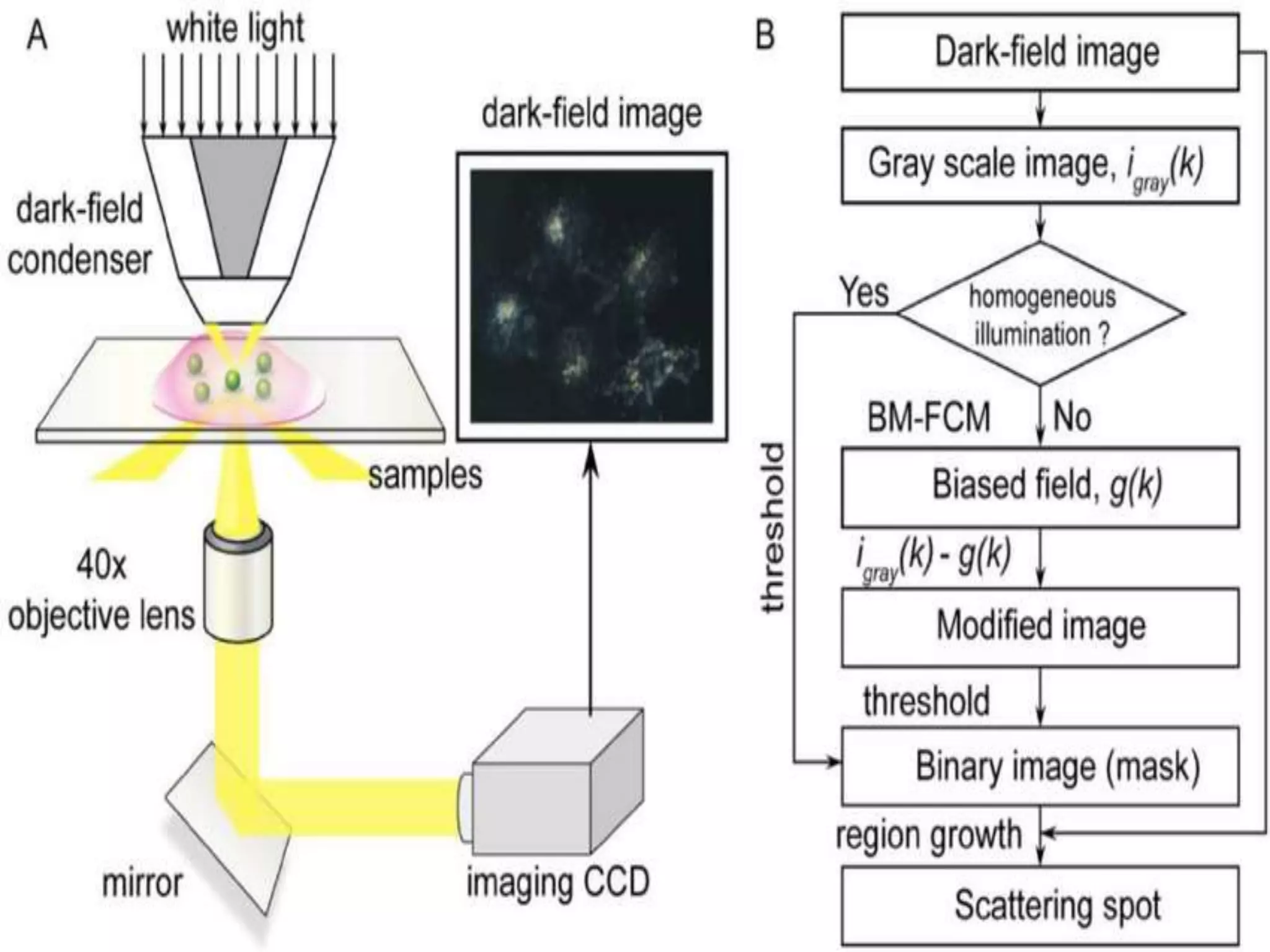
Bright field microscopy is the most commonly used technique where light passes through or reflects off a specimen. Biologists have used counterstaining for over 100 years to help differentiate tissues and organelles. The light path includes a light source, condenser lens to focus light, objective lens to collect and magnify the image, and oculars or camera to view the sample. It is mainly used for viewing stained specimens, pathological exams, blood tests, and inspections. Dark field microscopy uses oblique illumination to reveal fine detail, especially in bacteria. It illuminates specimens diagonally and observes light scattering in a darker field of view. It is useful for microbiology imaging and detecting scratches but lacks shading information.