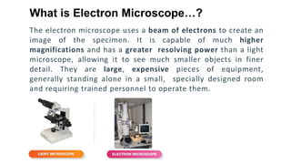
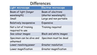
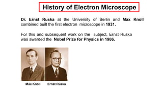
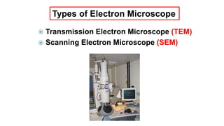
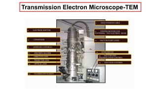
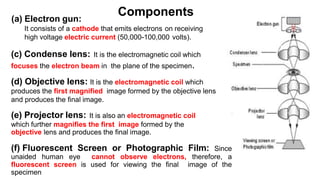
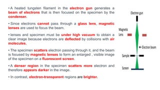
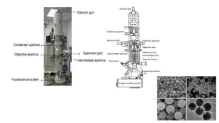
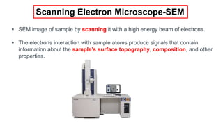
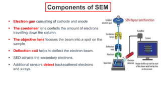
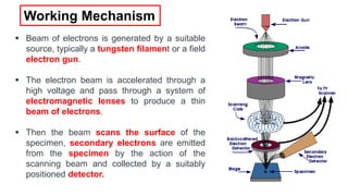
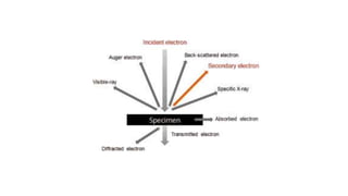
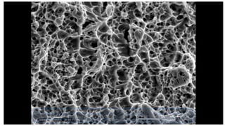
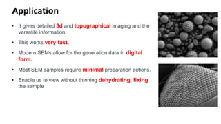
SlideShare a Scribd company logo
ELECTRON MICROSCOPE
Dr. Prafulla Katkar
Ph.D (Italy), PDF (Denmark)
In the 1920s it was discovered that accelerated electrons
(parts of the atom) behave in vacuum just like light. They
travel in straight lines and have a wavelength which is about
100 000 times smaller than that of light. Furthermore, it was
found that electric and magnetic fields have the same effect
on electrons as glass lenses and mirrors have on visible light.
Why use electrons instead of light ?
The electron microscope uses a beam of electrons to create an
image of the specimen. It is capable of much higher
magnifications and has a greater resolving power than a light
microscope, allowing it to see much smaller objects in finer
detail. They are large, expensive pieces of equipment,
generally standing alone in a small, specially designed room
and requiring trained personnel to operate them.
What is Electron Microscope…?
Dr. Ernst Ruska at the University of Berlin and Max Knoll
combined built the first electron microscope in 1931.
For this and subsequent work on the subject, Ernst Ruska
was awarded the Nobel Prize for Physics in 1986.
Max Knoll Ernst Ruska
History of Electron Microscope
Type of Electron Microscope
 Transmission Electron Microscope (TEM)
 Scanning Electron Microscope (SEM)
Types of Electron Microscope
Components
Transmission Electron Microscope-TEM
(a) Electron gun:
It consists of a cathode that emits electrons on receiving
high voltage electric current (50,000-100,000 volts).
(c) Condense lens: It is the electromagnetic coil which
focuses the electron beam in the plane of the specimen.
(d) Objective lens: It is the electromagnetic coil which
produces the first magnified image formed by the objective lens
and produces the final image.
(e) Projector lens: It is also an electromagnetic coil
which further magnifies the first image formed by the
objective lens and produces the final image.
(f) Fluorescent Screen or Photographic Film: Since
unaided human eye cannot observe electrons, therefore, a
fluorescent screen is used for viewing the final image of the
specimen
Components
• A heated tungsten filament in the electron gun generates a
beam of electrons that is then focused on the specimen by the
condenser.
• Since electrons cannot pass through a glass lens, magnetic
lenses are used to focus the beam.
• lenses and specimen must be under high vacuum to obtain a
clear image because electrons are deflected by collisions with air
molecules.
• The specimen scatters electron passing through it, and the beam
is focused by magnetic lenses to form an enlarged , visible image
of the specimen on a fluorescent screen.
• A denser region in the specimen scatters more electron and
therefore appears darker in the image.
• In contrast, electron-transparent regions are brighter.
Scanning Electron Microscope-SEM
 SEM image of sample by scanning it with a high energy beam of electrons.
 The electrons interaction with sample atoms produce signals that contain
information about the sample’s surface topography, composition, and other
properties.
Scanning Electron Microscope
 Electron gun consisting of cathode and anode
 The condenser lens controls the amount of electrons
travelling down the column.
 The objective lens focuses the beam into a spot on the
sample.
 Deflection coil helps to deflect the electron beam.
 SED attracts the secondary electrons.
 Additional sensors detect backscattered electrons
and x-rays.
Components of SEM
 Beam of electrons is generated by a suitable
source, typically a tungsten filament or a field
electron gun.
 The electron beam is accelerated through a
high voltage and pass through a system of
electromagnetic lenses to produce a thin
beam of electrons.
 Then the beam scans the surface of the
specimen, secondary electrons are emitted
from the specimen by the action of the
scanning beam and collected by a suitably
positioned detector.
Working Mechanism
Application
 It gives detailed 3d and topographical imaging and the
versatile information.
 This works very fast.
 Modern SEMs allow for the generation data in digital
form.
 Most SEM samples require minimal preparation actions.
 Enable us to view without thinning dehydrating, fixing
the sample
Electron microscope (SEM and TEM)

More Related Content

What's hot

Stem
StemStem
Stem
akshay567
 
Electron microscope
Electron microscopeElectron microscope
Electron microscope
sanjaysingh1256
 
Sem (2)
Sem (2)Sem (2)
Sem (2)
kanwalkayani1
 
Scanning Electron Microscope (SEM)
Scanning Electron Microscope (SEM)Scanning Electron Microscope (SEM)
Scanning Electron Microscope (SEM)
Amna Jalil
 
Sem
SemSem
Electron Microscope Shanthakumar
Electron Microscope ShanthakumarElectron Microscope Shanthakumar
Electron Microscope Shanthakumar
guestcd4662
 
ATOMIC FORCE MICROSCOPE MITHILESH CHOUDHARY
ATOMIC FORCE MICROSCOPE MITHILESH CHOUDHARYATOMIC FORCE MICROSCOPE MITHILESH CHOUDHARY
ATOMIC FORCE MICROSCOPE MITHILESH CHOUDHARY
Anjan Anant
 
Scanning Electron Microscope
Scanning Electron Microscope Scanning Electron Microscope
Scanning Electron Microscope
VrushankSalimath
 
SCANNING ELECTRON MICROSCOPE MITHILESH CHOUDHARY
SCANNING ELECTRON MICROSCOPE MITHILESH CHOUDHARYSCANNING ELECTRON MICROSCOPE MITHILESH CHOUDHARY
SCANNING ELECTRON MICROSCOPE MITHILESH CHOUDHARY
Anjan Anant
 
2018 HM-scanning electron microscope
2018 HM-scanning electron microscope2018 HM-scanning electron microscope
2018 HM-scanning electron microscope
Harsh Mohan
 
ELECTRON MICROSCOPY (TEM & SEM)
ELECTRON MICROSCOPY (TEM & SEM)ELECTRON MICROSCOPY (TEM & SEM)
ELECTRON MICROSCOPY (TEM & SEM)
ADITYA ARYA
 
Scanning electon microscope. Dr. GAURAV SALUNKHE
Scanning electon microscope. Dr. GAURAV SALUNKHEScanning electon microscope. Dr. GAURAV SALUNKHE
Scanning electon microscope. Dr. GAURAV SALUNKHE
Gaurav Salunkhe
 
Optical tweezers
Optical tweezersOptical tweezers
Optical tweezers
Harshit Srivastava
 
Scanning Electron Microscopy - SEM
Scanning Electron Microscopy - SEM Scanning Electron Microscopy - SEM
Scanning Electron Microscopy - SEM
Stephen Raj D
 
Chapter-3. Electron Microscopy.pptx
Chapter-3. Electron Microscopy.pptxChapter-3. Electron Microscopy.pptx
Chapter-3. Electron Microscopy.pptx
ShivayogiPatil2
 
SEM TEM NOTES.pptx
SEM TEM NOTES.pptxSEM TEM NOTES.pptx
SEM TEM NOTES.pptx
SivananditaLoganatha2
 
electron microscopy
electron microscopyelectron microscopy
electron microscopy
alagappa university karaikudi
 
ATOMIC FORCE MICROSCOPY.ppt
ATOMIC FORCE MICROSCOPY.pptATOMIC FORCE MICROSCOPY.ppt
ATOMIC FORCE MICROSCOPY.ppt
THE CREATORS ACADEMY
 
Composite/Nanocomposite fabrication by ball milling
Composite/Nanocomposite fabrication by ball milling  Composite/Nanocomposite fabrication by ball milling
Composite/Nanocomposite fabrication by ball milling
NaveenRoobaDossM
 
Practical skills in scanning electron microscope
Practical skills in scanning electron microscopePractical skills in scanning electron microscope
Practical skills in scanning electron microscope
Nawfal Aldujaily
 

What's hot (20)

Stem
StemStem
Stem
 
Electron microscope
Electron microscopeElectron microscope
Electron microscope
 
Sem (2)
Sem (2)Sem (2)
Sem (2)
 
Scanning Electron Microscope (SEM)
Scanning Electron Microscope (SEM)Scanning Electron Microscope (SEM)
Scanning Electron Microscope (SEM)
 
Sem
SemSem
Sem
 
Electron Microscope Shanthakumar
Electron Microscope ShanthakumarElectron Microscope Shanthakumar
Electron Microscope Shanthakumar
 
ATOMIC FORCE MICROSCOPE MITHILESH CHOUDHARY
ATOMIC FORCE MICROSCOPE MITHILESH CHOUDHARYATOMIC FORCE MICROSCOPE MITHILESH CHOUDHARY
ATOMIC FORCE MICROSCOPE MITHILESH CHOUDHARY
 
Scanning Electron Microscope
Scanning Electron Microscope Scanning Electron Microscope
Scanning Electron Microscope
 
SCANNING ELECTRON MICROSCOPE MITHILESH CHOUDHARY
SCANNING ELECTRON MICROSCOPE MITHILESH CHOUDHARYSCANNING ELECTRON MICROSCOPE MITHILESH CHOUDHARY
SCANNING ELECTRON MICROSCOPE MITHILESH CHOUDHARY
 
2018 HM-scanning electron microscope
2018 HM-scanning electron microscope2018 HM-scanning electron microscope
2018 HM-scanning electron microscope
 
ELECTRON MICROSCOPY (TEM & SEM)
ELECTRON MICROSCOPY (TEM & SEM)ELECTRON MICROSCOPY (TEM & SEM)
ELECTRON MICROSCOPY (TEM & SEM)
 
Scanning electon microscope. Dr. GAURAV SALUNKHE
Scanning electon microscope. Dr. GAURAV SALUNKHEScanning electon microscope. Dr. GAURAV SALUNKHE
Scanning electon microscope. Dr. GAURAV SALUNKHE
 
Optical tweezers
Optical tweezersOptical tweezers
Optical tweezers
 
Scanning Electron Microscopy - SEM
Scanning Electron Microscopy - SEM Scanning Electron Microscopy - SEM
Scanning Electron Microscopy - SEM
 
Chapter-3. Electron Microscopy.pptx
Chapter-3. Electron Microscopy.pptxChapter-3. Electron Microscopy.pptx
Chapter-3. Electron Microscopy.pptx
 
SEM TEM NOTES.pptx
SEM TEM NOTES.pptxSEM TEM NOTES.pptx
SEM TEM NOTES.pptx
 
electron microscopy
electron microscopyelectron microscopy
electron microscopy
 
ATOMIC FORCE MICROSCOPY.ppt
ATOMIC FORCE MICROSCOPY.pptATOMIC FORCE MICROSCOPY.ppt
ATOMIC FORCE MICROSCOPY.ppt
 
Composite/Nanocomposite fabrication by ball milling
Composite/Nanocomposite fabrication by ball milling  Composite/Nanocomposite fabrication by ball milling
Composite/Nanocomposite fabrication by ball milling
 
Practical skills in scanning electron microscope
Practical skills in scanning electron microscopePractical skills in scanning electron microscope
Practical skills in scanning electron microscope
 

Similar to Electron microscope (SEM and TEM)

Electron microscopy, M. Sc. Zoology, University of Mumbai
Electron microscopy, M. Sc. Zoology, University of MumbaiElectron microscopy, M. Sc. Zoology, University of Mumbai
Electron microscopy, M. Sc. Zoology, University of Mumbai
Royston Rogers
 
Sem md ppt
Sem md pptSem md ppt
Sem md ppt
Mulugeta Abera
 
ELECTRON MICROSCOPY-Scanning Electron MicroscopySEM.pptx
ELECTRON MICROSCOPY-Scanning Electron MicroscopySEM.pptxELECTRON MICROSCOPY-Scanning Electron MicroscopySEM.pptx
ELECTRON MICROSCOPY-Scanning Electron MicroscopySEM.pptx
SagarBhakare1
 
Sem
SemSem
Electron Microscopy
Electron MicroscopyElectron Microscopy
Electron Microscopy
Krishan Verma
 
Lecture_3_conventional-Microscope.pdf
Lecture_3_conventional-Microscope.pdfLecture_3_conventional-Microscope.pdf
Lecture_3_conventional-Microscope.pdf
yaleybro
 
Electron Microscope
Electron MicroscopeElectron Microscope
Electron Microscope
Ningaraj Belagall
 
Electron microscopy
Electron microscopyElectron microscopy
Electron microscopy
Najam ul Sehar Aiman
 
Electron microscopy
Electron microscopyElectron microscopy
Electron microscopy
PayalJain200
 
Scanning electron microscope
Scanning electron microscopeScanning electron microscope
Scanning electron microscope
SuganyaPaulraj
 
Scanning electron microscopy
Scanning electron microscopyScanning electron microscopy
Scanning electron microscopy
Mugilan Narayanasamy
 
Electron microscope - SEM and TEM
Electron microscope - SEM and TEMElectron microscope - SEM and TEM
Electron microscope - SEM and TEM
gaurav raja
 
SEM & FESEM.pptx
SEM & FESEM.pptxSEM & FESEM.pptx
SEM & FESEM.pptx
THE CREATORS ACADEMY
 
Scanning electron microscopy (sem)
Scanning electron microscopy (sem)Scanning electron microscopy (sem)
Scanning electron microscopy (sem)
Sivasangari Shanmugam
 
Scanning electron microscopy
Scanning electron microscopyScanning electron microscopy
Scanning electron microscopy
Ganesh Shinde
 
Presentation forensic micoscopy SEM microscope.pptx
Presentation forensic micoscopy SEM microscope.pptxPresentation forensic micoscopy SEM microscope.pptx
Presentation forensic micoscopy SEM microscope.pptx
HarishKumar330845
 
Electron Microscopy By Rimsha Khan.pptx
Electron Microscopy By Rimsha Khan.pptxElectron Microscopy By Rimsha Khan.pptx
Electron Microscopy By Rimsha Khan.pptx
RimshaKhan171320
 
Scanning electron microscopy mubbu
Scanning electron microscopy mubbuScanning electron microscopy mubbu
Scanning electron microscopy mubbu
Mubashshir Arif
 
Scanning and Transmission Electron Microscopy
Scanning and Transmission Electron MicroscopyScanning and Transmission Electron Microscopy
Scanning and Transmission Electron Microscopy
Dhruvil Hapani
 
ELECTRON MICROSCOPE
ELECTRON MICROSCOPEELECTRON MICROSCOPE
ELECTRON MICROSCOPE
prachann
 

Similar to Electron microscope (SEM and TEM) (20)

Electron microscopy, M. Sc. Zoology, University of Mumbai
Electron microscopy, M. Sc. Zoology, University of MumbaiElectron microscopy, M. Sc. Zoology, University of Mumbai
Electron microscopy, M. Sc. Zoology, University of Mumbai
 
Sem md ppt
Sem md pptSem md ppt
Sem md ppt
 
ELECTRON MICROSCOPY-Scanning Electron MicroscopySEM.pptx
ELECTRON MICROSCOPY-Scanning Electron MicroscopySEM.pptxELECTRON MICROSCOPY-Scanning Electron MicroscopySEM.pptx
ELECTRON MICROSCOPY-Scanning Electron MicroscopySEM.pptx
 
Sem
SemSem
Sem
 
Electron Microscopy
Electron MicroscopyElectron Microscopy
Electron Microscopy
 
Lecture_3_conventional-Microscope.pdf
Lecture_3_conventional-Microscope.pdfLecture_3_conventional-Microscope.pdf
Lecture_3_conventional-Microscope.pdf
 
Electron Microscope
Electron MicroscopeElectron Microscope
Electron Microscope
 
Electron microscopy
Electron microscopyElectron microscopy
Electron microscopy
 
Electron microscopy
Electron microscopyElectron microscopy
Electron microscopy
 
Scanning electron microscope
Scanning electron microscopeScanning electron microscope
Scanning electron microscope
 
Scanning electron microscopy
Scanning electron microscopyScanning electron microscopy
Scanning electron microscopy
 
Electron microscope - SEM and TEM
Electron microscope - SEM and TEMElectron microscope - SEM and TEM
Electron microscope - SEM and TEM
 
SEM & FESEM.pptx
SEM & FESEM.pptxSEM & FESEM.pptx
SEM & FESEM.pptx
 
Scanning electron microscopy (sem)
Scanning electron microscopy (sem)Scanning electron microscopy (sem)
Scanning electron microscopy (sem)
 
Scanning electron microscopy
Scanning electron microscopyScanning electron microscopy
Scanning electron microscopy
 
Presentation forensic micoscopy SEM microscope.pptx
Presentation forensic micoscopy SEM microscope.pptxPresentation forensic micoscopy SEM microscope.pptx
Presentation forensic micoscopy SEM microscope.pptx
 
Electron Microscopy By Rimsha Khan.pptx
Electron Microscopy By Rimsha Khan.pptxElectron Microscopy By Rimsha Khan.pptx
Electron Microscopy By Rimsha Khan.pptx
 
Scanning electron microscopy mubbu
Scanning electron microscopy mubbuScanning electron microscopy mubbu
Scanning electron microscopy mubbu
 
Scanning and Transmission Electron Microscopy
Scanning and Transmission Electron MicroscopyScanning and Transmission Electron Microscopy
Scanning and Transmission Electron Microscopy
 
ELECTRON MICROSCOPE
ELECTRON MICROSCOPEELECTRON MICROSCOPE
ELECTRON MICROSCOPE
 

More from Guru Nanak College of Science, Chandrapur MS india

Dark field and Phase Contrast Microscope
Dark field and Phase Contrast MicroscopeDark field and Phase Contrast Microscope
Dark field and Phase Contrast Microscope
Guru Nanak College of Science, Chandrapur MS india
 
DNA sequencing
DNA  sequencingDNA  sequencing
Dna fingerprinting
Dna fingerprintingDna fingerprinting
Topoisomerase inhibiton by Gold III
Topoisomerase inhibiton by Gold IIITopoisomerase inhibiton by Gold III
Topoisomerase inhibiton by Gold III
Guru Nanak College of Science, Chandrapur MS india
 
Topoisomerase inhibtion by thaspine
Topoisomerase inhibtion by thaspineTopoisomerase inhibtion by thaspine
Topoisomerase inhibtion by thaspine
Guru Nanak College of Science, Chandrapur MS india
 
Topoisomerase inhibitor
Topoisomerase inhibitor Topoisomerase inhibitor

More from Guru Nanak College of Science, Chandrapur MS india (6)

Dark field and Phase Contrast Microscope
Dark field and Phase Contrast MicroscopeDark field and Phase Contrast Microscope
Dark field and Phase Contrast Microscope
 
DNA sequencing
DNA  sequencingDNA  sequencing
DNA sequencing
 
Dna fingerprinting
Dna fingerprintingDna fingerprinting
Dna fingerprinting
 
Topoisomerase inhibiton by Gold III
Topoisomerase inhibiton by Gold IIITopoisomerase inhibiton by Gold III
Topoisomerase inhibiton by Gold III
 
Topoisomerase inhibtion by thaspine
Topoisomerase inhibtion by thaspineTopoisomerase inhibtion by thaspine
Topoisomerase inhibtion by thaspine
 
Topoisomerase inhibitor
Topoisomerase inhibitor Topoisomerase inhibitor
Topoisomerase inhibitor
 

Recently uploaded

Nutrition Inc FY 2024, 4 - Hour Training
Nutrition Inc FY 2024, 4 - Hour TrainingNutrition Inc FY 2024, 4 - Hour Training
Nutrition Inc FY 2024, 4 - Hour Training
melliereed
 
Pharmaceutics Pharmaceuticals best of brub
Pharmaceutics Pharmaceuticals best of brubPharmaceutics Pharmaceuticals best of brub
Pharmaceutics Pharmaceuticals best of brub
danielkiash986
 
RESULTS OF THE EVALUATION QUESTIONNAIRE.pptx
RESULTS OF THE EVALUATION QUESTIONNAIRE.pptxRESULTS OF THE EVALUATION QUESTIONNAIRE.pptx
RESULTS OF THE EVALUATION QUESTIONNAIRE.pptx
zuzanka
 
Mule event processing models | MuleSoft Mysore Meetup #47
Mule event processing models | MuleSoft Mysore Meetup #47Mule event processing models | MuleSoft Mysore Meetup #47
Mule event processing models | MuleSoft Mysore Meetup #47
MysoreMuleSoftMeetup
 
Film vocab for eal 3 students: Australia the movie
Film vocab for eal 3 students: Australia the movieFilm vocab for eal 3 students: Australia the movie
Film vocab for eal 3 students: Australia the movie
Nicholas Montgomery
 
HYPERTENSION - SLIDE SHARE PRESENTATION.
HYPERTENSION - SLIDE SHARE PRESENTATION.HYPERTENSION - SLIDE SHARE PRESENTATION.
HYPERTENSION - SLIDE SHARE PRESENTATION.
deepaannamalai16
 
writing about opinions about Australia the movie
writing about opinions about Australia the moviewriting about opinions about Australia the movie
writing about opinions about Australia the movie
Nicholas Montgomery
 
Bonku-Babus-Friend by Sathyajith Ray (9)
Bonku-Babus-Friend by Sathyajith Ray  (9)Bonku-Babus-Friend by Sathyajith Ray  (9)
Bonku-Babus-Friend by Sathyajith Ray (9)
nitinpv4ai
 
B. Ed Syllabus for babasaheb ambedkar education university.pdf
B. Ed Syllabus for babasaheb ambedkar education university.pdfB. Ed Syllabus for babasaheb ambedkar education university.pdf
B. Ed Syllabus for babasaheb ambedkar education university.pdf
BoudhayanBhattachari
 
BIOLOGY NATIONAL EXAMINATION COUNCIL (NECO) 2024 PRACTICAL MANUAL.pptx
BIOLOGY NATIONAL EXAMINATION COUNCIL (NECO) 2024 PRACTICAL MANUAL.pptxBIOLOGY NATIONAL EXAMINATION COUNCIL (NECO) 2024 PRACTICAL MANUAL.pptx
BIOLOGY NATIONAL EXAMINATION COUNCIL (NECO) 2024 PRACTICAL MANUAL.pptx
RidwanHassanYusuf
 
Temple of Asclepius in Thrace. Excavation results
Temple of Asclepius in Thrace. Excavation resultsTemple of Asclepius in Thrace. Excavation results
Temple of Asclepius in Thrace. Excavation results
Krassimira Luka
 
BÀI TẬP BỔ TRỢ TIẾNG ANH 8 CẢ NĂM - GLOBAL SUCCESS - NĂM HỌC 2023-2024 (CÓ FI...
BÀI TẬP BỔ TRỢ TIẾNG ANH 8 CẢ NĂM - GLOBAL SUCCESS - NĂM HỌC 2023-2024 (CÓ FI...BÀI TẬP BỔ TRỢ TIẾNG ANH 8 CẢ NĂM - GLOBAL SUCCESS - NĂM HỌC 2023-2024 (CÓ FI...
BÀI TẬP BỔ TRỢ TIẾNG ANH 8 CẢ NĂM - GLOBAL SUCCESS - NĂM HỌC 2023-2024 (CÓ FI...
Nguyen Thanh Tu Collection
 
SWOT analysis in the project Keeping the Memory @live.pptx
SWOT analysis in the project Keeping the Memory @live.pptxSWOT analysis in the project Keeping the Memory @live.pptx
SWOT analysis in the project Keeping the Memory @live.pptx
zuzanka
 
Standardized tool for Intelligence test.
Standardized tool for Intelligence test.Standardized tool for Intelligence test.
Standardized tool for Intelligence test.
deepaannamalai16
 
RHEOLOGY Physical pharmaceutics-II notes for B.pharm 4th sem students
RHEOLOGY Physical pharmaceutics-II notes for B.pharm 4th sem studentsRHEOLOGY Physical pharmaceutics-II notes for B.pharm 4th sem students
RHEOLOGY Physical pharmaceutics-II notes for B.pharm 4th sem students
Himanshu Rai
 
Présentationvvvvvvvvvvvvvvvvvvvvvvvvvvvv2.pptx
Présentationvvvvvvvvvvvvvvvvvvvvvvvvvvvv2.pptxPrésentationvvvvvvvvvvvvvvvvvvvvvvvvvvvv2.pptx
Présentationvvvvvvvvvvvvvvvvvvvvvvvvvvvv2.pptx
siemaillard
 
Wound healing PPT
Wound healing PPTWound healing PPT
Wound healing PPT
Jyoti Chand
 
math operations ued in python and all used
math operations ued in python and all usedmath operations ued in python and all used
math operations ued in python and all used
ssuser13ffe4
 
NEWSPAPERS - QUESTION 1 - REVISION POWERPOINT.pptx
NEWSPAPERS - QUESTION 1 - REVISION POWERPOINT.pptxNEWSPAPERS - QUESTION 1 - REVISION POWERPOINT.pptx
NEWSPAPERS - QUESTION 1 - REVISION POWERPOINT.pptx
iammrhaywood
 
Gender and Mental Health - Counselling and Family Therapy Applications and In...
Gender and Mental Health - Counselling and Family Therapy Applications and In...Gender and Mental Health - Counselling and Family Therapy Applications and In...
Gender and Mental Health - Counselling and Family Therapy Applications and In...
PsychoTech Services
 

Recently uploaded (20)

Nutrition Inc FY 2024, 4 - Hour Training
Nutrition Inc FY 2024, 4 - Hour TrainingNutrition Inc FY 2024, 4 - Hour Training
Nutrition Inc FY 2024, 4 - Hour Training
 
Pharmaceutics Pharmaceuticals best of brub
Pharmaceutics Pharmaceuticals best of brubPharmaceutics Pharmaceuticals best of brub
Pharmaceutics Pharmaceuticals best of brub
 
RESULTS OF THE EVALUATION QUESTIONNAIRE.pptx
RESULTS OF THE EVALUATION QUESTIONNAIRE.pptxRESULTS OF THE EVALUATION QUESTIONNAIRE.pptx
RESULTS OF THE EVALUATION QUESTIONNAIRE.pptx
 
Mule event processing models | MuleSoft Mysore Meetup #47
Mule event processing models | MuleSoft Mysore Meetup #47Mule event processing models | MuleSoft Mysore Meetup #47
Mule event processing models | MuleSoft Mysore Meetup #47
 
Film vocab for eal 3 students: Australia the movie
Film vocab for eal 3 students: Australia the movieFilm vocab for eal 3 students: Australia the movie
Film vocab for eal 3 students: Australia the movie
 
HYPERTENSION - SLIDE SHARE PRESENTATION.
HYPERTENSION - SLIDE SHARE PRESENTATION.HYPERTENSION - SLIDE SHARE PRESENTATION.
HYPERTENSION - SLIDE SHARE PRESENTATION.
 
writing about opinions about Australia the movie
writing about opinions about Australia the moviewriting about opinions about Australia the movie
writing about opinions about Australia the movie
 
Bonku-Babus-Friend by Sathyajith Ray (9)
Bonku-Babus-Friend by Sathyajith Ray  (9)Bonku-Babus-Friend by Sathyajith Ray  (9)
Bonku-Babus-Friend by Sathyajith Ray (9)
 
B. Ed Syllabus for babasaheb ambedkar education university.pdf
B. Ed Syllabus for babasaheb ambedkar education university.pdfB. Ed Syllabus for babasaheb ambedkar education university.pdf
B. Ed Syllabus for babasaheb ambedkar education university.pdf
 
BIOLOGY NATIONAL EXAMINATION COUNCIL (NECO) 2024 PRACTICAL MANUAL.pptx
BIOLOGY NATIONAL EXAMINATION COUNCIL (NECO) 2024 PRACTICAL MANUAL.pptxBIOLOGY NATIONAL EXAMINATION COUNCIL (NECO) 2024 PRACTICAL MANUAL.pptx
BIOLOGY NATIONAL EXAMINATION COUNCIL (NECO) 2024 PRACTICAL MANUAL.pptx
 
Temple of Asclepius in Thrace. Excavation results
Temple of Asclepius in Thrace. Excavation resultsTemple of Asclepius in Thrace. Excavation results
Temple of Asclepius in Thrace. Excavation results
 
BÀI TẬP BỔ TRỢ TIẾNG ANH 8 CẢ NĂM - GLOBAL SUCCESS - NĂM HỌC 2023-2024 (CÓ FI...
BÀI TẬP BỔ TRỢ TIẾNG ANH 8 CẢ NĂM - GLOBAL SUCCESS - NĂM HỌC 2023-2024 (CÓ FI...BÀI TẬP BỔ TRỢ TIẾNG ANH 8 CẢ NĂM - GLOBAL SUCCESS - NĂM HỌC 2023-2024 (CÓ FI...
BÀI TẬP BỔ TRỢ TIẾNG ANH 8 CẢ NĂM - GLOBAL SUCCESS - NĂM HỌC 2023-2024 (CÓ FI...
 
SWOT analysis in the project Keeping the Memory @live.pptx
SWOT analysis in the project Keeping the Memory @live.pptxSWOT analysis in the project Keeping the Memory @live.pptx
SWOT analysis in the project Keeping the Memory @live.pptx
 
Standardized tool for Intelligence test.
Standardized tool for Intelligence test.Standardized tool for Intelligence test.
Standardized tool for Intelligence test.
 
RHEOLOGY Physical pharmaceutics-II notes for B.pharm 4th sem students
RHEOLOGY Physical pharmaceutics-II notes for B.pharm 4th sem studentsRHEOLOGY Physical pharmaceutics-II notes for B.pharm 4th sem students
RHEOLOGY Physical pharmaceutics-II notes for B.pharm 4th sem students
 
Présentationvvvvvvvvvvvvvvvvvvvvvvvvvvvv2.pptx
Présentationvvvvvvvvvvvvvvvvvvvvvvvvvvvv2.pptxPrésentationvvvvvvvvvvvvvvvvvvvvvvvvvvvv2.pptx
Présentationvvvvvvvvvvvvvvvvvvvvvvvvvvvv2.pptx
 
Wound healing PPT
Wound healing PPTWound healing PPT
Wound healing PPT
 
math operations ued in python and all used
math operations ued in python and all usedmath operations ued in python and all used
math operations ued in python and all used
 
NEWSPAPERS - QUESTION 1 - REVISION POWERPOINT.pptx
NEWSPAPERS - QUESTION 1 - REVISION POWERPOINT.pptxNEWSPAPERS - QUESTION 1 - REVISION POWERPOINT.pptx
NEWSPAPERS - QUESTION 1 - REVISION POWERPOINT.pptx
 
Gender and Mental Health - Counselling and Family Therapy Applications and In...
Gender and Mental Health - Counselling and Family Therapy Applications and In...Gender and Mental Health - Counselling and Family Therapy Applications and In...
Gender and Mental Health - Counselling and Family Therapy Applications and In...
 

Electron microscope (SEM and TEM)

  • 1. ELECTRON MICROSCOPE Dr. Prafulla Katkar Ph.D (Italy), PDF (Denmark)
  • 2. In the 1920s it was discovered that accelerated electrons (parts of the atom) behave in vacuum just like light. They travel in straight lines and have a wavelength which is about 100 000 times smaller than that of light. Furthermore, it was found that electric and magnetic fields have the same effect on electrons as glass lenses and mirrors have on visible light. Why use electrons instead of light ?
  • 3. The electron microscope uses a beam of electrons to create an image of the specimen. It is capable of much higher magnifications and has a greater resolving power than a light microscope, allowing it to see much smaller objects in finer detail. They are large, expensive pieces of equipment, generally standing alone in a small, specially designed room and requiring trained personnel to operate them. What is Electron Microscope…?
  • 4.
  • 5.
  • 6. Dr. Ernst Ruska at the University of Berlin and Max Knoll combined built the first electron microscope in 1931. For this and subsequent work on the subject, Ernst Ruska was awarded the Nobel Prize for Physics in 1986. Max Knoll Ernst Ruska History of Electron Microscope
  • 7. Type of Electron Microscope  Transmission Electron Microscope (TEM)  Scanning Electron Microscope (SEM) Types of Electron Microscope
  • 9. (a) Electron gun: It consists of a cathode that emits electrons on receiving high voltage electric current (50,000-100,000 volts). (c) Condense lens: It is the electromagnetic coil which focuses the electron beam in the plane of the specimen. (d) Objective lens: It is the electromagnetic coil which produces the first magnified image formed by the objective lens and produces the final image. (e) Projector lens: It is also an electromagnetic coil which further magnifies the first image formed by the objective lens and produces the final image. (f) Fluorescent Screen or Photographic Film: Since unaided human eye cannot observe electrons, therefore, a fluorescent screen is used for viewing the final image of the specimen Components
  • 10. • A heated tungsten filament in the electron gun generates a beam of electrons that is then focused on the specimen by the condenser. • Since electrons cannot pass through a glass lens, magnetic lenses are used to focus the beam. • lenses and specimen must be under high vacuum to obtain a clear image because electrons are deflected by collisions with air molecules. • The specimen scatters electron passing through it, and the beam is focused by magnetic lenses to form an enlarged , visible image of the specimen on a fluorescent screen. • A denser region in the specimen scatters more electron and therefore appears darker in the image. • In contrast, electron-transparent regions are brighter.
  • 11.
  • 12.
  • 13. Scanning Electron Microscope-SEM  SEM image of sample by scanning it with a high energy beam of electrons.  The electrons interaction with sample atoms produce signals that contain information about the sample’s surface topography, composition, and other properties.
  • 14. Scanning Electron Microscope  Electron gun consisting of cathode and anode  The condenser lens controls the amount of electrons travelling down the column.  The objective lens focuses the beam into a spot on the sample.  Deflection coil helps to deflect the electron beam.  SED attracts the secondary electrons.  Additional sensors detect backscattered electrons and x-rays. Components of SEM
  • 15.  Beam of electrons is generated by a suitable source, typically a tungsten filament or a field electron gun.  The electron beam is accelerated through a high voltage and pass through a system of electromagnetic lenses to produce a thin beam of electrons.  Then the beam scans the surface of the specimen, secondary electrons are emitted from the specimen by the action of the scanning beam and collected by a suitably positioned detector. Working Mechanism
  • 16.
  • 17.
  • 18. Application  It gives detailed 3d and topographical imaging and the versatile information.  This works very fast.  Modern SEMs allow for the generation data in digital form.  Most SEM samples require minimal preparation actions.  Enable us to view without thinning dehydrating, fixing the sample