2
Introduction
• When Mendelcarried out his experiments nothing was
known about a possible substantial bearing of genetic
information in the germ cells.
• Chromosomes were identified, and mitosis and meiosis were
analyzed at about the end of the nineteenth century
3.
3
Introduction…
• In 1900the parallelism of Mendelian segregation and
chromosomal distribution during meiosis was realized, and
chromosomes were identified as bearers of the genetic
information.
• In 1902 Walter Sutton, an American medical student, and
Theodour Boveri, a German biologist, independently
proposed that chromosomes could be the bearers of
heredity
4.
Introduction…
• Waldeyer coinedthe term “chromosome.”
• The study of chromosomes and cell division is referred to as
cytogenetics
• Human cytogenetics deals with the study of human
chromosomes in health and disease
4
5.
5
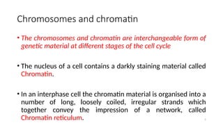
Chromosomes and chromatin
•The chromosomes and chromatin are interchangeable form of
genetic material at different stages of the cell cycle
• The nucleus of a cell contains a darkly staining material called
Chromatin.
• In an interphase cell the chromatin material is organised into a
number of long, loosely coiled, irregular strands which
together convey the impression of a network, called
Chromatin reticulum.
6.
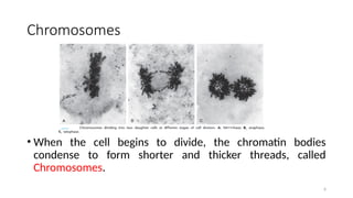
Chromosomes
• When thecell begins to divide, the chromatin bodies
condense to form shorter and thicker threads, called
Chromosomes.
6
7.
Chromosomes…
• Are thenucleoprotein structure which are generally more or
less rod-like during nuclear division.
• Chromosomes are the carriers of hereditary characters (genes),
which are passed from one generation to the next
• Genes are arranged on the chromosomes in a linear fashion.
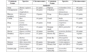
• Each species has a characteristic number of chromosomes (46
in humans) 7
8.
8
Chromosomes…
• Somatic cellsof organism contains two set of chromosomes,
forming homologous pairs and are called Diploid (2n)
• While gametes cell have only one set of chromosome and
are called haploid (n).
• This haploid set of chromosome is known as Genome.
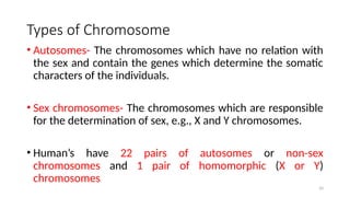
Types of Chromosome
•Autosomes- The chromosomes which have no relation with
the sex and contain the genes which determine the somatic
characters of the individuals.
• Sex chromosomes- The chromosomes which are responsible
for the determination of sex, e.g., X and Y chromosomes.
• Human’s have 22 pairs of autosomes or non-sex
chromosomes and 1 pair of homomorphic (X or Y)
chromosomes
10
11.
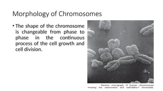
Morphology of Chromosomes
•The shape of the chromosome
is changeable from phase to
phase in the continuous
process of the cell growth and
cell division.
11
12.
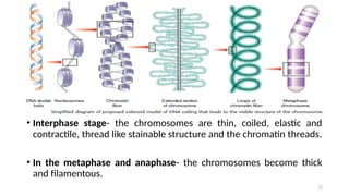
• Interphase stage-the chromosomes are thin, coiled, elastic and
contractile, thread like stainable structure and the chromatin threads.
• In the metaphase and anaphase- the chromosomes become thick
and filamentous.
12
13.
13
Morphology of Chromosomes…
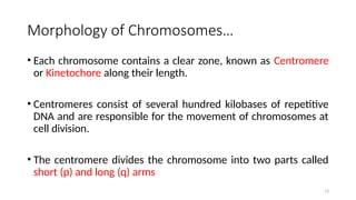
•Each chromosome contains a clear zone, known as Centromere
or Kinetochore along their length.
• Centromeres consist of several hundred kilobases of repetitive
DNA and are responsible for the movement of chromosomes at
cell division.
• The centromere divides the chromosome into two parts called
short (p) and long (q) arms
14.
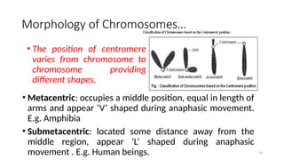
• Metacentric: occupiesa middle position, equal in length of
arms and appear ‘V’ shaped during anaphasic movement.
E.g. Amphibia
• Submetacentric: located some distance away from the
middle region, appear ‘L’ shaped during anaphasic
movement . E.g. Human beings. 14
• The position of centromere
varies from chromosome to
chromosome providing
different shapes.
Morphology of Chromosomes…
15.
15
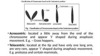
• Acrocentric: locateda little away from the end of the
chromosome and appear ‘J’ shaped during anaphasic
movement. E.g. – Grass hoppers.
• Telocentric: located at the tip and have only one long arm,
are very rare, appear ‘I’ shaped during anaphasic movement.
E.g. protozoa and certain mammals.
16.
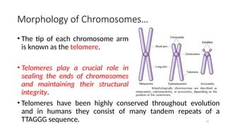
• The tipof each chromosome arm
is known as the telomere.
• Telomeres play a crucial role in
sealing the ends of chromosomes
and maintaining their structural
integrity.
16
Morphology of Chromosomes…
• Telomeres have been highly conserved throughout evolution
and in humans they consist of many tandem repeats of a
TTAGGG sequence.
17.
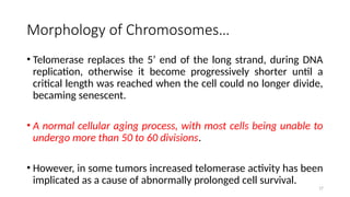
17
• Telomerase replacesthe 5’ end of the long strand, during DNA
replication, otherwise it become progressively shorter until a
critical length was reached when the cell could no longer divide,
becaming senescent.
• A normal cellular aging process, with most cells being unable to
undergo more than 50 to 60 divisions.
• However, in some tumors increased telomerase activity has been
implicated as a cause of abnormally prolonged cell survival.
Morphology of Chromosomes…
18.
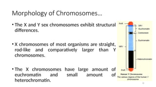
Morphology of Chromosomes…
•The X and Y sex chromosomes exhibit structural
differences.
• X chromosomes of most organisms are straight,
rod-like and comparatively larger than Y
chromosomes.
• The X chromosomes have large amount of
euchromatin and small amount of
heterochromatin. 18
19.
19
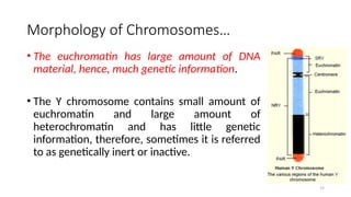
Morphology of Chromosomes…
•The euchromatin has large amount of DNA
material, hence, much genetic information.
• The Y chromosome contains small amount of
euchromatin and large amount of
heterochromatin and has little genetic
information, therefore, sometimes it is referred
to as genetically inert or inactive.
20.
20
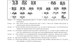
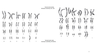
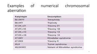
Karyotype
• A completeset of the entire metaphase chromosome in a
somatic cell is called karyotype.
• Chromosomes of a species are arranged according to their
shape, size and structure
• It helps to identify a particular chromosome.
• The study of complete chromosome complement is called
Karyotype analysis.
22
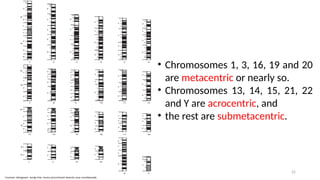
• Chromosomes 1,3, 16, 19 and 20
are metacentric or nearly so.
• Chromosomes 13, 14, 15, 21, 22
and Y are acrocentric, and
• the rest are submetacentric.
25
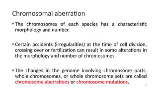
Chromosomal aberration
• Thechromosomes of each species has a characteristic
morphology and number.
• Certain accidents (irregularities) at the time of cell division,
crossing over or fertilization can result in some alterations in
the morphology and number of chromosomes.
• The changes in the genome involving chromosome parts,
whole chromosomes, or whole chromosome sets are called
chromosome aberrations or chromosome mutations.
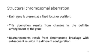
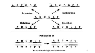
Structural chromosomal aberration
•Each gene is present at a fixed locus or position.
• This aberration results from changes in the definite
arrangement of the gene
• Rearrangements result from chromosome breakage with
subsequent reunion in a different configuration
27
28.
28
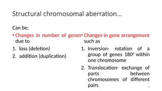
Structural chromosomal aberration…
Canbe;
• Changes in number of genes
due to
1. loss (deletion)
2. addition (duplication)
• Changes in gene arrangement
such as
1. Inversion- rotation of a
group of genes 1800
within
one chromosome
2. Translocation- exchange of
parts between
chromosomes of different
pairs
29.
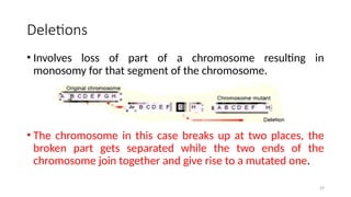
Deletions
• Involves lossof part of a chromosome resulting in
monosomy for that segment of the chromosome.
• The chromosome in this case breaks up at two places, the
broken part gets separated while the two ends of the
chromosome join together and give rise to a mutated one.
29
30.
30
Deletions…
• Very largedeletions are usually incompatible with survival to
term, and as a general rule any deletion resulting in loss of
more than 2% of the total haploid genome will have a lethal
outcome.
Examples
• Cri-du-chat syndrome-results from the loss of the short arm
of the 5th chromosome. The person is physically retarded
and produces a sound like the cry of a cat
• Others include Wolf-Hirschhorn, Prader-Willi and Angelman
syndromes
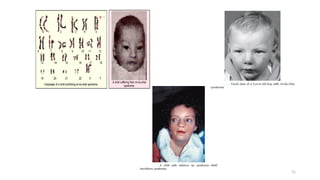
Deletions…
Angelman syndrome
• Theclinical features- developmental delay,
very poor speech, jerky movements,
paroxysms of inappropriate laughter, reduced
hair and skin pigmentation, facial
dysmorphisms and microcephaly
• The frequency is 1 in 20,000 and about 50% of
patients show a visible cytogenetic
microdeletion at 15q12.
• the deleted chromosome 15 is always
maternal in origin 32
33.
Deletions…
• Prader –Willi syndrome
• The frequency is 1 in 10,000 and in 50% a cytogenetic
microdeletion is apparent at 15q11 – 13.
• In contrast to Angelman syndrome, the deleted chromosome
in Prader–Willi syndrome is invariably paternal in origin.
Clinical features
• In the newborn, hypotonia and poor swallowing may be
marked, flat face with a tented upper lip, and the external
genitalia are hypoplastic.
33
34.
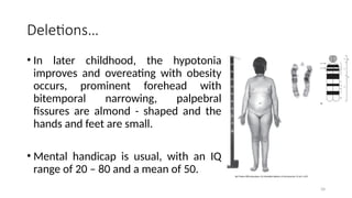
Deletions…
• In laterchildhood, the hypotonia
improves and overeating with obesity
occurs, prominent forehead with
bitemporal narrowing, palpebral
fissures are almond - shaped and the
hands and feet are small.
• Mental handicap is usual, with an IQ
range of 20 – 80 and a mean of 50.
34
35.
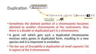
Duplication
• Sometimes thedeleted portion of a chromosome becomes
attached to another chromosome at the centromere, thus
there is a double or duplicated part in a chromosome.
• A germ cell which gets such a duplicated chromosome
receives extra genes in duplicated form, resulting in a new
species and it is important in evolution.
• The bar eye of Drosophila is duplication of small segment (16
A region) of the X chromosome. 35
36.
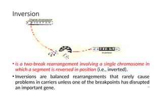
Inversion
• is atwo-break rearrangement involving a single chromosome in
which a segment is reversed in position (i.e., inverted).
• Inversions are balanced rearrangements that rarely cause
problems in carriers unless one of the breakpoints has disrupted
an important gene. 36
37.
37
Inversion…
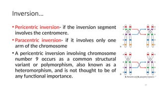
• Pericentric inversion-if the inversion segment
involves the centromere.
• Paracentric inversion- if it involves only one
arm of the chromosome
• A pericentric inversion involving chromosome
number 9 occurs as a common structural
variant or polymorphism, also known as a
heteromorphism, and is not thought to be of
any functional importance.
38.
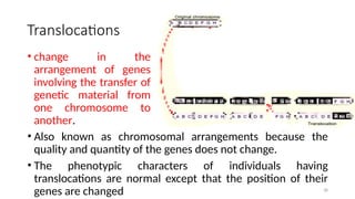
Translocations
• Also knownas chromosomal arrangements because the
quality and quantity of the genes does not change.
• The phenotypic characters of individuals having
translocations are normal except that the position of their
genes are changed 38
• change in the
arrangement of genes
involving the transfer of
genetic material from
one chromosome to
another.
39.
39
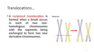
Translocations…
• A reciprocaltranslocation is
formed when a break occurs
in each of two non-
homologous chromosomes
with the segments being
exchanged to form two new
derivative chromosomes.
40.
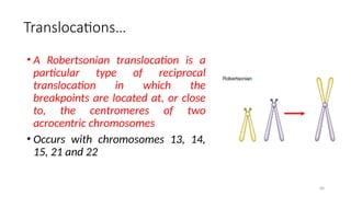
Translocations…
• A Robertsoniantranslocation is a
particular type of reciprocal
translocation in which the
breakpoints are located at, or close
to, the centromeres of two
acrocentric chromosomes
• Occurs with chromosomes 13, 14,
15, 21 and 22
40
41.
41
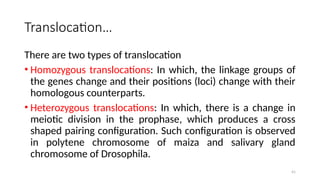
Translocation…
There are twotypes of translocation
• Homozygous translocations: In which, the linkage groups of
the genes change and their positions (loci) change with their
homologous counterparts.
• Heterozygous translocations: In which, there is a change in
meiotic division in the prophase, which produces a cross
shaped pairing configuration. Such configuration is observed
in polytene chromosome of maiza and salivary gland
chromosome of Drosophila.
42.
42
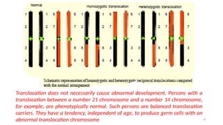
Translocation does notnecessarily cause abnormal development. Persons with a
translocation between a number 21 chromosome and a number 14 chromosome,
for example, are phenotypically normal. Such persons are balanced translocation
carriers. They have a tendency, independent of age, to produce germ cells with an
abnormal translocation chromosome
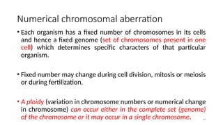
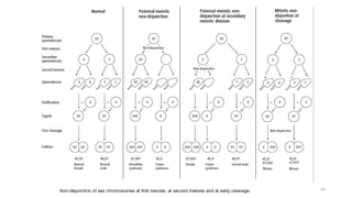
Numerical chromosomal aberration
•Each organism has a fixed number of chromosomes in its cells
and hence a fixed genome (set of chromosomes present in one
cell) which determines specific characters of that particular
organism.
• Fixed number may change during cell division, mitosis or meiosis
or during fertilization.
• A ploidy (variation in chromosome numbers or numerical change
in chromosome) can occur either in the complete set (genome)
of the chromosome or it may occur in a single chromosome. 44
45.
45
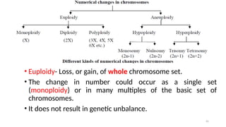
• Euploidy- Loss,or gain, of whole chromosome set.
• The change in number could occur as a single set
(monoploidy) or in many multiples of the basic set of
chromosomes.
• It does not result in genetic unbalance.
46.
46
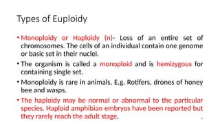
Types of Euploidy
•Monoploidy or Haploidy (n)- Loss of an entire set of
chromosomes. The cells of an individual contain one genome
or basic set in their nuclei.
• The organism is called a monoploid and is hemizygous for
containing single set.
• Monoploidy is rare in animals. E.g. Rotifers, drones of honey
bee and wasps.
• The haploidy may be normal or abnormal to the particular
species. Haploid amphibian embryos have been reported but
they rarely reach the adult stage.
47.
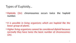
Types of Euploidy…
•Diploidy (2n): chromosomes occurs twice the haploid
number.
• It is possible in living organisms which are haploid like the
lower group of plants.
• Higher living organisms cannot be considered diploid because
normally they have twice the basic number of chromosomes
(2n).
47
48.
48
Types of Euploidy…
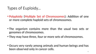
•Polyploidy (Multiple Set of Chromosomes): Addition of one
or more complete haploid sets of chromosomes.
• The organism contains more than the usual two sets or
genomes of chromosomes.
• They may have three, four or more sets of chromosomes.
• Occurs very rarely among animals and human beings and has
been observed only in cancer cells.
49.
49
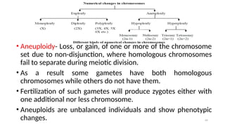
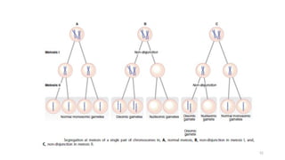
• Aneuploidy- Loss,or gain, of one or more of the chromosome
set due to non-disjunction, where homologous chromosomes
fail to separate during meiotic division.
• As a result some gametes have both homologous
chromosomes while others do not have them.
• Fertilization of such gametes will produce zygotes either with
one additional nor less chromosome.
• Aneuploids are unbalanced individuals and show phenotypic
changes.
50.
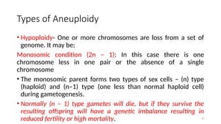
Types of Aneuploidy
•Hypoploidy- One or more chromosomes are loss from a set of
genome. It may be;
Monosomic condition (2n – 1): In this case there is one
chromosome less in one pair or the absence of a single
chromosome
• The monosomic parent forms two types of sex cells – (n) type
(haploid) and (n–1) type (one less than normal haploid cell)
during gametogenesis.
• Normally (n – 1) type gametes will die, but if they survive the
resulting offspring will have a genetic imbalance resulting in
reduced fertility or high mortality. 50
51.
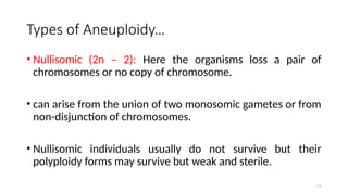
Types of Aneuploidy…
•Nullisomic (2n – 2): Here the organisms loss a pair of
chromosomes or no copy of chromosome.
• can arise from the union of two monosomic gametes or from
non-disjunction of chromosomes.
• Nullisomic individuals usually do not survive but their
polyploidy forms may survive but weak and sterile.
51
Types of Aneuploidy…
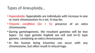
•Hyperploidy: hyperploids are individuals with increase in one
or more chromosomes to a set. It may be;
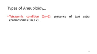
• Trisomic condition (2n + 1): presence of an extra
chromosome
• During gametogenesis, the resultant gametes will be two
types– (n) type gamete haploid sex cell and (n+1) type
gamete, containing an extra chromosome.
• In the human being trisomies can occur with any
chromosome, but often result in miscarriage.
53
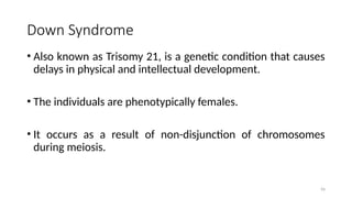
Down Syndrome
• Alsoknown as Trisomy 21, is a genetic condition that causes
delays in physical and intellectual development.
• The individuals are phenotypically females.
• It occurs as a result of non-disjunction of chromosomes
during meiosis.
56
57.
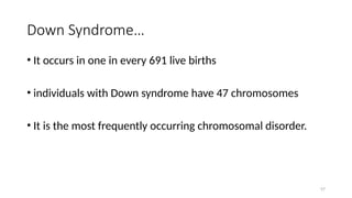
Down Syndrome…
• Itoccurs in one in every 691 live births
• individuals with Down syndrome have 47 chromosomes
• It is the most frequently occurring chromosomal disorder.
57
58.
Down Syndrome…
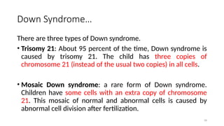
There arethree types of Down syndrome.
• Trisomy 21: About 95 percent of the time, Down syndrome is
caused by trisomy 21. The child has three copies of
chromosome 21 (instead of the usual two copies) in all cells.
• Mosaic Down syndrome: a rare form of Down syndrome.
Children have some cells with an extra copy of chromosome
21. This mosaic of normal and abnormal cells is caused by
abnormal cell division after fertilization.
58
59.
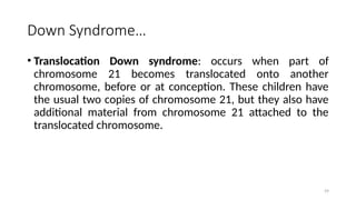
Down Syndrome…
• TranslocationDown syndrome: occurs when part of
chromosome 21 becomes translocated onto another
chromosome, before or at conception. These children have
the usual two copies of chromosome 21, but they also have
additional material from chromosome 21 attached to the
translocated chromosome.
59
60.
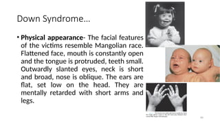
Down Syndrome…
• Physicalappearance- The facial features
of the victims resemble Mangolian race.
Flattened face, mouth is constantly open
and the tongue is protruded, teeth small.
Outwardly slanted eyes, neck is short
and broad, nose is oblique. The ears are
flat, set low on the head. They are
mentally retarded with short arms and
legs.
60
61.
Turner Syndrome
• Itoccurs in one in about 5000 people with the individuals being
phenotypically females with 45 (44 + XO) number of chromosomes.
• There is lack of contribution of an X or a Y chromosome resulting in
a 45, X karyotype that may arise from non - disjunction in either
parent.
• In 80% of patients, only the maternal X chromosome is present, thus
the error occurred in spermatogenesis or post-fertilisation.
• Individuals have the external appearance of a female but lack
ovaries or a degenerate ovary, are sterile.
61
62.
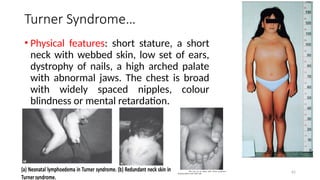
Turner Syndrome…
• Physicalfeatures: short stature, a short
neck with webbed skin, low set of ears,
dystrophy of nails, a high arched palate
with abnormal jaws. The chest is broad
with widely spaced nipples, colour
blindness or mental retardation.
62
63.
Klinefelter Syndrome
• Occursin phenotypically male individuals, 1 in 1000 males.
• The individuals have 47 chromosomes i.e. trisomy (XXY) to
chromosome number.
• The extra X chromosome is of maternal origin in 56% and
paternal in 44% of patients
• It is caused by non-disjunction of XX chromosomes at the
first (or occasionally the second) maternal meiotic division
and rarely as a mitotic error after fertilisation. 63
64.
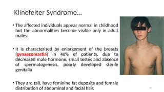
Klinefelter Syndrome…
• Theaffected individuals appear normal in childhood
but the abnormalities become visible only in adult
males.
• It is characterized by enlargement of the breasts
(gynaecomastia) in 40% of patients, due to
decreased male hormone, small testes and absence
of spermatogenesis, poorly developed sterile
genitalia
• They are tall, have feminine fat deposits and female
distribution of abdominal and facial hair. 64
65.
Klinefelter Syndrome…
• Canbe detected by mentally retarded with infertility and
develop a variety of psychiatric problems.
• About 15% of patients are mosaic 46,XY/47,XXY.
• Patients with 48,XXXY and 49,XXXXY have severe learning
difficulties and proximal radioulnar synostosis is a common
skeletal defect.
65
XXX Females
• approximately0.1% of all females have a 47,XXX karyotype.
• The birth frequency is 1 in 1000 females with a maternal age
effect
• In 95% of cases the additional X chromosome is of maternal
origin, usually arising from an error in meiosis I.
67
68.
68
XXX Females…
• usuallypresented with have no physical abnormalities, but can
show a mild reduction of between 10 and 20 points in
intellectual skills and sometimes quite oppositional behaviour.
• Adults are usually fertile and have children with normal
karyotypes.
• Women with more than three X chromosomes show a high
incidence of learning difficulties, the severity being directly
related to the number of X chromosomes.
69.
XYY Males
• Occursin about 1 : 1000 in males in newborn surveys but is
found in 2% to 3% of males who are in institutions because
of learning difficulties or antisocial criminal behaviour.
• However, it is important to stress that most 47,XYY men have
neither learning difficulty nor a criminal record, although
they can show emotional immaturity and impulsive
behaviour.
69
70.
70
XYY Males…
• Fertilityis normal.
• Physical appearance is normal and stature is usually above
average.
• Intelligence is mildly impaired, with an overall IQ score of 10
to 20 points below a control sample.
• The additional Y chromosome must arise either as a result of
non-disjunction in paternal meiosis II or as a post-zygotic
event.


![ONFH[AVN HIP] -TRIPLE REGIME -A NOVAL SURGICAL CONCEPT .pptx](https://cdn.slidesharecdn.com/ss_thumbnails/onfhavnhip2026koaconcalicutdrgokuldevdrmashraf-260210064517-213ec005-thumbnail.jpg?width=640&height=640&fit=bounds)

