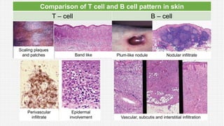
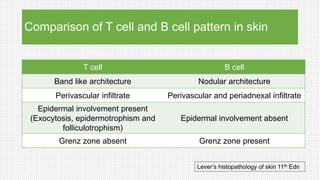
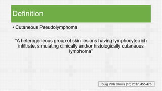
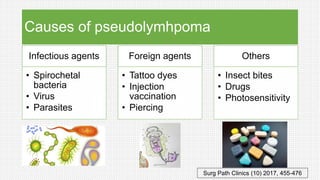
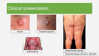
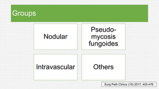
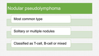
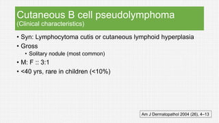
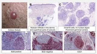
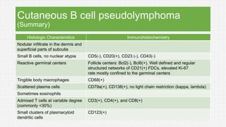
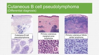
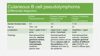
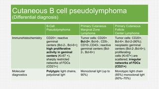
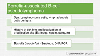
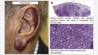
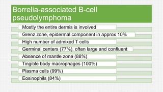
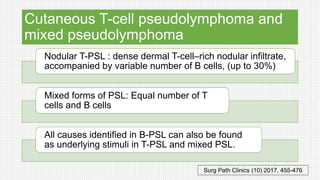
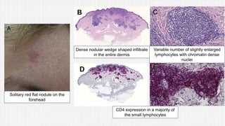
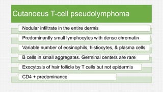
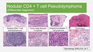
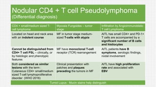
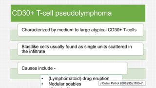
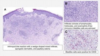
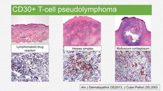
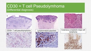
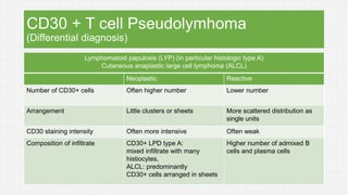
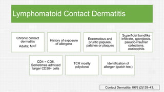
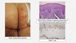
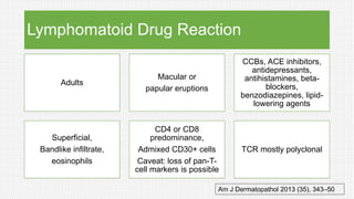
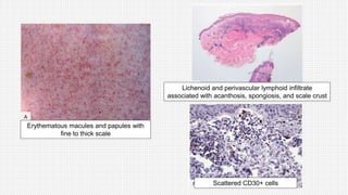
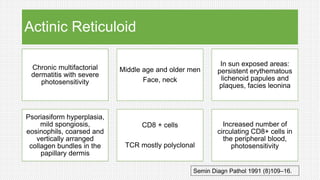
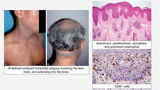
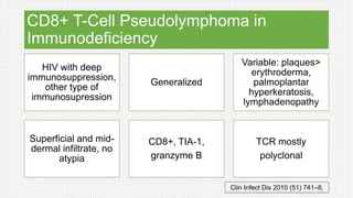
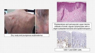
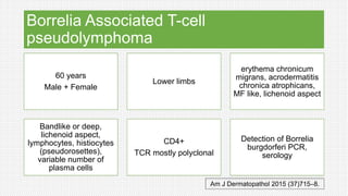
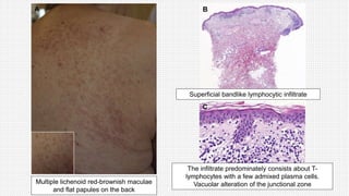
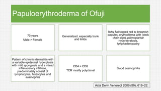
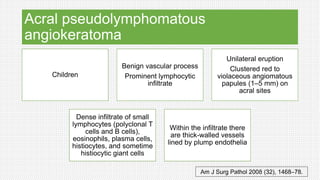
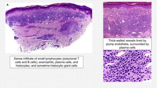
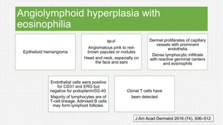
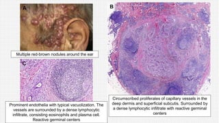
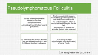
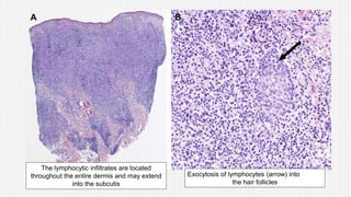
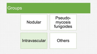
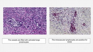
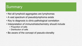
SlideShare a Scribd company logo
Cutaneous PSEUDO-lymphoma
Moderator – Prof Uma Nahar
Outline of presentation
• Pattern of B and T cell infiltration
• Define Cutaneous pseudolymphoma
• Classification
• Subtypes
• Important entities with clinico-pathological features
• Differential features from morphologically similar lesions
• Pseudoclonality
Comparison of T cell and B cell pattern in skin
T – cell B – cell
Scaling plaques
and patches Band like Plum-like nodule Nodular infiltrate
Perivascular
infiltrate
Epidermal
involvement Vascular, subcutis and interstitial infiltration
Comparison of T cell and B cell pattern in skin
T cell B cell
Band like architecture Nodular architecture
Perivascular infiltrate Perivascular and periadnexal infiltrate
Epidermal involvement present
(Exocytosis, epidermotrophism and
folliculotrophism)
Epidermal involvement absent
Grenz zone absent Grenz zone present
Lever’s histopathology of skin 11th Edn
Definition
• Cutaneous Pseudolymphoma
“A heterogeneous group of skin lesions having lymphocyte-rich
infiltrate, simulating clinically and/or histologically cutaneous
lymphoma”
Surg Path Clinics (10) 2017, 455-476
Most important ingredient for diagnosis
Clinicopathological co-relation
Causes of pseudolymhpoma
Infectious agents
• Spirochetal
bacteria
• Virus
• Parasites
Foreign agents
• Tattoo dyes
• Injection
vaccination
• Piercing
Others
• Insect bites
• Drugs
• Photosensitivity
Surg Path Clinics (10) 2017, 455-476
Clinical presentation
Nodule Clustered papules
Disseminated papules
Erythroderma
Surg Path Clinics (10) 2017, 455-476
Groups
Nodular
Pseudo-
mycosis
fungoides
Intravascular Others
Surg Path Clinics (10) 2017, 455-476
Groups
Nodular
Pseudo-
mycosis
fungoides
Intravascular Others
Nodular pseudolymphoma
Most common type
Solitary or multiple nodules
Classified as T-cell, B-cell or mixed
Cutaneous B cell pseudolymphoma
(Clinical characteristics)
• Syn: Lymphocytoma cutis or cutaneous lymphoid hyperplasia
• Gross
• Solitary nodule (most common)
• M: F :: 3:1
• <40 yrs, rare in children (<10%)
Am J Dermatopathol 2004 (26), 4–13
Solitary Nodule
Nodular infiltrate in the dermis
and superficial parts of subcutis
Small B cells, no nuclear atypia
Prominent germinal centers
Bcl6 positive Bcl2 negative CD21
Cutaneous B cell pseudolymphoma
(Summary)
Histologic Characteristics Immunohistochemistry
Nodular infiltrate in the dermis and
superficial parts of subcutis
Small B cells, no nuclear atypia CD5(-), CD20(+), CD23 (-), CD43(-)
Reactive germinal centers Follicle centers: Bcl2(-), Bcl6(+). Well defined and regular
structured networks of CD21(+) FDCs, elevated Ki-67
rate mostly confined to the germinal centers
Tingible body macrophages CD68(+)
Scattered plasma cells CD79a(+), CD138(+), no light chain restriction (kappa, lambda)
Sometimes eosinophils
Admixed T cells at variable degree
(commonly <30%)
CD3(+), CD4(+), and CD8(+)
Small clusters of plasmacytoid
dendritic cells
CD123(+)
Cutaneous B cell pseudolymphoma
(Differential diagnosis)
Cutaneous B cell
pseudolymphoma
Primary cutaneous
marginal zone
lymphoma
Primary cutaneous follicle
center lymphoma
Cutaneous B cell pseudolymphoma
(Differential diagnosis)
B-Cell
Pseudolymphoma
Primary Cutaneous
Marginal Zone
Lymphoma
Primary Cutaneous
Follicle
Center Lymphoma
Gender (female:male) 3:1 1:2 1:1.5
Age (y) 75% < 40 55 51
Localization Face > upper trunk >
arms
Upper trunk > upper
arms > face/head
Head/face > upper
trunk
Histology Dermal/superficial
subcutis, reactive
germinal centers,
tingible body
macrophages,
scattered
plasma cells
Dermal/superficial
subcutis, reactive
germinal centers can be
found, plasma cells in
the periphery of the
infiltrates and near by
the epidermis
Dermal/superficial
subcutis, neoplastic
irregular germinal
centers of different
size (CAVEAT: not
found in diffuse type)
Cutaneous B cell pseudolymphoma
(Differential diagnosis)
B-Cell
Pseudolymphoma
Primary Cutaneous
Marginal Zone
Lymphoma
Primary Cutaneous
Follicle
Center Lymphoma
Immunohistochemistry CD20+; reactive
germinal
centers (Bcl-2-, Bcl-6+);
high proliferative
activity in geminal
centers (Ki-67 +),
sharply restricted
networks of FDCs
(CD21+)
Tumor cells: CD20+
Bcl-2+; Bcl-6-, CD5-,
CD10-,CD43-; reactive
germinal centers (Bcl-
2-, Bcl-6+)
Tumor cells: CD20+,
Bcl-6+; Bcl-2-(90%);
neoplastic germinal
centers (Bcl-2-,Bcl-6+);
proliferating
cells (Ki-67+) are
scattered; irregular
networks of FDCs
(CD21+)
Molecular
diagnostics
Polytypic light chains,
polyclonal IgH
Monoclonal IgH (up to
90%)
Monotypic light chain
(85%) monoclonal IgH
(60%–70%)
Borrelia-associated B-cell
pseudolymphoma
Syn: Lymphocytoma cutis; lymphadenosis
cutis benigna
History of tick bite and localization at
predilection site (Earlobes, nipple, scrotum)
Borrelia burgdorferi - Serology; DNA PCR
J Cutan Pathol 2004 (31), 232–40
Blue Nodule
Dense dermal nodular infiltrate with reactive
germinal centers with small or completely lost
mantle zones
Germinal centers with multiple tingible body
macrophages
Borrelia-associated B-cell
pseudolymphoma
Mostly the entire dermis is involved
Grenz zone, epidermal component in approx 10%
High number of admixed T cells
Germinal centers (77%), often large and confluent
Absence of mantle zone (88%)
Tingible body macrophages (100%)
Plasma cells (99%)
Eosinophils (84%)
Cutaneous T-cell pseudolymphoma and
mixed pseudolymphoma
Nodular T-PSL : dense dermal T-cell–rich nodular infiltrate,
accompanied by variable number of B cells, (up to 30%)
Mixed forms of PSL: Equal number of T
cells and B cells
All causes identified in B-PSL can also be found
as underlying stimuli in T-PSL and mixed PSL.
Surg Path Clinics (10) 2017, 455-476
Solitary red flat nodule on the
forehead
CD4 expression in a majority of
the small lymphocytes
Variable number of slightly enlarged
lymphocytes with chromatin dense
nuclei
Dense nodular wedge shaped infiltrate
in the entire dermis
Cutanoeus T-cell pseudolymphoma
Nodular infiltrate in the entire dermis
Predominantly small lymphocytes with dense chromatin
Variable number of eosinophils, histiocytes, & plasma cells
B cells in small aggregates. Germinal centers are rare
Exocytosis of hair follicle by T cells but not epidermis
CD4 + predominance
Nodular CD4 + T cell Pseudolymphoma
(Differential diagnosis)
Nodular CD4+ T cell
pseudolymphoma
Tumor stage Mycosis
Fungoides
Angioimmunoblastic T-
cell lymphoma
Tumid lupus
Dermatology 2009 (219), 42–7
Nodular CD4 + T cell Pseudolymphoma
(Differential diagnosis)
CD4 + small/medium sized T-
cell lymphoma
Mycosis Fungoides – tumor
stage
Infiltration by Angioimmunoblstic
T cell Lymphoma
Located on head and neck area
with an indolent course
MF in tumor stage medium-
sized T-cells with atypia
AITL has small CD4+ and PD-1+
T cells are accompanied by a
significant number of B cells
and histiocytes
Cannot be distinguished from
CD4+ T cell PSL – clinically, or
by histologic and phenotypic
features
MF have monoclonal T-cell
receptor (TCR) rearrangement
AITL patients have B
symptoms, serologic findings,
nodal involvement
Both considered as similar
lesions with the term :
Cutaneous CD4+ small/medium
sized T-cell lymphoproliferative
disorder (WHO 2016)
Clinical presentation with
patches and plaques
preceding the tumors in MF
AITL have high proliferation
rate and associated with
EBV
Tumid Lupus : Mucin stains help distinguish
CD30+ T-cell pseudolymphoma
Characterized by medium to large atypical CD30+ T-cells
Blastlike cells usually found as single units scattered in
the infiltrate
Causes include -
• (Lymphomatoid) drug eruption
• Nodular scabies J Cutan Pathol 2008 (35),1100–7.
Arthropod bite reaction with a wedge shaped mixed infiltrate,
spongiotic dermatitis, and papillary edema
Infiltrate consists of lymphocytes,
histiocytes, and eosinophils.A few
(immuno-) blastlike cells are admixed
Blastlike cells were positive for CD30
CD30+ T-cell pseudolymphoma
Am J Dermatopathol (35)2013, J Cutan Pathol (35) 2003
Lymphomatoid drug
reaction
Herpes simplex Molluscum contagiosum
CD30 + T cell Pseudolymhoma
(Differential diagnosis)
CD30+ T cell pseudolymphoma Lymphomatoid papulosis Cutaneous anaplastic large cell
lymphoma
CD30 + T cell Pseudolymhoma
(Differential diagnosis)
Lymphomatoid papulosis (LYP) (in particular histologic type A)
Cutaneous anaplastic large cell lymphoma (ALCL)
Neoplastic Reactive
Number of CD30+ cells Often higher number Lower number
Arrangement Little clusters or sheets More scattered distribution as
single units
CD30 staining intensity Often more intensive Often weak
Composition of infiltrate CD30+ LPD type A:
mixed infiltrate with many
histiocytes,
ALCL: predominantly
CD30+ cells arranged in sheets
Higher number of admixed B
cells and plasma cells
Groups
Nodular
Pseudo-
mycosis
fungoides
Intravascular Others
Pseudo-mycosis fungoides
Group of disorders of different etiology, which histologically
mimic MF
Clinicopathologic correlation is crucial to avoid
misinterpretation
May exhibit subtle nuclear atypia
CD4+ or CD8 + with variable expression of CD30
Am J Dermatopathol 2013 (35), 343–50
Bandlike infiltrate of mostly small lymphocytes with exocytosis into
the epidermis. Many admixed eosinophils in the upper dermis
Pseudo-mycosis fungoides
(Differential diagnosis)
Differential diagnosis
• MF and Sezary syndrome
• Cutaneous CD8+
aggressive epidermotropic
cytotoxic T-cell lymphoma
• Primary cutaneous LYP
Features favouring
neoplasia
• Profound nuclear atypia,
• Predominance of medium-
sized to large cells
• Loss of pan T-cell markers
• Monoclonal rearrangement
of TCR genes
Pseudo-mycosis fungoides
Lymphomatoid Contact Dermatitis
Lymphomatoid Drug Reaction
Actinic Reticuloid
CD8+ T-cell Pseudolymphoma in Immunodeficiency
Borrelia-Associated Tcell Pseudolymphoma
Papuloerythroderma of Ofuji
Lymphomatoid Contact Dermatitis
Chronic contact
dermatitis
Adults; M=F
History of exposure
of allergens
Eczematous and
pruritic papules,
patches or plaques
Superficial bandlike
infiltrate, spongiosis,
pseudo-Pautrier
collections,
eosinophils
CD4 = CD8,
Sometimes admixed
larger CD30+ cells
TCR mostly
polyclonal
Identification of
allergen (patch test)
Contact Dermatitis 1976 (2)139–43.
Scaly, erythematous patches
CD8+ T cell
Inflammatory infiltrate of atypical lymphocytes in
the superficial dermis with epidermotropism
Lymphomatoid Drug Reaction
Adults
Macular or
papular eruptions
CCBs, ACE inhibitors,
antidepressants,
antihistamines, beta-
blockers,
benzodiazepines, lipid-
lowering agents
Superficial,
Bandlike infiltrate,
eosinophils
CD4 or CD8
predominance,
Admixed CD30+ cells
Caveat: loss of pan-T-
cell markers is possible
TCR mostly polyclonal
Am J Dermatopathol 2013 (35), 343–50
Erythematous macules and papules with
fine to thick scale
Lichenoid and perivascular lymphoid infiltrate
associated with acanthosis, spongiosis, and scale crust
Scattered CD30+ cells
Actinic Reticuloid
Chronic multifactorial
dermatitis with severe
photosensitivity
Middle age and older men
Face, neck
In sun exposed areas:
persistent erythematous
lichenoid papules and
plaques, facies leonina
Psoriasiform hyperplasia,
mild spongiosis,
eosinophils, coarsed and
vertically arranged
collagen bundles in the
papillary dermis
CD8 + cells
TCR mostly polyclonal
Increased number of
circulating CD8+ cells in
the peripheral blood,
photosensitivity
Semin Diagn Pathol 1991 (8)109–16.
Ill-defined confluent lichenified plaques involving the face,
neck, and extending into the torso.
Acanthosis, parakeratosis, spongiosis,
and prominent eosinophils
CD8+ cells
CD8+ T-Cell Pseudolymphoma in
Immunodeficiency
HIV with deep
immunosuppression,
other type of
immunosupression
Generalized
Variable: plaques>
erythroderma,
palmoplantar
hyperkeratosis,
lymphadenopathy
Superficial and mid-
dermal infiltrate, no
atypia
CD8+, TIA-1,
granzyme B
TCR mostly
polyclonal
Clin Infect Dis 2010 (51) 741–8.
Dry, scaly and pruriginous erythroderma
Parakeratosis and perivascular upper dermis
infiltrate of small, regular lymphocytes with a
slight subepidermal band and epidermotropism
CD3 + and CD8+ cells
Borrelia Associated T-cell
pseudolymphoma
60 years
Male + Female
Lower limbs
erythema chronicum
migrans, acrodermatitis
chronica atrophicans,
MF like, lichenoid aspect
Bandlike or deep,
lichenoid aspect,
lymphocytes, histiocytes
(pseudorosettes),
variable number of
plasma cells
CD4+
TCR mostly polyclonal
Detection of Borrelia
burgdorferi PCR,
serology
Am J Dermatopathol 2015 (37)715–8.
Multiple lichenoid red-brownish maculae
and flat papules on the back
Superficial bandlike lymphocytic infiltrate
The infiltrate predominately consists about T-
lymphocytes with a few admixed plasma cells.
Vacuolar alteration of the junctional zone
Papuloerythroderma of Ofuji
70 years
Male > Female
Generalized, especially trunk
and limbs
Itchy flat topped red to brownish
papules, erythoderma with (deck
chair sign), palmoplantar
hyperkeratosis,
lymphadenopathy
Pattern of chronic dermatitis with
a variable epidermal hyperplasia
with mild spongiosis and a mixed
inflammatory infiltrate,
predominately consist of
lymphocytes, histiocytes and
eosinophils
CD4 = CD8
TCR mostly polyclonal
Blood eosinophilia
Acta Derm Venereol 2009 (89), 618–22.
Erythroderma
Exocytosis. Perivascular and
periadnexal infiltrate.
Lymphocytes, esoinophils and plasma cells
Groups
Nodular
Pseudo-
mycosis
fungoides
Intravascular Others
Lymphocytic Infiltration of the Skin and
Palpable Arciforme Migratory Erythema
Assigned to the group of lupus
erythematosus
PAME: Infiltrated annular
erythema developing into large
migrating lesions (trunk)
LIS: sharply demarked often
symmetric infiltrated plaques
(face)
Dense perivascular and
periadnexal predominantly
lymphocytic infiltrate
Pattern of chronic dermatitis
with a variable epidermal
hyperplasia with mild
spongiosis
Mixed inflammatory infiltrate,
predominately consist of
lymphocytes, histiocytes and
eosinophils
TCR mostly polyclonal
J Am Acad Dermatol 2008 (58), 217–23.
Multiple erythematous plaques
Heavy chronic inflammatory cell infiltrate
cuffs the vessels in the superficial and mid
dermis
CD4+ lymphocytes
Arciform erythemas on the back
Dense lymphocytic infiltration about
vessels and appendages
Predominantly of lymphocytes
Infections as Simulators of Lymphomas
Cutaneous
leishmaniasis
Lymphocytes,
histiocytes, and plasma
cells
Detection of the agents
Herpes simplex virus
and varicella zoster
virus
Without the
pathognomonic epithelial
changes (herpes
incognito)
Lymphocytes enlarged
and atypical-appearing
chromatin dense nuclei.
CD30+
Detection of viral
antigens by IHC and/or
detection of viral DNA
Parapoxvirus
infections
Cytomorphological
changes and
expression of CD30 by
the infiltrating T cells
Inclusion bodies,
absence of loss of T-cell
markers, lack of
monoclonal
rearrangement of TCR
Surg Path Clinics (10) 2017, 455-476
Inflammatory Disorders as Simulators of
T-Cell Lymphoma
Lichen planus (6%) Lichen sclerosis et
atrophicus (13%)
Pigmented purpuric
dermatitis Pityriasis lichenoides (60%)
Interface
dermatitis
Clonal T-cell
populations have
been found
Surg Path Clinics (10) 2017, 455-476
Other ‘named’ pseudolymphomas
Acral pseudolymphomatous angiokeratoma
Angiolymphoid hyperplasia with eosinophilia
Cutaneous plasmacytosis
Lymphoplasmacytic plaque
Pseudolymphomatous Folliculitis
Acral pseudolymphomatous
angiokeratoma
Children
Benign vascular process
Prominent lymphocytic
infiltrate
Unilateral eruption
Clustered red to
violaceous angiomatous
papules (1–5 mm) on
acral sites
Dense infiltrate of small
lymphocytes (polyclonal T
cells and B cells),
eosinophils, plasma cells,
histiocytes, and sometime
histiocytic giant cells
Within the infiltrate there
are thick-walled vessels
lined by plump endothelia
Am J Surg Pathol 2008 (32), 1468–78.
Dense infiltrate of small lymphocytes (polyclonal T
cells and B cells), eosinophils, plasma cells, and
histiocytes, and sometime histiocytic giant cells
Thick-walled vessels lined by
plump endothelia, surrounded by
plasma cells
Angiolymphoid hyperplasia with
eosinophilia
Epithelioid hemangioma
M=F
Angiomatous pink to red-
brown papules or nodules
Head and neck, especially on
the face and ears
Dermal proliferates of capillary
vessels with prominent
endothelia
Dense lymphocytic infiltrate
with reactive germinal centers
and eosinophils
Endothelial cells were positive
for CD31 and ERG but
negative for podoplanin/D2-40.
Majority of lymphocytes are of
T-cell lineage. Admixed B cells
may form lymphoid follicles.
Clonal T cells have
been detected
J Am Acad Dermatol 2016 (74), 506–512
Prominent endothelia with typical vacuolization. The
vessels are surrounded by a dense lymphocytic
infiltrate, consisting eosinophils and plasma cell.
Reactive germinal centers
Multiple red-brown nodules around the ear
Circumscribed proliferates of capillary vessels in the
deep dermis and superficial subcutis. Surrounded by
a dense lymphocytic infiltrate with reactive germinal
centers
Cutaneous plasmacytosis
Rare disease,
Reported in Asian countries
Affects adults.
Multiple brownish small plaques
and nodules occurring all over
the body
Dermal infiltrates composed
predominantly of mature
polyclonal plasma cells.
Signs of a systemic involvement
(lymphadenopathy,
hepatospenomegaly,
hypergammaglobulinemia,
increased levels of interleukin 6
in the serum, and elevated
erythrocyte sedimentation rate)
J Am Acad Dermatol 2013 (68)978–85.
Reddish brown papules, patches and
plaques in the axilla
Epidermis shows acanthosis. There is a dense superficial and
mid perivascular infiltrate of plasma cells and lymphocytes
No light chain restriction is seen when stained with kappa and
lambda, respectively
Lymphoplasmacytic plaque
Rare disease
Children and adults
F> M
Pretibial, trunk and arms
Longstanding plaque or
circumscribed, often linear arranged
reddish and brownish papules and
plaques
Superficial, bandlike, or deep nodular
and interstitial infiltrate,
An epidermal hyperplasia is common.
Lymphocytes and histiocytes with
numerous polyclonal plasma cells
accounting for up to 25%
The interstitial histiocytes may form
granulomas around sclerotic collagen
bundles (so-called pseudorosettes)
Histiocytic giant cells and an
increased number of vessels
Arch Dermatol 2010 (146), 95–6.
Longstanding sharply demarcated reddish
plaque with scaling
The infiltrate consists of lymphocytes,
histiocytes, and plasma cells
Epidermal hyperplasia with a bandlike and
superficial perivascular infiltrate
Interstitial histiocytic granulomas around
sclerotic collagen bundles
Pseudolymphomatous Folliculitis
Solitary nodule preferentially
located on the face
T-cell predominance.
Admixed epithelioid histiocytes and
granulomas
The lymphocytic infiltrates are
located throughout the dermis and
may extend into the subcutis
The epidermis spared. There is
exocytosis of lymphocytes into the
hair
A hyperplasia of eccrine and
apocrine ducts is often observed
An admixture of numerous dendritic
cells with expression of CD1a and
S-100 was identified in all cases.
Unusual high number
(approximately 50% of the cases)
of clonal T cells
Am J Surg Pathol 1999 (23),1313–9.
The lymphocytic infiltrates are located
throughout the entire dermis and may extend
into the subcutis
Exocytosis of lymphocytes (arrow) into
the hair follicles
Groups
Nodular
Pseudo-
mycosis
fungoides
Intravascular Others
Intravascular pseudolymphoma
Intravascular proliferation of blasts with or without expression of CD30
Inflammatory skin diseases or trauma to the skin
Obstruction of lymphatic vessels due to inflammation with disrupted immune cell trafficking may
result in the accumulation of activated CD30+ lymphocytes
Express T-cell markers (CD3 and CD4) and in some cases CD30.
No association with Epstein-Barr virus infection. Clonality studies reveal the
polyclonal nature of the process
Am J Dermatopathol 2013 (35), 143–50.
The vessels are filled with activated large
lymphocytes
The intravascular lymphocytes are positive for
CD30
Pseudo-clonality
“Refers to detection of a clone that on double or triple testing
appears at a different position”
Br J Dermatol (159)2008
Pseudolymphomatous drug eruption - pseudoclonal IgH rearrangement
Pseudolymphomatous folliculotrophic lesion - pseudoclonal TCR rearrangement
Pseudo-clonality
Artefact of PCR as it is very sensitive
Picks up small groups of cells
Carry out analyses in duplicate and/or analyze
multiple or alternative targets
Integrate the results with histopathological
information
Ther Adv Hematol (5) 2014
Case of SLE presenting with Mycosis Fungoides
Summary
• Not all lymphoid aggregates are lymphomas
• A vast spectrum of pseudolymphoma exists
• Key to diagnosis is clinic-pathological correlation
• Interpretation of immunohistochemistry should include
• Proportion of cells
• Distribution of cells
• Be aware of the concept of pseudo-clonality
THANKYOU

More Related Content

What's hot

Mastocytosis (Cutaneous and Systemic)
Mastocytosis (Cutaneous and Systemic)Mastocytosis (Cutaneous and Systemic)
Mastocytosis (Cutaneous and Systemic)
Jerriton Brewin
 
Cutaneous Sarcoidosis, Sarcoidosis, Approach to cutaneous sarcoidosis, Manage...
Cutaneous Sarcoidosis, Sarcoidosis, Approach to cutaneous sarcoidosis, Manage...Cutaneous Sarcoidosis, Sarcoidosis, Approach to cutaneous sarcoidosis, Manage...
Cutaneous Sarcoidosis, Sarcoidosis, Approach to cutaneous sarcoidosis, Manage...
sanjay singh
 
Chronic actinic dermatitis
Chronic actinic dermatitisChronic actinic dermatitis
Chronic actinic dermatitisChandan N
 
Cutaneous lymphomas
Cutaneous lymphomasCutaneous lymphomas
Cutaneous lymphomas
Archer Review USMLE and NCLEX
 
Mastocytosis
MastocytosisMastocytosis
Morphea
MorpheaMorphea
Morphea
Mostafa Sanad
 
Bacteriology & Immunology of Leprosy
Bacteriology & Immunology of LeprosyBacteriology & Immunology of Leprosy
Bacteriology & Immunology of Leprosy
Jerriton Brewin
 
Cicatricisial alopecia
Cicatricisial alopeciaCicatricisial alopecia
Cicatricisial alopecia
Dr Daulatram Dhaked
 
Introduction To Dermatopathology
Introduction To DermatopathologyIntroduction To Dermatopathology
Introduction To Dermatopathology
Ibrahim Farag
 
Adnexal Neoplasms
Adnexal NeoplasmsAdnexal Neoplasms
Adnexal Neoplasms
Ibrahim Farag
 
Immunofluorescence in dermatopathology
Immunofluorescence in dermatopathologyImmunofluorescence in dermatopathology
Immunofluorescence in dermatopathology
Sumit Mandal
 
Introduction to dermatopathology
Introduction to dermatopathologyIntroduction to dermatopathology
Introduction to dermatopathology
dermlogic
 
Dermatology 5th year, 3rd lecture (Dr. Kazhan)
Dermatology 5th year, 3rd lecture (Dr. Kazhan)Dermatology 5th year, 3rd lecture (Dr. Kazhan)
Dermatology 5th year, 3rd lecture (Dr. Kazhan)
College of Medicine, Sulaymaniyah
 
Cutaneous mucinoses
Cutaneous mucinosesCutaneous mucinoses
Cutaneous mucinosesheera sanju
 
Cutaneous amyloidosis
Cutaneous amyloidosisCutaneous amyloidosis
Cutaneous amyloidosis
Dr. Saba Niyazee
 
Lichenoid dermatoses.potx
Lichenoid dermatoses.potxLichenoid dermatoses.potx
Lichenoid dermatoses.potx
Ibrahim Farag
 
Cutaneous T Cell Lymphomas
Cutaneous T Cell LymphomasCutaneous T Cell Lymphomas
Cutaneous T Cell Lymphomas
Jerriton Brewin
 
Dyschromatosis and Reticulate pigmentary disorders
Dyschromatosis and Reticulate pigmentary disordersDyschromatosis and Reticulate pigmentary disorders
Dyschromatosis and Reticulate pigmentary disorders
sanjay singh
 
Panniculitis. Nathan C. Walk
Panniculitis. Nathan C. WalkPanniculitis. Nathan C. Walk
Panniculitis. Nathan C. Walk
Mario Fernando Dueñas Patólogo.
 
Immunofluorescence in Dermatopathology
Immunofluorescence in DermatopathologyImmunofluorescence in Dermatopathology
Immunofluorescence in Dermatopathology
hitesh mahato
 

What's hot (20)

Mastocytosis (Cutaneous and Systemic)
Mastocytosis (Cutaneous and Systemic)Mastocytosis (Cutaneous and Systemic)
Mastocytosis (Cutaneous and Systemic)
 
Cutaneous Sarcoidosis, Sarcoidosis, Approach to cutaneous sarcoidosis, Manage...
Cutaneous Sarcoidosis, Sarcoidosis, Approach to cutaneous sarcoidosis, Manage...Cutaneous Sarcoidosis, Sarcoidosis, Approach to cutaneous sarcoidosis, Manage...
Cutaneous Sarcoidosis, Sarcoidosis, Approach to cutaneous sarcoidosis, Manage...
 
Chronic actinic dermatitis
Chronic actinic dermatitisChronic actinic dermatitis
Chronic actinic dermatitis
 
Cutaneous lymphomas
Cutaneous lymphomasCutaneous lymphomas
Cutaneous lymphomas
 
Mastocytosis
MastocytosisMastocytosis
Mastocytosis
 
Morphea
MorpheaMorphea
Morphea
 
Bacteriology & Immunology of Leprosy
Bacteriology & Immunology of LeprosyBacteriology & Immunology of Leprosy
Bacteriology & Immunology of Leprosy
 
Cicatricisial alopecia
Cicatricisial alopeciaCicatricisial alopecia
Cicatricisial alopecia
 
Introduction To Dermatopathology
Introduction To DermatopathologyIntroduction To Dermatopathology
Introduction To Dermatopathology
 
Adnexal Neoplasms
Adnexal NeoplasmsAdnexal Neoplasms
Adnexal Neoplasms
 
Immunofluorescence in dermatopathology
Immunofluorescence in dermatopathologyImmunofluorescence in dermatopathology
Immunofluorescence in dermatopathology
 
Introduction to dermatopathology
Introduction to dermatopathologyIntroduction to dermatopathology
Introduction to dermatopathology
 
Dermatology 5th year, 3rd lecture (Dr. Kazhan)
Dermatology 5th year, 3rd lecture (Dr. Kazhan)Dermatology 5th year, 3rd lecture (Dr. Kazhan)
Dermatology 5th year, 3rd lecture (Dr. Kazhan)
 
Cutaneous mucinoses
Cutaneous mucinosesCutaneous mucinoses
Cutaneous mucinoses
 
Cutaneous amyloidosis
Cutaneous amyloidosisCutaneous amyloidosis
Cutaneous amyloidosis
 
Lichenoid dermatoses.potx
Lichenoid dermatoses.potxLichenoid dermatoses.potx
Lichenoid dermatoses.potx
 
Cutaneous T Cell Lymphomas
Cutaneous T Cell LymphomasCutaneous T Cell Lymphomas
Cutaneous T Cell Lymphomas
 
Dyschromatosis and Reticulate pigmentary disorders
Dyschromatosis and Reticulate pigmentary disordersDyschromatosis and Reticulate pigmentary disorders
Dyschromatosis and Reticulate pigmentary disorders
 
Panniculitis. Nathan C. Walk
Panniculitis. Nathan C. WalkPanniculitis. Nathan C. Walk
Panniculitis. Nathan C. Walk
 
Immunofluorescence in Dermatopathology
Immunofluorescence in DermatopathologyImmunofluorescence in Dermatopathology
Immunofluorescence in Dermatopathology
 

Similar to Cutaneous pseudolymphoma

Cutaneous Lymphomas
Cutaneous Lymphomas Cutaneous Lymphomas
Cutaneous Lymphomas
Dr.Shriniwas Bhawana Rishi
 
Tumour and tumour like lesions of spleen
Tumour and tumour like lesions of spleenTumour and tumour like lesions of spleen
Tumour and tumour like lesions of spleen
Ashwini Gowda
 
Mature T/NK cell Neoplasms
Mature T/NK cell NeoplasmsMature T/NK cell Neoplasms
Mature T/NK cell Neoplasms
Ahmed Makboul
 
Q72.cut t cell lymphom,as
Q72.cut t cell lymphom,as Q72.cut t cell lymphom,as
Q72.cut t cell lymphom,as
DR RML DELHI
 
Q72.cut t cell lymphom,as
Q72.cut t cell lymphom,as Q72.cut t cell lymphom,as
Q72.cut t cell lymphom,as
DR RML DELHI
 
Thymoma
ThymomaThymoma
Thymoma
Rawa Muhsin
 
nodular T cell.pptx
nodular T cell.pptxnodular T cell.pptx
nodular T cell.pptx
Swarnimc
 
Non hodgkin lymphoma
Non hodgkin lymphomaNon hodgkin lymphoma
Non hodgkin lymphoma
Vijay Shankar
 
Lymphoma lecture(1)
Lymphoma lecture(1)Lymphoma lecture(1)
Lymphoma lecture(1)
Hayelom kassaye
 
T cell lymphomas - By Dr MULUKALA SWETHA
T cell lymphomas - By Dr MULUKALA SWETHAT cell lymphomas - By Dr MULUKALA SWETHA
T cell lymphomas - By Dr MULUKALA SWETHA
Swetha Mulukala
 
NHL.pptx
NHL.pptxNHL.pptx
Thymic epithelial tumours and recent updates
Thymic epithelial tumours and recent updatesThymic epithelial tumours and recent updates
Thymic epithelial tumours and recent updates
Argha Baruah
 
Malt lymphoma
Malt lymphomaMalt lymphoma
Malt lymphoma
Reema Agarwal
 
Cutaneous lymphoproliferative disorders
Cutaneous lymphoproliferative disordersCutaneous lymphoproliferative disorders
Cutaneous lymphoproliferative disorders
kanwalpreet15
 
Molecular pathology of lymphoma by dr ramesh
Molecular pathology of  lymphoma by dr ramesh Molecular pathology of  lymphoma by dr ramesh
Molecular pathology of lymphoma by dr ramesh Ramesh Purohit
 
NHL.pptx
NHL.pptxNHL.pptx
Indolent NK/T cell lymphoproliferative disorders
Indolent NK/T cell lymphoproliferative disordersIndolent NK/T cell lymphoproliferative disorders
Indolent NK/T cell lymphoproliferative disorders
C. Jeff Chang, MD, PhD
 
WHO Bone and soft tissue tumor UPDATE 2020
WHO Bone and soft tissue tumor UPDATE 2020WHO Bone and soft tissue tumor UPDATE 2020
WHO Bone and soft tissue tumor UPDATE 2020
mlahori
 
Myeloid sarcoma.pptx
Myeloid sarcoma.pptxMyeloid sarcoma.pptx
Myeloid sarcoma.pptx
SnehvarshaBhagat
 

Similar to Cutaneous pseudolymphoma (20)

Cutaneous Lymphomas
Cutaneous Lymphomas Cutaneous Lymphomas
Cutaneous Lymphomas
 
Tumour and tumour like lesions of spleen
Tumour and tumour like lesions of spleenTumour and tumour like lesions of spleen
Tumour and tumour like lesions of spleen
 
Mature T/NK cell Neoplasms
Mature T/NK cell NeoplasmsMature T/NK cell Neoplasms
Mature T/NK cell Neoplasms
 
Q72.cut t cell lymphom,as
Q72.cut t cell lymphom,as Q72.cut t cell lymphom,as
Q72.cut t cell lymphom,as
 
Q72.cut t cell lymphom,as
Q72.cut t cell lymphom,as Q72.cut t cell lymphom,as
Q72.cut t cell lymphom,as
 
Thymoma
ThymomaThymoma
Thymoma
 
nodular T cell.pptx
nodular T cell.pptxnodular T cell.pptx
nodular T cell.pptx
 
Non hodgkin lymphoma
Non hodgkin lymphomaNon hodgkin lymphoma
Non hodgkin lymphoma
 
Lymphoma lecture(1)
Lymphoma lecture(1)Lymphoma lecture(1)
Lymphoma lecture(1)
 
T cell lymphomas - By Dr MULUKALA SWETHA
T cell lymphomas - By Dr MULUKALA SWETHAT cell lymphomas - By Dr MULUKALA SWETHA
T cell lymphomas - By Dr MULUKALA SWETHA
 
NHL.pptx
NHL.pptxNHL.pptx
NHL.pptx
 
Lymphoma
LymphomaLymphoma
Lymphoma
 
Thymic epithelial tumours and recent updates
Thymic epithelial tumours and recent updatesThymic epithelial tumours and recent updates
Thymic epithelial tumours and recent updates
 
Malt lymphoma
Malt lymphomaMalt lymphoma
Malt lymphoma
 
Cutaneous lymphoproliferative disorders
Cutaneous lymphoproliferative disordersCutaneous lymphoproliferative disorders
Cutaneous lymphoproliferative disorders
 
Molecular pathology of lymphoma by dr ramesh
Molecular pathology of  lymphoma by dr ramesh Molecular pathology of  lymphoma by dr ramesh
Molecular pathology of lymphoma by dr ramesh
 
NHL.pptx
NHL.pptxNHL.pptx
NHL.pptx
 
Indolent NK/T cell lymphoproliferative disorders
Indolent NK/T cell lymphoproliferative disordersIndolent NK/T cell lymphoproliferative disorders
Indolent NK/T cell lymphoproliferative disorders
 
WHO Bone and soft tissue tumor UPDATE 2020
WHO Bone and soft tissue tumor UPDATE 2020WHO Bone and soft tissue tumor UPDATE 2020
WHO Bone and soft tissue tumor UPDATE 2020
 
Myeloid sarcoma.pptx
Myeloid sarcoma.pptxMyeloid sarcoma.pptx
Myeloid sarcoma.pptx
 

More from Manoj Madakshira Gopal

Normal karyotype and cytogenetics disorders
Normal karyotype and cytogenetics disordersNormal karyotype and cytogenetics disorders
Normal karyotype and cytogenetics disorders
Manoj Madakshira Gopal
 
Non neoplastic lesions of lymph node
Non neoplastic lesions of lymph nodeNon neoplastic lesions of lymph node
Non neoplastic lesions of lymph node
Manoj Madakshira Gopal
 
Non neoplastic breast lesions
Non neoplastic breast lesionsNon neoplastic breast lesions
Non neoplastic breast lesions
Manoj Madakshira Gopal
 
Necrosis and gangrene
Necrosis and gangreneNecrosis and gangrene
Necrosis and gangrene
Manoj Madakshira Gopal
 
Myopathies
MyopathiesMyopathies
Why change to liquid based cytology
Why change to liquid based cytologyWhy change to liquid based cytology
Why change to liquid based cytology
Manoj Madakshira Gopal
 
Lesions of oral cavity and salivary gland
Lesions of oral cavity and salivary glandLesions of oral cavity and salivary gland
Lesions of oral cavity and salivary gland
Manoj Madakshira Gopal
 
Laboratory diagnosis of diabetes mellitus
Laboratory diagnosis of diabetes mellitusLaboratory diagnosis of diabetes mellitus
Laboratory diagnosis of diabetes mellitus
Manoj Madakshira Gopal
 
Interpretation of endometrial biopsy
Interpretation of endometrial biopsyInterpretation of endometrial biopsy
Interpretation of endometrial biopsy
Manoj Madakshira Gopal
 
Coronary artery by pass graft
Coronary artery by pass graftCoronary artery by pass graft
Coronary artery by pass graft
Manoj Madakshira Gopal
 
Congenital heart disease
Congenital heart diseaseCongenital heart disease
Congenital heart disease
Manoj Madakshira Gopal
 
Chronic inflammation
Chronic inflammationChronic inflammation
Chronic inflammation
Manoj Madakshira Gopal
 
Chemical mediators of inflammation
Chemical mediators of inflammationChemical mediators of inflammation
Chemical mediators of inflammation
Manoj Madakshira Gopal
 
Cellular adaptations
Cellular adaptationsCellular adaptations
Cellular adaptations
Manoj Madakshira Gopal
 
Bone healing
Bone healingBone healing
Blood groups
Blood groupsBlood groups
Approach to hemolytic anemia
Approach to hemolytic anemiaApproach to hemolytic anemia
Approach to hemolytic anemia
Manoj Madakshira Gopal
 
Approach to bleeding disorders
Approach to bleeding disordersApproach to bleeding disorders
Approach to bleeding disorders
Manoj Madakshira Gopal
 
Mmp in cvd
Mmp in cvdMmp in cvd
Pet guided biopsy a pathologist perspective
Pet guided biopsy   a pathologist perspectivePet guided biopsy   a pathologist perspective
Pet guided biopsy a pathologist perspective
Manoj Madakshira Gopal
 

More from Manoj Madakshira Gopal (20)

Normal karyotype and cytogenetics disorders
Normal karyotype and cytogenetics disordersNormal karyotype and cytogenetics disorders
Normal karyotype and cytogenetics disorders
 
Non neoplastic lesions of lymph node
Non neoplastic lesions of lymph nodeNon neoplastic lesions of lymph node
Non neoplastic lesions of lymph node
 
Non neoplastic breast lesions
Non neoplastic breast lesionsNon neoplastic breast lesions
Non neoplastic breast lesions
 
Necrosis and gangrene
Necrosis and gangreneNecrosis and gangrene
Necrosis and gangrene
 
Myopathies
MyopathiesMyopathies
Myopathies
 
Why change to liquid based cytology
Why change to liquid based cytologyWhy change to liquid based cytology
Why change to liquid based cytology
 
Lesions of oral cavity and salivary gland
Lesions of oral cavity and salivary glandLesions of oral cavity and salivary gland
Lesions of oral cavity and salivary gland
 
Laboratory diagnosis of diabetes mellitus
Laboratory diagnosis of diabetes mellitusLaboratory diagnosis of diabetes mellitus
Laboratory diagnosis of diabetes mellitus
 
Interpretation of endometrial biopsy
Interpretation of endometrial biopsyInterpretation of endometrial biopsy
Interpretation of endometrial biopsy
 
Coronary artery by pass graft
Coronary artery by pass graftCoronary artery by pass graft
Coronary artery by pass graft
 
Congenital heart disease
Congenital heart diseaseCongenital heart disease
Congenital heart disease
 
Chronic inflammation
Chronic inflammationChronic inflammation
Chronic inflammation
 
Chemical mediators of inflammation
Chemical mediators of inflammationChemical mediators of inflammation
Chemical mediators of inflammation
 
Cellular adaptations
Cellular adaptationsCellular adaptations
Cellular adaptations
 
Bone healing
Bone healingBone healing
Bone healing
 
Blood groups
Blood groupsBlood groups
Blood groups
 
Approach to hemolytic anemia
Approach to hemolytic anemiaApproach to hemolytic anemia
Approach to hemolytic anemia
 
Approach to bleeding disorders
Approach to bleeding disordersApproach to bleeding disorders
Approach to bleeding disorders
 
Mmp in cvd
Mmp in cvdMmp in cvd
Mmp in cvd
 
Pet guided biopsy a pathologist perspective
Pet guided biopsy   a pathologist perspectivePet guided biopsy   a pathologist perspective
Pet guided biopsy a pathologist perspective
 

Recently uploaded

Superficial & Deep Fascia of the NECK.pptx
Superficial & Deep Fascia of the NECK.pptxSuperficial & Deep Fascia of the NECK.pptx
Superficial & Deep Fascia of the NECK.pptx
Dr. Rabia Inam Gandapore
 
Non-respiratory Functions of the Lungs.pdf
Non-respiratory Functions of the Lungs.pdfNon-respiratory Functions of the Lungs.pdf
Non-respiratory Functions of the Lungs.pdf
MedicoseAcademics
 
micro teaching on communication m.sc nursing.pdf
micro teaching on communication m.sc nursing.pdfmicro teaching on communication m.sc nursing.pdf
micro teaching on communication m.sc nursing.pdf
Anurag Sharma
 
Thyroid Gland- Gross Anatomy by Dr. Rabia Inam Gandapore.pptx
Thyroid Gland- Gross Anatomy by Dr. Rabia Inam Gandapore.pptxThyroid Gland- Gross Anatomy by Dr. Rabia Inam Gandapore.pptx
Thyroid Gland- Gross Anatomy by Dr. Rabia Inam Gandapore.pptx
Dr. Rabia Inam Gandapore
 
New Drug Discovery and Development .....
New Drug Discovery and Development .....New Drug Discovery and Development .....
New Drug Discovery and Development .....
NEHA GUPTA
 
Light House Retreats: Plant Medicine Retreat Europe
Light House Retreats: Plant Medicine Retreat EuropeLight House Retreats: Plant Medicine Retreat Europe
Light House Retreats: Plant Medicine Retreat Europe
Lighthouse Retreat
 
Gram Stain introduction, principle, Procedure
Gram Stain introduction, principle, ProcedureGram Stain introduction, principle, Procedure
Gram Stain introduction, principle, Procedure
Suraj Goswami
 
Are There Any Natural Remedies To Treat Syphilis.pdf
Are There Any Natural Remedies To Treat Syphilis.pdfAre There Any Natural Remedies To Treat Syphilis.pdf
Are There Any Natural Remedies To Treat Syphilis.pdf
Little Cross Family Clinic
 
Novas diretrizes da OMS para os cuidados perinatais de mais qualidade
Novas diretrizes da OMS para os cuidados perinatais de mais qualidadeNovas diretrizes da OMS para os cuidados perinatais de mais qualidade
Novas diretrizes da OMS para os cuidados perinatais de mais qualidade
Prof. Marcus Renato de Carvalho
 
Pharma Pcd Franchise in Jharkhand - Yodley Lifesciences
Pharma Pcd Franchise in Jharkhand - Yodley LifesciencesPharma Pcd Franchise in Jharkhand - Yodley Lifesciences
Pharma Pcd Franchise in Jharkhand - Yodley Lifesciences
Yodley Lifesciences
 
Triangles of Neck and Clinical Correlation by Dr. RIG.pptx
Triangles of Neck and Clinical Correlation by Dr. RIG.pptxTriangles of Neck and Clinical Correlation by Dr. RIG.pptx
Triangles of Neck and Clinical Correlation by Dr. RIG.pptx
Dr. Rabia Inam Gandapore
 
Flu Vaccine Alert in Bangalore Karnataka
Flu Vaccine Alert in Bangalore KarnatakaFlu Vaccine Alert in Bangalore Karnataka
Flu Vaccine Alert in Bangalore Karnataka
addon Scans
 
Physiology of Special Chemical Sensation of Taste
Physiology of Special Chemical Sensation of TastePhysiology of Special Chemical Sensation of Taste
Physiology of Special Chemical Sensation of Taste
MedicoseAcademics
 
How to Give Better Lectures: Some Tips for Doctors
How to Give Better Lectures: Some Tips for DoctorsHow to Give Better Lectures: Some Tips for Doctors
How to Give Better Lectures: Some Tips for Doctors
LanceCatedral
 
Knee anatomy and clinical tests 2024.pdf
Knee anatomy and clinical tests 2024.pdfKnee anatomy and clinical tests 2024.pdf
Knee anatomy and clinical tests 2024.pdf
vimalpl1234
 
Sex determination from mandible pelvis and skull
Sex determination from mandible pelvis and skullSex determination from mandible pelvis and skull
Sex determination from mandible pelvis and skull
ShashankRoodkee
 
Basavarajeeyam - Ayurvedic heritage book of Andhra pradesh
Basavarajeeyam - Ayurvedic heritage book of Andhra pradeshBasavarajeeyam - Ayurvedic heritage book of Andhra pradesh
Basavarajeeyam - Ayurvedic heritage book of Andhra pradesh
Dr. Madduru Muni Haritha
 
Vision-1.pptx, Eye structure, basics of optics
Vision-1.pptx, Eye structure, basics of opticsVision-1.pptx, Eye structure, basics of optics
Vision-1.pptx, Eye structure, basics of optics
Sai Sailesh Kumar Goothy
 
ARTHROLOGY PPT NCISM SYLLABUS AYURVEDA STUDENTS
ARTHROLOGY PPT NCISM SYLLABUS AYURVEDA STUDENTSARTHROLOGY PPT NCISM SYLLABUS AYURVEDA STUDENTS
ARTHROLOGY PPT NCISM SYLLABUS AYURVEDA STUDENTS
Dr. Vinay Pareek
 
ANATOMY AND PHYSIOLOGY OF URINARY SYSTEM.pptx
ANATOMY AND PHYSIOLOGY OF URINARY SYSTEM.pptxANATOMY AND PHYSIOLOGY OF URINARY SYSTEM.pptx
ANATOMY AND PHYSIOLOGY OF URINARY SYSTEM.pptx
Swetaba Besh
 

Recently uploaded (20)

Superficial & Deep Fascia of the NECK.pptx
Superficial & Deep Fascia of the NECK.pptxSuperficial & Deep Fascia of the NECK.pptx
Superficial & Deep Fascia of the NECK.pptx
 
Non-respiratory Functions of the Lungs.pdf
Non-respiratory Functions of the Lungs.pdfNon-respiratory Functions of the Lungs.pdf
Non-respiratory Functions of the Lungs.pdf
 
micro teaching on communication m.sc nursing.pdf
micro teaching on communication m.sc nursing.pdfmicro teaching on communication m.sc nursing.pdf
micro teaching on communication m.sc nursing.pdf
 
Thyroid Gland- Gross Anatomy by Dr. Rabia Inam Gandapore.pptx
Thyroid Gland- Gross Anatomy by Dr. Rabia Inam Gandapore.pptxThyroid Gland- Gross Anatomy by Dr. Rabia Inam Gandapore.pptx
Thyroid Gland- Gross Anatomy by Dr. Rabia Inam Gandapore.pptx
 
New Drug Discovery and Development .....
New Drug Discovery and Development .....New Drug Discovery and Development .....
New Drug Discovery and Development .....
 
Light House Retreats: Plant Medicine Retreat Europe
Light House Retreats: Plant Medicine Retreat EuropeLight House Retreats: Plant Medicine Retreat Europe
Light House Retreats: Plant Medicine Retreat Europe
 
Gram Stain introduction, principle, Procedure
Gram Stain introduction, principle, ProcedureGram Stain introduction, principle, Procedure
Gram Stain introduction, principle, Procedure
 
Are There Any Natural Remedies To Treat Syphilis.pdf
Are There Any Natural Remedies To Treat Syphilis.pdfAre There Any Natural Remedies To Treat Syphilis.pdf
Are There Any Natural Remedies To Treat Syphilis.pdf
 
Novas diretrizes da OMS para os cuidados perinatais de mais qualidade
Novas diretrizes da OMS para os cuidados perinatais de mais qualidadeNovas diretrizes da OMS para os cuidados perinatais de mais qualidade
Novas diretrizes da OMS para os cuidados perinatais de mais qualidade
 
Pharma Pcd Franchise in Jharkhand - Yodley Lifesciences
Pharma Pcd Franchise in Jharkhand - Yodley LifesciencesPharma Pcd Franchise in Jharkhand - Yodley Lifesciences
Pharma Pcd Franchise in Jharkhand - Yodley Lifesciences
 
Triangles of Neck and Clinical Correlation by Dr. RIG.pptx
Triangles of Neck and Clinical Correlation by Dr. RIG.pptxTriangles of Neck and Clinical Correlation by Dr. RIG.pptx
Triangles of Neck and Clinical Correlation by Dr. RIG.pptx
 
Flu Vaccine Alert in Bangalore Karnataka
Flu Vaccine Alert in Bangalore KarnatakaFlu Vaccine Alert in Bangalore Karnataka
Flu Vaccine Alert in Bangalore Karnataka
 
Physiology of Special Chemical Sensation of Taste
Physiology of Special Chemical Sensation of TastePhysiology of Special Chemical Sensation of Taste
Physiology of Special Chemical Sensation of Taste
 
How to Give Better Lectures: Some Tips for Doctors
How to Give Better Lectures: Some Tips for DoctorsHow to Give Better Lectures: Some Tips for Doctors
How to Give Better Lectures: Some Tips for Doctors
 
Knee anatomy and clinical tests 2024.pdf
Knee anatomy and clinical tests 2024.pdfKnee anatomy and clinical tests 2024.pdf
Knee anatomy and clinical tests 2024.pdf
 
Sex determination from mandible pelvis and skull
Sex determination from mandible pelvis and skullSex determination from mandible pelvis and skull
Sex determination from mandible pelvis and skull
 
Basavarajeeyam - Ayurvedic heritage book of Andhra pradesh
Basavarajeeyam - Ayurvedic heritage book of Andhra pradeshBasavarajeeyam - Ayurvedic heritage book of Andhra pradesh
Basavarajeeyam - Ayurvedic heritage book of Andhra pradesh
 
Vision-1.pptx, Eye structure, basics of optics
Vision-1.pptx, Eye structure, basics of opticsVision-1.pptx, Eye structure, basics of optics
Vision-1.pptx, Eye structure, basics of optics
 
ARTHROLOGY PPT NCISM SYLLABUS AYURVEDA STUDENTS
ARTHROLOGY PPT NCISM SYLLABUS AYURVEDA STUDENTSARTHROLOGY PPT NCISM SYLLABUS AYURVEDA STUDENTS
ARTHROLOGY PPT NCISM SYLLABUS AYURVEDA STUDENTS
 
ANATOMY AND PHYSIOLOGY OF URINARY SYSTEM.pptx
ANATOMY AND PHYSIOLOGY OF URINARY SYSTEM.pptxANATOMY AND PHYSIOLOGY OF URINARY SYSTEM.pptx
ANATOMY AND PHYSIOLOGY OF URINARY SYSTEM.pptx
 

Cutaneous pseudolymphoma

  • 2. Outline of presentation • Pattern of B and T cell infiltration • Define Cutaneous pseudolymphoma • Classification • Subtypes • Important entities with clinico-pathological features • Differential features from morphologically similar lesions • Pseudoclonality
  • 3. Comparison of T cell and B cell pattern in skin T – cell B – cell Scaling plaques and patches Band like Plum-like nodule Nodular infiltrate Perivascular infiltrate Epidermal involvement Vascular, subcutis and interstitial infiltration
  • 4. Comparison of T cell and B cell pattern in skin T cell B cell Band like architecture Nodular architecture Perivascular infiltrate Perivascular and periadnexal infiltrate Epidermal involvement present (Exocytosis, epidermotrophism and folliculotrophism) Epidermal involvement absent Grenz zone absent Grenz zone present Lever’s histopathology of skin 11th Edn
  • 5. Definition • Cutaneous Pseudolymphoma “A heterogeneous group of skin lesions having lymphocyte-rich infiltrate, simulating clinically and/or histologically cutaneous lymphoma” Surg Path Clinics (10) 2017, 455-476
  • 6. Most important ingredient for diagnosis Clinicopathological co-relation
  • 7. Causes of pseudolymhpoma Infectious agents • Spirochetal bacteria • Virus • Parasites Foreign agents • Tattoo dyes • Injection vaccination • Piercing Others • Insect bites • Drugs • Photosensitivity Surg Path Clinics (10) 2017, 455-476
  • 8. Clinical presentation Nodule Clustered papules Disseminated papules Erythroderma Surg Path Clinics (10) 2017, 455-476
  • 11. Nodular pseudolymphoma Most common type Solitary or multiple nodules Classified as T-cell, B-cell or mixed
  • 12. Cutaneous B cell pseudolymphoma (Clinical characteristics) • Syn: Lymphocytoma cutis or cutaneous lymphoid hyperplasia • Gross • Solitary nodule (most common) • M: F :: 3:1 • <40 yrs, rare in children (<10%) Am J Dermatopathol 2004 (26), 4–13
  • 13. Solitary Nodule Nodular infiltrate in the dermis and superficial parts of subcutis Small B cells, no nuclear atypia Prominent germinal centers Bcl6 positive Bcl2 negative CD21
  • 14. Cutaneous B cell pseudolymphoma (Summary) Histologic Characteristics Immunohistochemistry Nodular infiltrate in the dermis and superficial parts of subcutis Small B cells, no nuclear atypia CD5(-), CD20(+), CD23 (-), CD43(-) Reactive germinal centers Follicle centers: Bcl2(-), Bcl6(+). Well defined and regular structured networks of CD21(+) FDCs, elevated Ki-67 rate mostly confined to the germinal centers Tingible body macrophages CD68(+) Scattered plasma cells CD79a(+), CD138(+), no light chain restriction (kappa, lambda) Sometimes eosinophils Admixed T cells at variable degree (commonly <30%) CD3(+), CD4(+), and CD8(+) Small clusters of plasmacytoid dendritic cells CD123(+)
  • 15. Cutaneous B cell pseudolymphoma (Differential diagnosis) Cutaneous B cell pseudolymphoma Primary cutaneous marginal zone lymphoma Primary cutaneous follicle center lymphoma
  • 16. Cutaneous B cell pseudolymphoma (Differential diagnosis) B-Cell Pseudolymphoma Primary Cutaneous Marginal Zone Lymphoma Primary Cutaneous Follicle Center Lymphoma Gender (female:male) 3:1 1:2 1:1.5 Age (y) 75% < 40 55 51 Localization Face > upper trunk > arms Upper trunk > upper arms > face/head Head/face > upper trunk Histology Dermal/superficial subcutis, reactive germinal centers, tingible body macrophages, scattered plasma cells Dermal/superficial subcutis, reactive germinal centers can be found, plasma cells in the periphery of the infiltrates and near by the epidermis Dermal/superficial subcutis, neoplastic irregular germinal centers of different size (CAVEAT: not found in diffuse type)
  • 17. Cutaneous B cell pseudolymphoma (Differential diagnosis) B-Cell Pseudolymphoma Primary Cutaneous Marginal Zone Lymphoma Primary Cutaneous Follicle Center Lymphoma Immunohistochemistry CD20+; reactive germinal centers (Bcl-2-, Bcl-6+); high proliferative activity in geminal centers (Ki-67 +), sharply restricted networks of FDCs (CD21+) Tumor cells: CD20+ Bcl-2+; Bcl-6-, CD5-, CD10-,CD43-; reactive germinal centers (Bcl- 2-, Bcl-6+) Tumor cells: CD20+, Bcl-6+; Bcl-2-(90%); neoplastic germinal centers (Bcl-2-,Bcl-6+); proliferating cells (Ki-67+) are scattered; irregular networks of FDCs (CD21+) Molecular diagnostics Polytypic light chains, polyclonal IgH Monoclonal IgH (up to 90%) Monotypic light chain (85%) monoclonal IgH (60%–70%)
  • 18. Borrelia-associated B-cell pseudolymphoma Syn: Lymphocytoma cutis; lymphadenosis cutis benigna History of tick bite and localization at predilection site (Earlobes, nipple, scrotum) Borrelia burgdorferi - Serology; DNA PCR J Cutan Pathol 2004 (31), 232–40
  • 19. Blue Nodule Dense dermal nodular infiltrate with reactive germinal centers with small or completely lost mantle zones Germinal centers with multiple tingible body macrophages
  • 20. Borrelia-associated B-cell pseudolymphoma Mostly the entire dermis is involved Grenz zone, epidermal component in approx 10% High number of admixed T cells Germinal centers (77%), often large and confluent Absence of mantle zone (88%) Tingible body macrophages (100%) Plasma cells (99%) Eosinophils (84%)
  • 21.
  • 22. Cutaneous T-cell pseudolymphoma and mixed pseudolymphoma Nodular T-PSL : dense dermal T-cell–rich nodular infiltrate, accompanied by variable number of B cells, (up to 30%) Mixed forms of PSL: Equal number of T cells and B cells All causes identified in B-PSL can also be found as underlying stimuli in T-PSL and mixed PSL. Surg Path Clinics (10) 2017, 455-476
  • 23. Solitary red flat nodule on the forehead CD4 expression in a majority of the small lymphocytes Variable number of slightly enlarged lymphocytes with chromatin dense nuclei Dense nodular wedge shaped infiltrate in the entire dermis
  • 24. Cutanoeus T-cell pseudolymphoma Nodular infiltrate in the entire dermis Predominantly small lymphocytes with dense chromatin Variable number of eosinophils, histiocytes, & plasma cells B cells in small aggregates. Germinal centers are rare Exocytosis of hair follicle by T cells but not epidermis CD4 + predominance
  • 25. Nodular CD4 + T cell Pseudolymphoma (Differential diagnosis) Nodular CD4+ T cell pseudolymphoma Tumor stage Mycosis Fungoides Angioimmunoblastic T- cell lymphoma Tumid lupus Dermatology 2009 (219), 42–7
  • 26. Nodular CD4 + T cell Pseudolymphoma (Differential diagnosis) CD4 + small/medium sized T- cell lymphoma Mycosis Fungoides – tumor stage Infiltration by Angioimmunoblstic T cell Lymphoma Located on head and neck area with an indolent course MF in tumor stage medium- sized T-cells with atypia AITL has small CD4+ and PD-1+ T cells are accompanied by a significant number of B cells and histiocytes Cannot be distinguished from CD4+ T cell PSL – clinically, or by histologic and phenotypic features MF have monoclonal T-cell receptor (TCR) rearrangement AITL patients have B symptoms, serologic findings, nodal involvement Both considered as similar lesions with the term : Cutaneous CD4+ small/medium sized T-cell lymphoproliferative disorder (WHO 2016) Clinical presentation with patches and plaques preceding the tumors in MF AITL have high proliferation rate and associated with EBV Tumid Lupus : Mucin stains help distinguish
  • 27. CD30+ T-cell pseudolymphoma Characterized by medium to large atypical CD30+ T-cells Blastlike cells usually found as single units scattered in the infiltrate Causes include - • (Lymphomatoid) drug eruption • Nodular scabies J Cutan Pathol 2008 (35),1100–7.
  • 28. Arthropod bite reaction with a wedge shaped mixed infiltrate, spongiotic dermatitis, and papillary edema Infiltrate consists of lymphocytes, histiocytes, and eosinophils.A few (immuno-) blastlike cells are admixed Blastlike cells were positive for CD30
  • 29. CD30+ T-cell pseudolymphoma Am J Dermatopathol (35)2013, J Cutan Pathol (35) 2003 Lymphomatoid drug reaction Herpes simplex Molluscum contagiosum
  • 30. CD30 + T cell Pseudolymhoma (Differential diagnosis) CD30+ T cell pseudolymphoma Lymphomatoid papulosis Cutaneous anaplastic large cell lymphoma
  • 31. CD30 + T cell Pseudolymhoma (Differential diagnosis) Lymphomatoid papulosis (LYP) (in particular histologic type A) Cutaneous anaplastic large cell lymphoma (ALCL) Neoplastic Reactive Number of CD30+ cells Often higher number Lower number Arrangement Little clusters or sheets More scattered distribution as single units CD30 staining intensity Often more intensive Often weak Composition of infiltrate CD30+ LPD type A: mixed infiltrate with many histiocytes, ALCL: predominantly CD30+ cells arranged in sheets Higher number of admixed B cells and plasma cells
  • 33. Pseudo-mycosis fungoides Group of disorders of different etiology, which histologically mimic MF Clinicopathologic correlation is crucial to avoid misinterpretation May exhibit subtle nuclear atypia CD4+ or CD8 + with variable expression of CD30 Am J Dermatopathol 2013 (35), 343–50
  • 34. Bandlike infiltrate of mostly small lymphocytes with exocytosis into the epidermis. Many admixed eosinophils in the upper dermis
  • 35. Pseudo-mycosis fungoides (Differential diagnosis) Differential diagnosis • MF and Sezary syndrome • Cutaneous CD8+ aggressive epidermotropic cytotoxic T-cell lymphoma • Primary cutaneous LYP Features favouring neoplasia • Profound nuclear atypia, • Predominance of medium- sized to large cells • Loss of pan T-cell markers • Monoclonal rearrangement of TCR genes
  • 36. Pseudo-mycosis fungoides Lymphomatoid Contact Dermatitis Lymphomatoid Drug Reaction Actinic Reticuloid CD8+ T-cell Pseudolymphoma in Immunodeficiency Borrelia-Associated Tcell Pseudolymphoma Papuloerythroderma of Ofuji
  • 37. Lymphomatoid Contact Dermatitis Chronic contact dermatitis Adults; M=F History of exposure of allergens Eczematous and pruritic papules, patches or plaques Superficial bandlike infiltrate, spongiosis, pseudo-Pautrier collections, eosinophils CD4 = CD8, Sometimes admixed larger CD30+ cells TCR mostly polyclonal Identification of allergen (patch test) Contact Dermatitis 1976 (2)139–43.
  • 38. Scaly, erythematous patches CD8+ T cell Inflammatory infiltrate of atypical lymphocytes in the superficial dermis with epidermotropism
  • 39. Lymphomatoid Drug Reaction Adults Macular or papular eruptions CCBs, ACE inhibitors, antidepressants, antihistamines, beta- blockers, benzodiazepines, lipid- lowering agents Superficial, Bandlike infiltrate, eosinophils CD4 or CD8 predominance, Admixed CD30+ cells Caveat: loss of pan-T- cell markers is possible TCR mostly polyclonal Am J Dermatopathol 2013 (35), 343–50
  • 40. Erythematous macules and papules with fine to thick scale Lichenoid and perivascular lymphoid infiltrate associated with acanthosis, spongiosis, and scale crust Scattered CD30+ cells
  • 41. Actinic Reticuloid Chronic multifactorial dermatitis with severe photosensitivity Middle age and older men Face, neck In sun exposed areas: persistent erythematous lichenoid papules and plaques, facies leonina Psoriasiform hyperplasia, mild spongiosis, eosinophils, coarsed and vertically arranged collagen bundles in the papillary dermis CD8 + cells TCR mostly polyclonal Increased number of circulating CD8+ cells in the peripheral blood, photosensitivity Semin Diagn Pathol 1991 (8)109–16.
  • 42. Ill-defined confluent lichenified plaques involving the face, neck, and extending into the torso. Acanthosis, parakeratosis, spongiosis, and prominent eosinophils CD8+ cells
  • 43. CD8+ T-Cell Pseudolymphoma in Immunodeficiency HIV with deep immunosuppression, other type of immunosupression Generalized Variable: plaques> erythroderma, palmoplantar hyperkeratosis, lymphadenopathy Superficial and mid- dermal infiltrate, no atypia CD8+, TIA-1, granzyme B TCR mostly polyclonal Clin Infect Dis 2010 (51) 741–8.
  • 44. Dry, scaly and pruriginous erythroderma Parakeratosis and perivascular upper dermis infiltrate of small, regular lymphocytes with a slight subepidermal band and epidermotropism CD3 + and CD8+ cells
  • 45. Borrelia Associated T-cell pseudolymphoma 60 years Male + Female Lower limbs erythema chronicum migrans, acrodermatitis chronica atrophicans, MF like, lichenoid aspect Bandlike or deep, lichenoid aspect, lymphocytes, histiocytes (pseudorosettes), variable number of plasma cells CD4+ TCR mostly polyclonal Detection of Borrelia burgdorferi PCR, serology Am J Dermatopathol 2015 (37)715–8.
  • 46. Multiple lichenoid red-brownish maculae and flat papules on the back Superficial bandlike lymphocytic infiltrate The infiltrate predominately consists about T- lymphocytes with a few admixed plasma cells. Vacuolar alteration of the junctional zone
  • 47. Papuloerythroderma of Ofuji 70 years Male > Female Generalized, especially trunk and limbs Itchy flat topped red to brownish papules, erythoderma with (deck chair sign), palmoplantar hyperkeratosis, lymphadenopathy Pattern of chronic dermatitis with a variable epidermal hyperplasia with mild spongiosis and a mixed inflammatory infiltrate, predominately consist of lymphocytes, histiocytes and eosinophils CD4 = CD8 TCR mostly polyclonal Blood eosinophilia Acta Derm Venereol 2009 (89), 618–22.
  • 48. Erythroderma Exocytosis. Perivascular and periadnexal infiltrate. Lymphocytes, esoinophils and plasma cells
  • 50. Lymphocytic Infiltration of the Skin and Palpable Arciforme Migratory Erythema Assigned to the group of lupus erythematosus PAME: Infiltrated annular erythema developing into large migrating lesions (trunk) LIS: sharply demarked often symmetric infiltrated plaques (face) Dense perivascular and periadnexal predominantly lymphocytic infiltrate Pattern of chronic dermatitis with a variable epidermal hyperplasia with mild spongiosis Mixed inflammatory infiltrate, predominately consist of lymphocytes, histiocytes and eosinophils TCR mostly polyclonal J Am Acad Dermatol 2008 (58), 217–23.
  • 51. Multiple erythematous plaques Heavy chronic inflammatory cell infiltrate cuffs the vessels in the superficial and mid dermis CD4+ lymphocytes
  • 52. Arciform erythemas on the back Dense lymphocytic infiltration about vessels and appendages Predominantly of lymphocytes
  • 53. Infections as Simulators of Lymphomas Cutaneous leishmaniasis Lymphocytes, histiocytes, and plasma cells Detection of the agents Herpes simplex virus and varicella zoster virus Without the pathognomonic epithelial changes (herpes incognito) Lymphocytes enlarged and atypical-appearing chromatin dense nuclei. CD30+ Detection of viral antigens by IHC and/or detection of viral DNA Parapoxvirus infections Cytomorphological changes and expression of CD30 by the infiltrating T cells Inclusion bodies, absence of loss of T-cell markers, lack of monoclonal rearrangement of TCR Surg Path Clinics (10) 2017, 455-476
  • 54. Inflammatory Disorders as Simulators of T-Cell Lymphoma Lichen planus (6%) Lichen sclerosis et atrophicus (13%) Pigmented purpuric dermatitis Pityriasis lichenoides (60%) Interface dermatitis Clonal T-cell populations have been found Surg Path Clinics (10) 2017, 455-476
  • 55. Other ‘named’ pseudolymphomas Acral pseudolymphomatous angiokeratoma Angiolymphoid hyperplasia with eosinophilia Cutaneous plasmacytosis Lymphoplasmacytic plaque Pseudolymphomatous Folliculitis
  • 56. Acral pseudolymphomatous angiokeratoma Children Benign vascular process Prominent lymphocytic infiltrate Unilateral eruption Clustered red to violaceous angiomatous papules (1–5 mm) on acral sites Dense infiltrate of small lymphocytes (polyclonal T cells and B cells), eosinophils, plasma cells, histiocytes, and sometime histiocytic giant cells Within the infiltrate there are thick-walled vessels lined by plump endothelia Am J Surg Pathol 2008 (32), 1468–78.
  • 57. Dense infiltrate of small lymphocytes (polyclonal T cells and B cells), eosinophils, plasma cells, and histiocytes, and sometime histiocytic giant cells Thick-walled vessels lined by plump endothelia, surrounded by plasma cells
  • 58. Angiolymphoid hyperplasia with eosinophilia Epithelioid hemangioma M=F Angiomatous pink to red- brown papules or nodules Head and neck, especially on the face and ears Dermal proliferates of capillary vessels with prominent endothelia Dense lymphocytic infiltrate with reactive germinal centers and eosinophils Endothelial cells were positive for CD31 and ERG but negative for podoplanin/D2-40. Majority of lymphocytes are of T-cell lineage. Admixed B cells may form lymphoid follicles. Clonal T cells have been detected J Am Acad Dermatol 2016 (74), 506–512
  • 59. Prominent endothelia with typical vacuolization. The vessels are surrounded by a dense lymphocytic infiltrate, consisting eosinophils and plasma cell. Reactive germinal centers Multiple red-brown nodules around the ear Circumscribed proliferates of capillary vessels in the deep dermis and superficial subcutis. Surrounded by a dense lymphocytic infiltrate with reactive germinal centers
  • 60. Cutaneous plasmacytosis Rare disease, Reported in Asian countries Affects adults. Multiple brownish small plaques and nodules occurring all over the body Dermal infiltrates composed predominantly of mature polyclonal plasma cells. Signs of a systemic involvement (lymphadenopathy, hepatospenomegaly, hypergammaglobulinemia, increased levels of interleukin 6 in the serum, and elevated erythrocyte sedimentation rate) J Am Acad Dermatol 2013 (68)978–85.
  • 61. Reddish brown papules, patches and plaques in the axilla Epidermis shows acanthosis. There is a dense superficial and mid perivascular infiltrate of plasma cells and lymphocytes No light chain restriction is seen when stained with kappa and lambda, respectively
  • 62. Lymphoplasmacytic plaque Rare disease Children and adults F> M Pretibial, trunk and arms Longstanding plaque or circumscribed, often linear arranged reddish and brownish papules and plaques Superficial, bandlike, or deep nodular and interstitial infiltrate, An epidermal hyperplasia is common. Lymphocytes and histiocytes with numerous polyclonal plasma cells accounting for up to 25% The interstitial histiocytes may form granulomas around sclerotic collagen bundles (so-called pseudorosettes) Histiocytic giant cells and an increased number of vessels Arch Dermatol 2010 (146), 95–6.
  • 63. Longstanding sharply demarcated reddish plaque with scaling The infiltrate consists of lymphocytes, histiocytes, and plasma cells Epidermal hyperplasia with a bandlike and superficial perivascular infiltrate Interstitial histiocytic granulomas around sclerotic collagen bundles
  • 64. Pseudolymphomatous Folliculitis Solitary nodule preferentially located on the face T-cell predominance. Admixed epithelioid histiocytes and granulomas The lymphocytic infiltrates are located throughout the dermis and may extend into the subcutis The epidermis spared. There is exocytosis of lymphocytes into the hair A hyperplasia of eccrine and apocrine ducts is often observed An admixture of numerous dendritic cells with expression of CD1a and S-100 was identified in all cases. Unusual high number (approximately 50% of the cases) of clonal T cells Am J Surg Pathol 1999 (23),1313–9.
  • 65. The lymphocytic infiltrates are located throughout the entire dermis and may extend into the subcutis Exocytosis of lymphocytes (arrow) into the hair follicles
  • 67. Intravascular pseudolymphoma Intravascular proliferation of blasts with or without expression of CD30 Inflammatory skin diseases or trauma to the skin Obstruction of lymphatic vessels due to inflammation with disrupted immune cell trafficking may result in the accumulation of activated CD30+ lymphocytes Express T-cell markers (CD3 and CD4) and in some cases CD30. No association with Epstein-Barr virus infection. Clonality studies reveal the polyclonal nature of the process Am J Dermatopathol 2013 (35), 143–50.
  • 68. The vessels are filled with activated large lymphocytes The intravascular lymphocytes are positive for CD30
  • 69. Pseudo-clonality “Refers to detection of a clone that on double or triple testing appears at a different position” Br J Dermatol (159)2008
  • 70. Pseudolymphomatous drug eruption - pseudoclonal IgH rearrangement
  • 71. Pseudolymphomatous folliculotrophic lesion - pseudoclonal TCR rearrangement
  • 72. Pseudo-clonality Artefact of PCR as it is very sensitive Picks up small groups of cells Carry out analyses in duplicate and/or analyze multiple or alternative targets Integrate the results with histopathological information Ther Adv Hematol (5) 2014
  • 73. Case of SLE presenting with Mycosis Fungoides
  • 74. Summary • Not all lymphoid aggregates are lymphomas • A vast spectrum of pseudolymphoma exists • Key to diagnosis is clinic-pathological correlation • Interpretation of immunohistochemistry should include • Proportion of cells • Distribution of cells • Be aware of the concept of pseudo-clonality