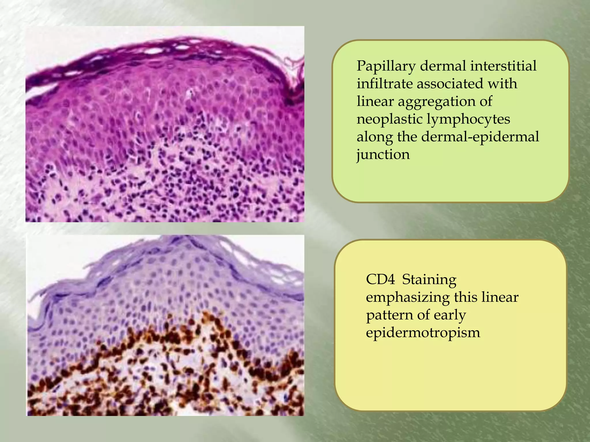
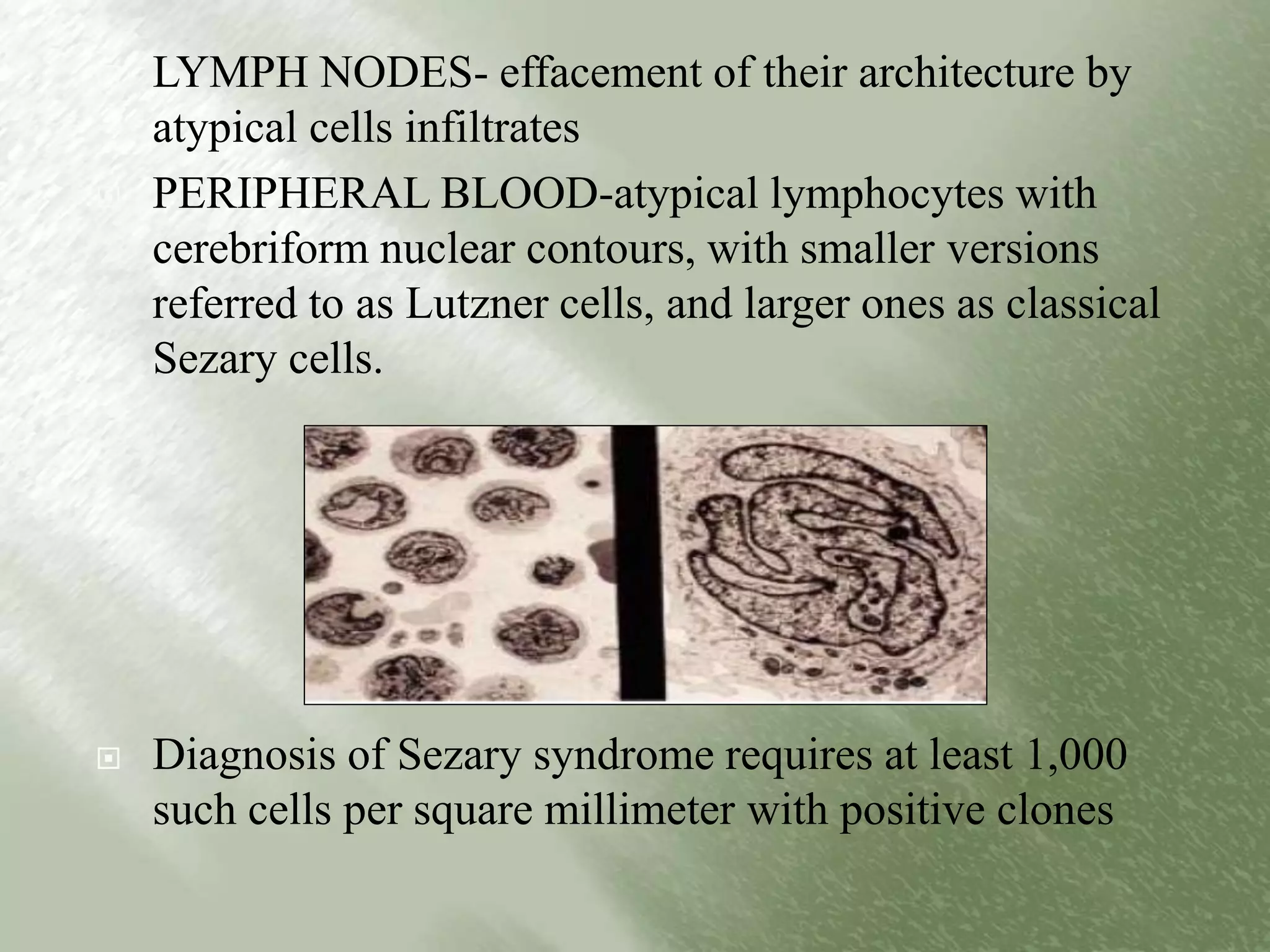
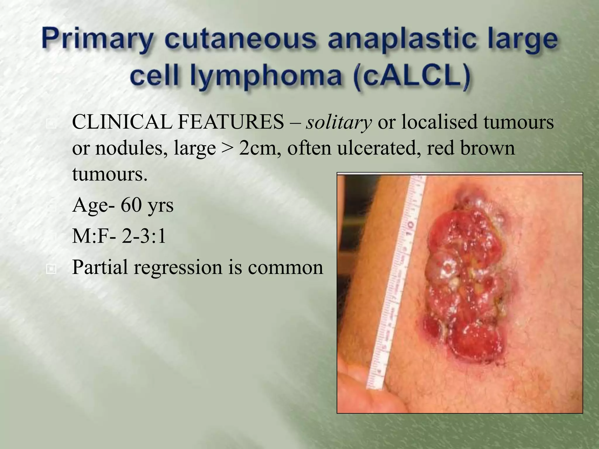
By- Dr. Kanwalpreet Kaur
Moderator- Dr. Karuna Gupta
 PRIMARY LYMPHOMAS- that present in the skin
with no evidence of extracutaneous disease at the time
of diagnosis
 SECONDARY LYMPHOMAS- systemic lymphomas
that secondarily involve the skin.
 Differences in adhesive ligands between T and B
cells
 Differential affinity for different compartments
• Benign and malignant T cells migrate to
epidermis and its adnexa.
• This phenomena is known as
EXOCYTOSIS in BENIGN CONDITIONS;
EPIDERMOTROPISM in MALIGNANT
CONDITIONS.
• This pattern may be blunted as tumour
evolves into more aggressive and poorly
differentiated state.
• B cells spare epidermis and
papillary dermis
• Sparing of papillary dermis produces
grenz zone
• innate proclivity of B cells to
aggregate into follicular structures, B-
cell infiltrates in the skin often (but
not invariably) show a nodular
architecture
T CELL PATTERN
B CELL PATTERN- GRENZ ZONE
AND GERMINAL CENTRE
FORMATION
 In both T- and B-cell responses in skin
circulating lymphocytes initially adhere to activated
endothelium lining dermal postcapillary venules.
PERIVASCULAR/
ANGIOCENTRIC DERMATITIS
Further migration may be
Stunted
e.g. erythema chornium migrans
 Migration away from the vessel wall is presumably swift
enough that in the case of T cells
papillary dermal accumulation and epidermotropism, in addition
to a persistent angiocentric pattern
IMMUNOLOGICALLY ACTIVE
T CELLS
1.Produce cytokines and growth factors
so T cell exocytosis is associated with
epithelial spongiosis, acanthosis or
apoptosis
2. seen in contact hypersensitivity,
psoriasis, cytotoxic dermatitis
MALIGNANT EPIDERMOTROPIC
T CELL INFILTRATE
1.Epidermis is passive
2.No reactive epithelial alteration
3.No spongiosis or apoptosis
4.However, epidermal injury in the form
of mucinous degeneration may occur.
5.The epidermis becomes more
extensively infiltrated, impaired
maturation often manifests as clinical
abnormalities in scale production
A: Abnormal epidermotropism into a “passive” epidermis.
B: Scaling patches and plaques as a consequence of altered epidermal maturation
associated with T-cell epidermotropism (MF).
REACTIVE B CELLS
1) perivascular accumulation of
cells
2) No significant involvement of
papillary dermis or epidermis
3) Grenz zone of papillary
sparing
4) Preservation of archietecture
MALIGNANT B CELLS
1) Destructive relationship to
pre-exsisting structures
2) Infiltatre in interstitial pattern
causing
3) Splaying of collagen bundles
and mesenchymal cells within
reticular dermis
4) This progressive dermal
infiltration+ epidermal
sparing red to plum colored
nodules covered by
nonkeratotic epidermis
Normal and abnormal trafficking patterns. A: B-cell pattern showing nondestructive infiltration
of superficial and deep dermis.
B: B-cell lymphomas showing vascular invasion , replacement of subcutis , and diffuse
interstitial pattern .
C: Plum-colored nodule covered by thinned, glistening epidermis due to B-cell dermal
infiltration.
 Heterogenous spectrum of lymphoid infiltrates that can
be similar histologically to malignant lymphomas but do
not have clinical presentation or biological outcome of
lymphoma.
 Inflammatory infiltrate is band like, nodular or diffuse
and composed of lymphocytes predominantly, but other
inflammatory cells are also present.
 Depending on the predominant cell type in the infiltrate,
they are divided into T and B cell types.
CUTANEOUS T- cell and NK- cell Lymphomas
1. MYCOSIS FUNGOIDES (MF)
2. MYCOSIS FUNGOIDES VARIANTS AND SUBTYPES:
a) Folliculotropic MF
b) Pagetoid reticulois
c) Granulomatous slack skin
3. SEAZARY SYNDROME
4. ADULT T- cell LYMPHOMA
5. PRIMARY CUTANEOUS CD30+ LYMPHOPROLIFERATIVE DISORDERS
a) Primary cutaneous anaplastic large cell lymphoma (cALCL)
b) Lymphomatoid papulosis
6. SUBCUTANEOUS PANNICULITIS LIKE T CELL LYMPHOMA
7. EXTRANODAL NK/T- cell LYMPHOMA, NASAL TYPE
8. PRIMARY CUTANEOUS PERIPHERAL T- cell LYMPHOMA, UNSPECIFIED
a) Primary Cutaneous Aggressive Epidermotropic CD8+ T- Cell Lymphoma
b) Cutaneous γ/ δ T- Cell Lymphoma
c) Primary Cutaneous CD4+ Small/Medium Sized Pleomorphic T- Cell Lymphoma
CUTANEOUS B-cell LYMPHOMAS
1. Primary cutaneous follicle centre lymphoma
2. Primary cutaneous marginal zone B-cell lymphoma
3. Primary cutaneous diffuse large B-cell lymphoma, leg type
4. Primary cutaneous diffuse large B-cell lymphoma , other
5. Intravascular large B-cell lymphoma
PRECURSOR HEMATOLOGIC NEOPLASM
CD4+/CD56+ hematodermic neoplasm (blastic NK cell lymphoma)
 Low grade epidermotropic T-cell lymphoma
 Mycosis fungoides is relatively rare, accounting for less than
1% of non-Hodgkin lymphomas. However, of lymphomas
that arise primarily in skin, it is the most common
 Peak age 55-60
 Male: female 2:1
 Typically begins as slowly progressive dermatitis-like patches
and plaques on non sun-exposed skin, and when untreated
evolves to nodules and eventual systemic dissemination
Lesions initially -erythematous scaling
patches and plaques that typically are
resistant to antiinflammatory therapy;
Large with polygonal or arcuate
configurations;
Plaques and nodules of
advanced disease which may
eventually ulcerate
• When extracutaneous spread eventuates, lymph nodes, spleen, liver,
and lungs are often involved, in addition to the peripheral blood.
• Bone marrow involvement is rare
• Lymph node enlargement at such advanced stages must be
differentiated from nodal hyperplasia resulting from chronic
drainage of involved sites (dermatopathic lymphadenopathy)
• Progression to Extracutaneous disease correlates with extent of skin
disease
• A) Limited patch or plaque- very rare
• B) Generalized plaque- 8 %
• C) Tumorous or generalized erythroderma-30-40% Hence,
extracutaneous is more commonly seen in Sezary syndrome.
 Atypical SMALL to MEDIUM sized mononuclear
neoplastic T cells with cerebriform nuclei infiltrating the
upper dermis among epidermal keratinocytes
(epidermotropism) forming intra epidermal aggregates
(Pautrier microabscesses), or arranged in linear fashion
just above the basement membrane.
 Pautriers abscesses – pathognomonic but present only in
38 % cases of MF
 Minimal epidermal spongiosis typically present
 Lymphocytes aligned within the basal layer
1.Patch stage: superficial band-like or lichenoid infiltrate,
mainly lymphocytes and histiocytes, but also a few atypical cells
(small/medium-sized, cerebriform nuclei, halo) usually confined
to the epidermis (basal layer)= EPIDERMOTROPISM
2.Plaque stage: more pronounced epidermotropism with
occasional characteristic intraepidermal collections of atypical
cells (Pautrier microabscesses)
3. Tumor stage: more dense nodular or diffuse dermal infiltrate,
may lose epidermotropism, more size and shape variability of
tumor cells, histologic transformation to large cell phenotype
may occur (>25% large lymphoid cells in the dermal infiltrate)
Papillary dermal interstitial
infiltrate associated with
linear aggregation of
neoplastic lymphocytes
along the dermal-epidermal
junction
CD4 Staining
emphasizing this linear
pattern of early
epidermotropism
Plaque-stage disease -prominent clusters of
atypical lymphocytes within the epidermis
(Pautrier microabscesses)
 CD2+, CD 3+ , CD 5+, CD4+, CD8-, CD 7- and usually
CD 30-

CD 4 STAINING CD 8 STAINING
 Clonal T-cell receptor( TCR) gene rearrangements
have been detected (mostly alpha/beta, rarely
gamma/delta receptors)
 Increased rate of aberrations involving multiple
chromosomes 1,6,11,8,17,15,13, and 9 in advanced
stage disease
 ISCL/EORTC Diagnostic algorithm :
 Point based system
 A total of 4 points is required for the diagnosis of
MF based on any combination of points from the
clinical, histopathologic, molecular biological, and
immunopathologic criteria.
CRITERIA MAJOR
(2points)
MINOR
(1 point)
CLINICAL
Persistent and/or progressive patches and plaques
plus
Any2 Any 1
a) Non sun exposed areas
b)size/shape variation
c)Poikiloderma
HISTOPATHOLOGIC
Superficial lymphoid infiltrate plus Both Either
a)Epidermotropism without spongiosis
b) Lymphoid atypia
MOLECULAR BIOLOGICAL:
clonal TCR gene rearrangement
present
IMMUNOPATHOLOGIC:
a) CD 2,3,5 < 50% of T cells Any one
b) CD7< 10% T cells
c) Epidermal discordance from expression of
CD2,3,5 or CD 7 on dermal T cells
SKIN
T1- limited patches, plaques and /or plaques covering < 10% of skin surface
T2- patches, papules or plaques covering 10% or more skin surface
T3- one or more tumours (>1cm)
T4- confluence of erythema covering 80% or more of body surface area
NODE
N0- no clinically abnormal peripheral lymph nodes
N1- clinically abnormal peripheral lymph nodes; histopathologically Dutch
grade 1 or NC1 LN0-2
N2- clinically abnormal peripheral lymph nodes; histopathologically Dutch
grade 1 or NC1 LN0-2
N3- clinically abnormal peripheral lymph nodes histopathology dutch
grades 3-4
VISCERAL
M0- no visceral organ involvement
M1- visceral involvement
PERIPHERAL BLOOD INVOLVEMENT
B0- absence of significant blood involvement:5% or less of peripheral
blood lymphocytes are atypical
B1- low blood tumour burden:>5% of peripheral blood lymphocytes are
atypical
B2- high blood tumour burden: 1000/microl sezary cells
 It is characterised by – usual type of cells seen in
epidermotropic forms of mycosis fungoides but is
confined to hair follicles and is associated with
follicular mucinosis.
 CLINICAL FEATURES- slowly enlarging follicular
patches or plaques on head and neck ; may be associated
with alopecia
 WORSE PROGNOSIS compared to other variants and
require aggressive therapy
Folliculocentric infiltrate with
mucinous expansion of the follicle.
Infiltrate is usually monomorphic containing cerebriform CD4+ Lymphocytes
 Rare and peculiar form of mycosis fungoides
 CLINICAL FEATURES- solitary acral lesion in the
form of verrucous plaque that does not progress
clinically
 If multiple lesions are present / widely disseminated in
skin- KETRON-GOODMAN type  should not be
considered pagetoid reticulosis but primary cutaneous
aggressive epidermotropic CD8+ cytotoxic T-cell
lymphoma
-exclusively epidermal
infiltration
-INFILTRATE-
mononuclear convoluted T
cells
-pagetoid pattern with or
without intraepidermal
nests
- Epidermal hyperplasia
- hyerkeratosis
Mixed infiltrate of non neoplastic lymphocytes and histiocytes may be seen in
superficial dermis
 CD3+, CD4+, CD8- or CD3+, CD4-, CD8+
 MOLECULAR GENETICS
 Clonal TCR gene rearrangements
 Extremely rare subtype
 CLINICAL FEATURES- slowly developing zones
of lax skin in the axillae and inguinal regions;
occurs predominantly in men
 Good prognosis
Diffuse mononuclear infiltrate+
macrophages+multinucleate giant
cells+ T cells with convoluted
nucleus. Elastolysis seen in
dermis.
 Potentially aggressive form
 These malignant T cells have phenotypic and antigenic
overlap with those of mycosis fungoides, Sezary
syndrome is sometimes regarded as a mycosis fungoides
variant. However, important clinical and
histopathologic differences exist between classical
mycosis fungoides and Sezary syndrome
Generalized redness and scaling of the skin (erythroderma)
+lymphadenopathy + presence of malignant convoluted T cells
(sezary cells) in the skin, lymph nodes and peripheral blood
 Diffuse skin erythema and lymphadenopathy. There may also
be itching, hair loss, nail dystrophy, and hyperkeratosis
affecting the skin of palms and soles

 In addition to lymph node, skin, and blood involvement,
malignant T cells may also infiltrate viscera in late stages,
although bone marrow is often spared
band-like papillary dermal
lymphoid infiltrate;
Lack epidermotropism:
More monomorphic cells
dense perivascular and superficial
dermal atypical lymphoid
infiltrate with minimal focal
epidermotropism.
 LYMPH NODES- effacement of their architecture by
atypical cells infiltrates
 PERIPHERAL BLOOD-atypical lymphocytes with
cerebriform nuclear contours, with smaller versions
referred to as Lutzner cells, and larger ones as classical
Sezary cells.
 Diagnosis of Sezary syndrome requires at least 1,000
such cells per square millimeter with positive clones
A mixed population of small
and large tumor lymphocytes
with cerebriform nuclear
morphology are noted (arrow
on typical Sèzary cell).
Eosinophilia (top cell) is also
frequently present in Sezary
Syndrome and may contribute
to the pruritus and atopy.
 * Diagnosis is made when there is a clonal rearrangement
of the T-cell receptor (TCR) in the blood (identified by
PCR or southern blot analysis) plus
 *either
 an absolute Sezary cell count of at least 1000 cells/microL
 *or
one of the following two criteria
 1) Increased CD4+ or CD3+ cells with a CD4 to CD8
ratio of 10 or more.
 2) Increased CD4+ cells with an abnormal phenotype
(such as a CD4+ to CD7- ratio ≥40 percent or a
CD4+ to CD26- ratio ≥30 percent)
1)Other causes of erythroderma: atopic dermatitis,
seborrheic dermatitis, pityriasis rubra piliaris, psoriasis
2) Advanced mycosis fungoides
3) Adult T cell lymphoma/leukemia
 Spectrum of related, relatively low-grade disorders that
primarily affect skin and develop from an activated or
transformed large T cell that expresses the CD30 antigen.
 CD30 antigen is a cytokine receptor belonging to the
tumor necrosis factor receptor superfamily
 Second most common form of cutaneous T-cell lymphomas
 It includes:
a)primary cutaneous anaplastic large cell lymphoma (cALCL),
 b)lymphomatoid papulosis and
 c)borderline cases
 CLINICAL FEATURES – solitary or localised tumours
or nodules, large > 2cm, often ulcerated, red brown
tumours.
 Age- 60 yrs
 M:F- 2-3:1
 Partial regression is common
Nodular or diffuse dermal infiltrate
sheets of cohesive CD30+
atypical cells; may also involve
superficial cutis
A non neoplastic mixed
inflammatory infiltrate of small
reactive lymphocytes, macrophages,
eosinophils is present
ATYPICAL CELLS- large
rounded or irregular
vesicular nuclei; prominent
centrally placed nucleoli;
ample clear to amphophilic
cytoplasm
CD 30 IMMUNOSTAINING : strong
cytoplasmic membrane staining of tumor cells
According to the WHO-
EORTC system, cALCL is
classified by the expression of
CD30 in more than 75% of
large atypical cells
Cells may also be CD4+;
LOSS OF PAN-T MARKERS
(CD2,CD3,CD5) may occur
EMA and ALK are not
expressed in C-ALCL, but
these two markers are positive
in systemic (nodal) anaplastic
large cell lymphoma
(ALCL).
 Clonal rearrangement of TCR genes is detected in
90% of cases
 Translocation (t2;5) (p23;q35) charateristic of
systemic ALCL is rarely, if ever, found in primary
cALCL
 CLINICAL FEATURES- presence of a
self-healing skin eruption of erythematous
papules and nodules that may become
haemorrhagic, necrotic, and/or ulcerative
 While individual lesions take weeks to
several months to resolve, the disease may
recur for decades and is highly variable
 Patients should be monitored lifelong since
2-25% of patients develop second
lymphoma such as Hodgkin lymphoma or
anaplastic large cell lymphoma
Type A lymphomatoid papulosis
 Borderline cases- those in which a difference between the
clinical and histologic appearance exists.
 Include cases with the clinical presentation of a
CD30+ CALCL but histologic features suggestive of LyP,
and, conversely, cases with a recurrent, self-healing skin
eruption that shows histologic features characteristic of a
CD30+ CALCL.
 The distinction between LyP and CD30+ CALCL is not
always possible based on histologic criteria.
 LyP type C has been described as a borderline lesion of
CD30+ CALCL.
 Thus, the clinical appearance and the clinical course over
time are used as decisive criteria for the definitive diagnosis
and choice of treatment.
 Provisional category in WHO-EORTC clasification
 Characterised by CD8+ cytotoxic epidermotropic T
cell infiltrates
 CLINICAL FEATURES- mean age- 53 yrs
 ulcerated and necrotic eruptive papules, nodules, and
tumors with frequent dissemination to other visceral
sites- lungs,testis,CNS,oral mucosa
 Lymph nodes – typically spared
 Aggressive course. Mean survival – 32 months
 MICROSCOPIC FEATURES- Medium to large
pleomorphic cells; pronounced epidermotropism.
 Epidermis often ulcerated + necrotic keratinocytes+
spongiosis
 IMMUNOPHENOTYPICALLY- βF1+, CD3+,
CD8+, granzyme B+, perforin+,TIA-1+ and CD4-.
 MOLECULAR GENETICS-clonal TCR gene
rearrangements are present in most of cases
 Provisional category
 Small to medium sized pleomorphic CD4+ CTCL
without a history of classic patches and plaques of MF
and a favorable clinical course
 CLINICAL FEATURES-mean age- 69 yrs
solitary nodule on the face, upper arms, or trunk is
common. Patients may also present with multiple papules
or nodules, but lack patches or plaques.
 MICROSCOPIC FEATURES- diffuse growth
pattern in dermis or subcutis; epidermotropism focal
and inconspicous; neoplastic infiltrate is
accompanied by mixed inflammatory infiltrate.
 IMMUNOPHENOTYPICALLY- CD4+ CD8-
Loss of CD2, CD3, CD5. CD30-
 Subcutaneous panniculitis-like T-cell lymphoma
(SPTCL) is defined as a rare lymphoma that
(a) primarily infiltrates subcutaneous tissue;
(b) shows high-grade cytologic features;
(c) is composed of T cells with a cytotoxic phenotype
It only consists of cases with α β + T cell phenotype; cases
with γδ T cell phenotype are included in separate category
 CLINICAL FEATURES- subcutaneous nodules
 and plaques, usually on the legs or trunk.
 Systemic symptoms and signs, when present, may relate to
hemophagocytic syndrome with pancytopenia, fever, and
hepatosplenomegaly usually in the absence of
lymphadenopathy
diffuse, variably cellular infiltrate
involving both septae and lobules
within the subcutis; sparing of
dermis; INFILTRATE- smaller
lymphocytes with visible cytoplasm
admixed with larger transformed cells
with hyperchromatic nuclei
Rimming of individual adipocytes by neoplastic T cells -characteristic,
but not specific, feature
CD3+, CD4-, CD8+,CD5+,TIA+,granzyme B +
MOLECULAR GENETICS- clonal TCR rearrangement in 50%
cases
TIA-1 immunohistochemistry showing characteristic
rimming about individual adipocytes.
 Provisional category
 CLINICAL FEATURES- eroded to ulcerated patches,
deep tumours located on extremities of adults.
 Constitutional symptoms usually present
 Mucosal and extranodal sites frequently involved
 Bone marrow, lymph nodes, and spleen are spared
 Highly aggressive course
 MICROSCOPIC FEATURES-can be predominantly
epidermotropic, dermal, or subcutaneous.
 Subcutaneous infiltrates show rimming of neoplastic
cells around individual adipocytes, similar to
subcutaneous panniculitis–like T-cell lymphoma, but
dermis and/or epidermis also are involved
 IMMUNOPHENOTYPE CD3+, CD56+,
granzymeB+, TIA-1+, CD4 −, CD8−, CD5−, and βF1−
 MOLECULAR GENETICS- Clonal rearrangement of
the TCR-γ gene occurs
 Develops in a small minority individuals with prolonged
infection with human T-cell leukemia virus type 1
(HTLV-1)
 MICROSCOPIC FEATURES-a superficial or diffuse
infiltrate of pleomorphic multinucleate T lymphocytes
with prominent epidermotropism is seen.
 Lesion may be indistinguishable from mycosis
fungoides
 IMMUNOPHENOTYPE- CD3+,CD4+,CD8-, CD25+
 MOLECULAR GENETICS- Clonal rearrangement of
the TCR genes and clonal integration of HTLV-1 occur
 Rare variant
 Neoplastic cells are NK cell phenotype, can be cytotoxic T cell derived
and are always EBV positive.
 CLINICAL FEATURES-multiple ulcerated plaques and nodules typically
distributed on the trunk or extremities. Accompanying systemic
symptoms are common.
 MICROSCOPIC FEATURES-Medium-sized pleomorphic lymphocytic
infiltrates are present in the dermis and subcutis and show prominent
localization and destruction of blood vessels. Apoptosis and necrosis are
conspicuous.
 IMMUNOPHENOTYPE-CD2+, CD56+, surfaceCD3−, TIA-1+,
granzyme B+, perforin+, and EBV+
CUTANEOUS T- cell and NK- cell Lymphomas
1. MYCOSIS FUNGOIDES (MF)
2. MYCOSIS FUNGOIDES VARIANTS AND SUBTYPES:
a) Folliculotropic MF
b) Pagetoid reticulois
c) Granulomatous slack skin
3. SEAZARY SYNDROME
4. ADULT T- cell LYMPHOMA
5. PRIMARY CUTANEOUS CD30+ LYMPHOPROLIFERATIVE DISORDERS
a) Primary cutaneous anaplastic large cell lymphoma (cALCL)
b) Lymphomatoid papulosis
6. SUBCUTANEOUS PANNICULITIS LIKE T CELL LYMPHOMA
7. EXTRANODAL NK/T- cell LYMPHOMA, NASAL TYPE
8. PRIMARY CUTANEOUS PERIPHERAL T- cell LYMPHOMA, UNSPECIFIED
a) Primary Cutaneous Aggressive Epidermotropic CD8+ T- Cell Lymphoma
b) Cutaneous γ/ δ T- Cell Lymphoma
c) Primary Cutaneous CD4+ Small/Medium Sized Pleomorphic T- Cell Lymphoma
CUTANEOUS B-cell LYMPHOMAS
1. Primary cutaneous follicle centre lymphoma
2. Primary cutaneous marginal zone B-cell lymphoma
3. Primary cutaneous diffuse large B-cell lymphoma, leg type
4. Primary cutaneous diffuse large B-cell lymphoma , other
5. Intravascular large B-cell lymphoma
PRECURSOR HEMATOLOGIC NEOPLASM
CD4+/CD56+ hematodermic neoplasm (blastic NK cell lymphoma)
Neoplasm derived from follicular centre B cells including
centrocytes (cleaved follicular centre cells) and centroblasts
(non cleaved follicular centre cells)
Most common type
CLINICAL FEATURES – middle age men
one to several red- to plum-colored
plaques, nodules or tumours;
Head and neck or upper trunk;
cutaneous infiltrates are nodular or diffuse, usually sparing the epidermis.
nodules are similar to germinal centers
of lymph nodes; monomorphic
neoplastic cells; devoid of mantle zone;
no tingible body macrophages are
present
Early lesions are mostly centrocytic,
containing cells with small to large
cleaved nuclei, with fewer
centroblastic cells, which are large and
contain prominent nucleoli
 IMMUNOPHENOTYPING- CD20, CD79a, bcl-6
CD 20 STAINING BCL-6 STAINING NEGATIVE FOR MUM-1
Weak heterogeneous expression of Bcl-2 CD 21 highlighting FOLLICULAR
DENDRITIC CELLS meshwork
 Predominance of centroblasts and immunoblasts.
 CLINICAL FEATURES- usually women of 70 years
 Red to blue nodules on leg
 Has poor prognosis
Diffuse dense non epidermotropic monotonous infiltrates of predominantly
medium to large cells with round nuclei, prominent nucleoli and frequent
mitosis resembling centroblasts, large centrocytes, immunoblasts.
CD 20 STAINING IRF4/MUM1 STAINING + BCL2 STAINING +
These are in contrast to follicular centre cell
lymphoma of head and neck
 Lymphoma of small lymphoplasmacytoid cells, small
lymphocytes, and plasma cells with monotypic
cytoplasmic Ig.
 In 2008 WHO classification, marginal zone lymphomas
of skin are grouped into broader category of extranodal
marginal zone lymphoma of mucosa associated
lymphoid tissues(MALT lymphomas)
 CLINICAL FEATURES- solitary or multiple cutaneous
or subcutaneous tumors on the trunk and extremities
nodular or diffuse infiltrates of
small lymphoplasmacytoid
cells, small lymphocytes, and
plasma cells are present that
can be associated with
centrocytes and centroblasts.
Cell of origin – post germinal centre marginal zone B cell
Reactive B cells follicles
Neoplastic cells infiltration in
marginal zone distribution
Atypical neoplastic cells have irregular
nuclear contours and slightly dispersed
chromatin
Some proliferations are composed of
cells with abundant clear cytoplasm
imparting a monocytoid appearance
CD 20+ BCL 2+
NEGATIVE FOR CD5, CD 10 and BCL 6
 Intravascular B-cell lymphoma with a relatively poor
survival rate at 5 years, especially if sites other than skin
are involved.
 CLINICAL FEATURES-violet patches and plaques on
the legs and trunk, often thought to be subcutaneous
 MICROSCOPIC FEATURES-dermal and subcutaneous
blood vessels are dilated and stuffed with tumor cells
that are somewhat pleomorphic
 IMMUNOPHENOTYPICALLY-cells are CD20+ and
CD79a+, with monotypic surface Ig
Lymphoblastic lymphomas may be of either B or T cell
lineage
Although majority of systemic lymphoblastic lymphomas
are of T cell lineage, but its B cell lineage lymphomas
which show tendency for cutaneous involvement
CLINICAL FEATURES- <35 years
head and neck
Nodular or diffuse dermal
infiltrate; mitotically active
lymphoblasts with convoluted
nuclei containing finely
stippled chromatin and
inconspicious nucleoli
 Precursor B cell
 Neoplastic cells lack surface immnoglobulin expression
 May harbour cytoplasmic μ chain
 +  TdT, CD79a, CD19, CD 10

 MOLECULAR GENETICS
 Favorable prognosis: hyperdiploidy >50 chromosomes
 Unfavorable prognosis: hypodiploidy, t(9;22), t(4;11)
 t(1;19)
 Most common mature B cell neoplasm to secondarily
involve skin- leukaemia cutis
 CLINICAL FEATURES- older adults; M:F- 2:1
 Leukaemia cutis occurs in patients with established
CLL/SLL
 May produce localized or disseminated erythematous
plaques, papules or nodules
 May be angiocentric pattern, interstitial pattern and
mixed nodular/diffuse pattern
 Epidermis and papillary dermis uninvolved- typical
grenz zone seen
Diffuse, cellular infiltrate
composed of uniform populations
of small lymphocytes containing
rounded, hyperchromatic nuclei
with inconspicuous nucleoli.
 + ve  CD 19, CD5, CD79a, CD23, CD43
 -ve  CD 10 , cyclin D1
 Low grade neoplasm of centrocytes and centroblasts
(follicular centre cells)
 CLINICAL FEATURES- >59 years
 head and neck
The dermis is replaced by nodules of neoplastic cells that resemble
germinal centers but lack mantle zones.
 Cell of origin – germinal B cell
 +  surface immunoglobulin, BCL2 , CD 10, CD 19,
CD 20, CD22
 -ve  CD5 , CD43

 MOLECULAR GENETICS-
t(14;18)
B cell neoplasm of uniform populations of small to
medium sized lymphocytes – resemble centrocytes
CLINICAL FEATURES- middle aged to older
 Disease is usually advanced at time of detection (stage III or
IV) and generally affects lymph nodes, spleen, bone marrow
with or without peripheral blood involvement, and
gastrointestinal tract
 Involvement of skin – rare
 Cutaneous MCL more aggressive than PCFCL or
plasmacytoid B-CLL/SLL . Median survival – 3-5 years
vaguely nodular to diffuse
infiltrate composed of small- to
medium-sized lymphocytes.
 Peripheral B cell of inner mantle zone
CD 20 +
+VE  CD5, CD43, BCL2, Cyclin D1
-ve  CD 10 , CD23
MOLECULAR GENETICS-
t(11;14) involving cyclin D1 gene and
immunoglobulin heavy chain locus
 Aggressive lymphoma
 Diffuse tissue infiltration by monotonous populations of
large histiocytoid malignant lymphocytes
 Associated with EBV
 CLINICAL FEATURES- median age 70 years
 occasionally children also affected
 red to purpuric nodules
Dermis is diffusely effaced by a
destructive infiltrate causing effacement
of pre-existing structures
Infiltrate is composed of large
neoplastic lymphocytes
resembling centroblasts
 Peripheral germinal centre or post germinal centre B
cell
 CD19, CD20, CD22, CD79a
 Considered rare subtype of DLBCL
 Intraluminal aggregates of large malignant B cells
within vessels of involved tissues
 CLINICAL FEATURES- adults
 Widely disseminated with involvement of extranodal
tissues including skin  red plaques and nodules
Superficial dermal vessels are
partially occluded by hyperchromatic
malignant cells.
malignant-appearing lymphocytes
partially adherent to the endothelial
surface of involved vessels.
 Peripheral germinal centre or post germinal centre
origindefective homing receptors CD11a/ CD18
impaired diapedesis  intravascular accumulation
 +ve  CD20, CD5, CD10

 Rare aggressive neoplasm
 Once thought to have an NK cell origin, but now it
is believed to be derived from plasmacytoid
dendritic cells.The neoplastic cells characteristically
coexpress CD4 and CD56 without B-, T-, and NK-
cell or myeloid lineage-specific markers
 CLINICAL FEATURES-cutaneous lesions that
typically are solitary or localized nodules or tumors
in elderly men.
 dermal infiltrate of monotonous medium-sized cells
with blastoid morphology is present regular nuclear
contours, dispersed chromatin, and small, distinct
nucleoli
 Mitoses are frequent, but angiodestruction and necrosis
are not seen
CD 4+ CD 56 + CD 123 +
MPO –ve
Also –ve for CD3, CD33
THANK
YOU….

Cutaneous lymphoproliferative disorders

  • 1.
    By- Dr. KanwalpreetKaur Moderator- Dr. Karuna Gupta
  • 2.
     PRIMARY LYMPHOMAS-that present in the skin with no evidence of extracutaneous disease at the time of diagnosis  SECONDARY LYMPHOMAS- systemic lymphomas that secondarily involve the skin.
  • 3.
     Differences inadhesive ligands between T and B cells  Differential affinity for different compartments • Benign and malignant T cells migrate to epidermis and its adnexa. • This phenomena is known as EXOCYTOSIS in BENIGN CONDITIONS; EPIDERMOTROPISM in MALIGNANT CONDITIONS. • This pattern may be blunted as tumour evolves into more aggressive and poorly differentiated state. • B cells spare epidermis and papillary dermis • Sparing of papillary dermis produces grenz zone • innate proclivity of B cells to aggregate into follicular structures, B- cell infiltrates in the skin often (but not invariably) show a nodular architecture
  • 4.
    T CELL PATTERN BCELL PATTERN- GRENZ ZONE AND GERMINAL CENTRE FORMATION
  • 5.
     In bothT- and B-cell responses in skin circulating lymphocytes initially adhere to activated endothelium lining dermal postcapillary venules. PERIVASCULAR/ ANGIOCENTRIC DERMATITIS Further migration may be Stunted e.g. erythema chornium migrans
  • 6.
     Migration awayfrom the vessel wall is presumably swift enough that in the case of T cells papillary dermal accumulation and epidermotropism, in addition to a persistent angiocentric pattern IMMUNOLOGICALLY ACTIVE T CELLS 1.Produce cytokines and growth factors so T cell exocytosis is associated with epithelial spongiosis, acanthosis or apoptosis 2. seen in contact hypersensitivity, psoriasis, cytotoxic dermatitis MALIGNANT EPIDERMOTROPIC T CELL INFILTRATE 1.Epidermis is passive 2.No reactive epithelial alteration 3.No spongiosis or apoptosis 4.However, epidermal injury in the form of mucinous degeneration may occur. 5.The epidermis becomes more extensively infiltrated, impaired maturation often manifests as clinical abnormalities in scale production
  • 7.
    A: Abnormal epidermotropisminto a “passive” epidermis. B: Scaling patches and plaques as a consequence of altered epidermal maturation associated with T-cell epidermotropism (MF).
  • 8.
    REACTIVE B CELLS 1)perivascular accumulation of cells 2) No significant involvement of papillary dermis or epidermis 3) Grenz zone of papillary sparing 4) Preservation of archietecture MALIGNANT B CELLS 1) Destructive relationship to pre-exsisting structures 2) Infiltatre in interstitial pattern causing 3) Splaying of collagen bundles and mesenchymal cells within reticular dermis 4) This progressive dermal infiltration+ epidermal sparing red to plum colored nodules covered by nonkeratotic epidermis
  • 9.
    Normal and abnormaltrafficking patterns. A: B-cell pattern showing nondestructive infiltration of superficial and deep dermis. B: B-cell lymphomas showing vascular invasion , replacement of subcutis , and diffuse interstitial pattern . C: Plum-colored nodule covered by thinned, glistening epidermis due to B-cell dermal infiltration.
  • 10.
     Heterogenous spectrumof lymphoid infiltrates that can be similar histologically to malignant lymphomas but do not have clinical presentation or biological outcome of lymphoma.  Inflammatory infiltrate is band like, nodular or diffuse and composed of lymphocytes predominantly, but other inflammatory cells are also present.  Depending on the predominant cell type in the infiltrate, they are divided into T and B cell types.
  • 12.
    CUTANEOUS T- celland NK- cell Lymphomas 1. MYCOSIS FUNGOIDES (MF) 2. MYCOSIS FUNGOIDES VARIANTS AND SUBTYPES: a) Folliculotropic MF b) Pagetoid reticulois c) Granulomatous slack skin 3. SEAZARY SYNDROME 4. ADULT T- cell LYMPHOMA 5. PRIMARY CUTANEOUS CD30+ LYMPHOPROLIFERATIVE DISORDERS a) Primary cutaneous anaplastic large cell lymphoma (cALCL) b) Lymphomatoid papulosis 6. SUBCUTANEOUS PANNICULITIS LIKE T CELL LYMPHOMA 7. EXTRANODAL NK/T- cell LYMPHOMA, NASAL TYPE 8. PRIMARY CUTANEOUS PERIPHERAL T- cell LYMPHOMA, UNSPECIFIED a) Primary Cutaneous Aggressive Epidermotropic CD8+ T- Cell Lymphoma b) Cutaneous γ/ δ T- Cell Lymphoma c) Primary Cutaneous CD4+ Small/Medium Sized Pleomorphic T- Cell Lymphoma
  • 13.
    CUTANEOUS B-cell LYMPHOMAS 1.Primary cutaneous follicle centre lymphoma 2. Primary cutaneous marginal zone B-cell lymphoma 3. Primary cutaneous diffuse large B-cell lymphoma, leg type 4. Primary cutaneous diffuse large B-cell lymphoma , other 5. Intravascular large B-cell lymphoma PRECURSOR HEMATOLOGIC NEOPLASM CD4+/CD56+ hematodermic neoplasm (blastic NK cell lymphoma)
  • 14.
     Low gradeepidermotropic T-cell lymphoma  Mycosis fungoides is relatively rare, accounting for less than 1% of non-Hodgkin lymphomas. However, of lymphomas that arise primarily in skin, it is the most common  Peak age 55-60  Male: female 2:1
  • 15.
     Typically beginsas slowly progressive dermatitis-like patches and plaques on non sun-exposed skin, and when untreated evolves to nodules and eventual systemic dissemination Lesions initially -erythematous scaling patches and plaques that typically are resistant to antiinflammatory therapy; Large with polygonal or arcuate configurations;
  • 16.
    Plaques and nodulesof advanced disease which may eventually ulcerate
  • 17.
    • When extracutaneousspread eventuates, lymph nodes, spleen, liver, and lungs are often involved, in addition to the peripheral blood. • Bone marrow involvement is rare • Lymph node enlargement at such advanced stages must be differentiated from nodal hyperplasia resulting from chronic drainage of involved sites (dermatopathic lymphadenopathy) • Progression to Extracutaneous disease correlates with extent of skin disease • A) Limited patch or plaque- very rare • B) Generalized plaque- 8 % • C) Tumorous or generalized erythroderma-30-40% Hence, extracutaneous is more commonly seen in Sezary syndrome.
  • 18.
     Atypical SMALLto MEDIUM sized mononuclear neoplastic T cells with cerebriform nuclei infiltrating the upper dermis among epidermal keratinocytes (epidermotropism) forming intra epidermal aggregates (Pautrier microabscesses), or arranged in linear fashion just above the basement membrane.  Pautriers abscesses – pathognomonic but present only in 38 % cases of MF  Minimal epidermal spongiosis typically present  Lymphocytes aligned within the basal layer
  • 19.
    1.Patch stage: superficialband-like or lichenoid infiltrate, mainly lymphocytes and histiocytes, but also a few atypical cells (small/medium-sized, cerebriform nuclei, halo) usually confined to the epidermis (basal layer)= EPIDERMOTROPISM 2.Plaque stage: more pronounced epidermotropism with occasional characteristic intraepidermal collections of atypical cells (Pautrier microabscesses) 3. Tumor stage: more dense nodular or diffuse dermal infiltrate, may lose epidermotropism, more size and shape variability of tumor cells, histologic transformation to large cell phenotype may occur (>25% large lymphoid cells in the dermal infiltrate)
  • 20.
    Papillary dermal interstitial infiltrateassociated with linear aggregation of neoplastic lymphocytes along the dermal-epidermal junction CD4 Staining emphasizing this linear pattern of early epidermotropism
  • 21.
    Plaque-stage disease -prominentclusters of atypical lymphocytes within the epidermis (Pautrier microabscesses)
  • 22.
     CD2+, CD3+ , CD 5+, CD4+, CD8-, CD 7- and usually CD 30-  CD 4 STAINING CD 8 STAINING
  • 23.
     Clonal T-cellreceptor( TCR) gene rearrangements have been detected (mostly alpha/beta, rarely gamma/delta receptors)  Increased rate of aberrations involving multiple chromosomes 1,6,11,8,17,15,13, and 9 in advanced stage disease
  • 25.
     ISCL/EORTC Diagnosticalgorithm :  Point based system  A total of 4 points is required for the diagnosis of MF based on any combination of points from the clinical, histopathologic, molecular biological, and immunopathologic criteria.
  • 26.
    CRITERIA MAJOR (2points) MINOR (1 point) CLINICAL Persistentand/or progressive patches and plaques plus Any2 Any 1 a) Non sun exposed areas b)size/shape variation c)Poikiloderma HISTOPATHOLOGIC Superficial lymphoid infiltrate plus Both Either a)Epidermotropism without spongiosis b) Lymphoid atypia MOLECULAR BIOLOGICAL: clonal TCR gene rearrangement present IMMUNOPATHOLOGIC: a) CD 2,3,5 < 50% of T cells Any one b) CD7< 10% T cells c) Epidermal discordance from expression of CD2,3,5 or CD 7 on dermal T cells
  • 27.
    SKIN T1- limited patches,plaques and /or plaques covering < 10% of skin surface T2- patches, papules or plaques covering 10% or more skin surface T3- one or more tumours (>1cm) T4- confluence of erythema covering 80% or more of body surface area NODE N0- no clinically abnormal peripheral lymph nodes N1- clinically abnormal peripheral lymph nodes; histopathologically Dutch grade 1 or NC1 LN0-2 N2- clinically abnormal peripheral lymph nodes; histopathologically Dutch grade 1 or NC1 LN0-2 N3- clinically abnormal peripheral lymph nodes histopathology dutch grades 3-4
  • 28.
    VISCERAL M0- no visceralorgan involvement M1- visceral involvement PERIPHERAL BLOOD INVOLVEMENT B0- absence of significant blood involvement:5% or less of peripheral blood lymphocytes are atypical B1- low blood tumour burden:>5% of peripheral blood lymphocytes are atypical B2- high blood tumour burden: 1000/microl sezary cells
  • 29.
     It ischaracterised by – usual type of cells seen in epidermotropic forms of mycosis fungoides but is confined to hair follicles and is associated with follicular mucinosis.  CLINICAL FEATURES- slowly enlarging follicular patches or plaques on head and neck ; may be associated with alopecia  WORSE PROGNOSIS compared to other variants and require aggressive therapy
  • 30.
    Folliculocentric infiltrate with mucinousexpansion of the follicle. Infiltrate is usually monomorphic containing cerebriform CD4+ Lymphocytes
  • 31.
     Rare andpeculiar form of mycosis fungoides  CLINICAL FEATURES- solitary acral lesion in the form of verrucous plaque that does not progress clinically  If multiple lesions are present / widely disseminated in skin- KETRON-GOODMAN type  should not be considered pagetoid reticulosis but primary cutaneous aggressive epidermotropic CD8+ cytotoxic T-cell lymphoma
  • 32.
    -exclusively epidermal infiltration -INFILTRATE- mononuclear convolutedT cells -pagetoid pattern with or without intraepidermal nests - Epidermal hyperplasia - hyerkeratosis Mixed infiltrate of non neoplastic lymphocytes and histiocytes may be seen in superficial dermis
  • 33.
     CD3+, CD4+,CD8- or CD3+, CD4-, CD8+  MOLECULAR GENETICS  Clonal TCR gene rearrangements
  • 34.
     Extremely raresubtype  CLINICAL FEATURES- slowly developing zones of lax skin in the axillae and inguinal regions; occurs predominantly in men  Good prognosis
  • 35.
    Diffuse mononuclear infiltrate+ macrophages+multinucleategiant cells+ T cells with convoluted nucleus. Elastolysis seen in dermis.
  • 36.
     Potentially aggressiveform  These malignant T cells have phenotypic and antigenic overlap with those of mycosis fungoides, Sezary syndrome is sometimes regarded as a mycosis fungoides variant. However, important clinical and histopathologic differences exist between classical mycosis fungoides and Sezary syndrome Generalized redness and scaling of the skin (erythroderma) +lymphadenopathy + presence of malignant convoluted T cells (sezary cells) in the skin, lymph nodes and peripheral blood
  • 37.
     Diffuse skinerythema and lymphadenopathy. There may also be itching, hair loss, nail dystrophy, and hyperkeratosis affecting the skin of palms and soles   In addition to lymph node, skin, and blood involvement, malignant T cells may also infiltrate viscera in late stages, although bone marrow is often spared
  • 38.
    band-like papillary dermal lymphoidinfiltrate; Lack epidermotropism: More monomorphic cells
  • 39.
    dense perivascular andsuperficial dermal atypical lymphoid infiltrate with minimal focal epidermotropism.
  • 40.
     LYMPH NODES-effacement of their architecture by atypical cells infiltrates  PERIPHERAL BLOOD-atypical lymphocytes with cerebriform nuclear contours, with smaller versions referred to as Lutzner cells, and larger ones as classical Sezary cells.  Diagnosis of Sezary syndrome requires at least 1,000 such cells per square millimeter with positive clones
  • 41.
    A mixed populationof small and large tumor lymphocytes with cerebriform nuclear morphology are noted (arrow on typical Sèzary cell). Eosinophilia (top cell) is also frequently present in Sezary Syndrome and may contribute to the pruritus and atopy.
  • 42.
     * Diagnosisis made when there is a clonal rearrangement of the T-cell receptor (TCR) in the blood (identified by PCR or southern blot analysis) plus  *either  an absolute Sezary cell count of at least 1000 cells/microL  *or one of the following two criteria  1) Increased CD4+ or CD3+ cells with a CD4 to CD8 ratio of 10 or more.  2) Increased CD4+ cells with an abnormal phenotype (such as a CD4+ to CD7- ratio ≥40 percent or a CD4+ to CD26- ratio ≥30 percent)
  • 43.
    1)Other causes oferythroderma: atopic dermatitis, seborrheic dermatitis, pityriasis rubra piliaris, psoriasis 2) Advanced mycosis fungoides 3) Adult T cell lymphoma/leukemia
  • 44.
     Spectrum ofrelated, relatively low-grade disorders that primarily affect skin and develop from an activated or transformed large T cell that expresses the CD30 antigen.  CD30 antigen is a cytokine receptor belonging to the tumor necrosis factor receptor superfamily  Second most common form of cutaneous T-cell lymphomas  It includes: a)primary cutaneous anaplastic large cell lymphoma (cALCL),  b)lymphomatoid papulosis and  c)borderline cases
  • 45.
     CLINICAL FEATURES– solitary or localised tumours or nodules, large > 2cm, often ulcerated, red brown tumours.  Age- 60 yrs  M:F- 2-3:1  Partial regression is common
  • 46.
    Nodular or diffusedermal infiltrate sheets of cohesive CD30+ atypical cells; may also involve superficial cutis A non neoplastic mixed inflammatory infiltrate of small reactive lymphocytes, macrophages, eosinophils is present
  • 47.
    ATYPICAL CELLS- large roundedor irregular vesicular nuclei; prominent centrally placed nucleoli; ample clear to amphophilic cytoplasm
  • 48.
    CD 30 IMMUNOSTAINING: strong cytoplasmic membrane staining of tumor cells According to the WHO- EORTC system, cALCL is classified by the expression of CD30 in more than 75% of large atypical cells Cells may also be CD4+; LOSS OF PAN-T MARKERS (CD2,CD3,CD5) may occur EMA and ALK are not expressed in C-ALCL, but these two markers are positive in systemic (nodal) anaplastic large cell lymphoma (ALCL).
  • 49.
     Clonal rearrangementof TCR genes is detected in 90% of cases  Translocation (t2;5) (p23;q35) charateristic of systemic ALCL is rarely, if ever, found in primary cALCL
  • 50.
     CLINICAL FEATURES-presence of a self-healing skin eruption of erythematous papules and nodules that may become haemorrhagic, necrotic, and/or ulcerative  While individual lesions take weeks to several months to resolve, the disease may recur for decades and is highly variable  Patients should be monitored lifelong since 2-25% of patients develop second lymphoma such as Hodgkin lymphoma or anaplastic large cell lymphoma
  • 52.
  • 53.
     Borderline cases-those in which a difference between the clinical and histologic appearance exists.  Include cases with the clinical presentation of a CD30+ CALCL but histologic features suggestive of LyP, and, conversely, cases with a recurrent, self-healing skin eruption that shows histologic features characteristic of a CD30+ CALCL.  The distinction between LyP and CD30+ CALCL is not always possible based on histologic criteria.  LyP type C has been described as a borderline lesion of CD30+ CALCL.  Thus, the clinical appearance and the clinical course over time are used as decisive criteria for the definitive diagnosis and choice of treatment.
  • 55.
     Provisional categoryin WHO-EORTC clasification  Characterised by CD8+ cytotoxic epidermotropic T cell infiltrates  CLINICAL FEATURES- mean age- 53 yrs  ulcerated and necrotic eruptive papules, nodules, and tumors with frequent dissemination to other visceral sites- lungs,testis,CNS,oral mucosa  Lymph nodes – typically spared  Aggressive course. Mean survival – 32 months
  • 56.
     MICROSCOPIC FEATURES-Medium to large pleomorphic cells; pronounced epidermotropism.  Epidermis often ulcerated + necrotic keratinocytes+ spongiosis  IMMUNOPHENOTYPICALLY- βF1+, CD3+, CD8+, granzyme B+, perforin+,TIA-1+ and CD4-.  MOLECULAR GENETICS-clonal TCR gene rearrangements are present in most of cases
  • 57.
     Provisional category Small to medium sized pleomorphic CD4+ CTCL without a history of classic patches and plaques of MF and a favorable clinical course  CLINICAL FEATURES-mean age- 69 yrs solitary nodule on the face, upper arms, or trunk is common. Patients may also present with multiple papules or nodules, but lack patches or plaques.
  • 58.
     MICROSCOPIC FEATURES-diffuse growth pattern in dermis or subcutis; epidermotropism focal and inconspicous; neoplastic infiltrate is accompanied by mixed inflammatory infiltrate.  IMMUNOPHENOTYPICALLY- CD4+ CD8- Loss of CD2, CD3, CD5. CD30-
  • 59.
     Subcutaneous panniculitis-likeT-cell lymphoma (SPTCL) is defined as a rare lymphoma that (a) primarily infiltrates subcutaneous tissue; (b) shows high-grade cytologic features; (c) is composed of T cells with a cytotoxic phenotype It only consists of cases with α β + T cell phenotype; cases with γδ T cell phenotype are included in separate category
  • 60.
     CLINICAL FEATURES-subcutaneous nodules  and plaques, usually on the legs or trunk.  Systemic symptoms and signs, when present, may relate to hemophagocytic syndrome with pancytopenia, fever, and hepatosplenomegaly usually in the absence of lymphadenopathy
  • 61.
    diffuse, variably cellularinfiltrate involving both septae and lobules within the subcutis; sparing of dermis; INFILTRATE- smaller lymphocytes with visible cytoplasm admixed with larger transformed cells with hyperchromatic nuclei Rimming of individual adipocytes by neoplastic T cells -characteristic, but not specific, feature
  • 62.
    CD3+, CD4-, CD8+,CD5+,TIA+,granzymeB + MOLECULAR GENETICS- clonal TCR rearrangement in 50% cases TIA-1 immunohistochemistry showing characteristic rimming about individual adipocytes.
  • 63.
     Provisional category CLINICAL FEATURES- eroded to ulcerated patches, deep tumours located on extremities of adults.  Constitutional symptoms usually present  Mucosal and extranodal sites frequently involved  Bone marrow, lymph nodes, and spleen are spared  Highly aggressive course
  • 64.
     MICROSCOPIC FEATURES-canbe predominantly epidermotropic, dermal, or subcutaneous.  Subcutaneous infiltrates show rimming of neoplastic cells around individual adipocytes, similar to subcutaneous panniculitis–like T-cell lymphoma, but dermis and/or epidermis also are involved  IMMUNOPHENOTYPE CD3+, CD56+, granzymeB+, TIA-1+, CD4 −, CD8−, CD5−, and βF1−  MOLECULAR GENETICS- Clonal rearrangement of the TCR-γ gene occurs
  • 65.
     Develops ina small minority individuals with prolonged infection with human T-cell leukemia virus type 1 (HTLV-1)  MICROSCOPIC FEATURES-a superficial or diffuse infiltrate of pleomorphic multinucleate T lymphocytes with prominent epidermotropism is seen.  Lesion may be indistinguishable from mycosis fungoides  IMMUNOPHENOTYPE- CD3+,CD4+,CD8-, CD25+  MOLECULAR GENETICS- Clonal rearrangement of the TCR genes and clonal integration of HTLV-1 occur
  • 66.
     Rare variant Neoplastic cells are NK cell phenotype, can be cytotoxic T cell derived and are always EBV positive.  CLINICAL FEATURES-multiple ulcerated plaques and nodules typically distributed on the trunk or extremities. Accompanying systemic symptoms are common.  MICROSCOPIC FEATURES-Medium-sized pleomorphic lymphocytic infiltrates are present in the dermis and subcutis and show prominent localization and destruction of blood vessels. Apoptosis and necrosis are conspicuous.  IMMUNOPHENOTYPE-CD2+, CD56+, surfaceCD3−, TIA-1+, granzyme B+, perforin+, and EBV+
  • 68.
    CUTANEOUS T- celland NK- cell Lymphomas 1. MYCOSIS FUNGOIDES (MF) 2. MYCOSIS FUNGOIDES VARIANTS AND SUBTYPES: a) Folliculotropic MF b) Pagetoid reticulois c) Granulomatous slack skin 3. SEAZARY SYNDROME 4. ADULT T- cell LYMPHOMA 5. PRIMARY CUTANEOUS CD30+ LYMPHOPROLIFERATIVE DISORDERS a) Primary cutaneous anaplastic large cell lymphoma (cALCL) b) Lymphomatoid papulosis 6. SUBCUTANEOUS PANNICULITIS LIKE T CELL LYMPHOMA 7. EXTRANODAL NK/T- cell LYMPHOMA, NASAL TYPE 8. PRIMARY CUTANEOUS PERIPHERAL T- cell LYMPHOMA, UNSPECIFIED a) Primary Cutaneous Aggressive Epidermotropic CD8+ T- Cell Lymphoma b) Cutaneous γ/ δ T- Cell Lymphoma c) Primary Cutaneous CD4+ Small/Medium Sized Pleomorphic T- Cell Lymphoma
  • 69.
    CUTANEOUS B-cell LYMPHOMAS 1.Primary cutaneous follicle centre lymphoma 2. Primary cutaneous marginal zone B-cell lymphoma 3. Primary cutaneous diffuse large B-cell lymphoma, leg type 4. Primary cutaneous diffuse large B-cell lymphoma , other 5. Intravascular large B-cell lymphoma PRECURSOR HEMATOLOGIC NEOPLASM CD4+/CD56+ hematodermic neoplasm (blastic NK cell lymphoma)
  • 72.
    Neoplasm derived fromfollicular centre B cells including centrocytes (cleaved follicular centre cells) and centroblasts (non cleaved follicular centre cells) Most common type CLINICAL FEATURES – middle age men one to several red- to plum-colored plaques, nodules or tumours; Head and neck or upper trunk;
  • 73.
    cutaneous infiltrates arenodular or diffuse, usually sparing the epidermis.
  • 74.
    nodules are similarto germinal centers of lymph nodes; monomorphic neoplastic cells; devoid of mantle zone; no tingible body macrophages are present Early lesions are mostly centrocytic, containing cells with small to large cleaved nuclei, with fewer centroblastic cells, which are large and contain prominent nucleoli
  • 75.
     IMMUNOPHENOTYPING- CD20,CD79a, bcl-6 CD 20 STAINING BCL-6 STAINING NEGATIVE FOR MUM-1 Weak heterogeneous expression of Bcl-2 CD 21 highlighting FOLLICULAR DENDRITIC CELLS meshwork
  • 76.
     Predominance ofcentroblasts and immunoblasts.  CLINICAL FEATURES- usually women of 70 years  Red to blue nodules on leg  Has poor prognosis
  • 77.
    Diffuse dense nonepidermotropic monotonous infiltrates of predominantly medium to large cells with round nuclei, prominent nucleoli and frequent mitosis resembling centroblasts, large centrocytes, immunoblasts.
  • 78.
    CD 20 STAININGIRF4/MUM1 STAINING + BCL2 STAINING + These are in contrast to follicular centre cell lymphoma of head and neck
  • 79.
     Lymphoma ofsmall lymphoplasmacytoid cells, small lymphocytes, and plasma cells with monotypic cytoplasmic Ig.  In 2008 WHO classification, marginal zone lymphomas of skin are grouped into broader category of extranodal marginal zone lymphoma of mucosa associated lymphoid tissues(MALT lymphomas)  CLINICAL FEATURES- solitary or multiple cutaneous or subcutaneous tumors on the trunk and extremities
  • 80.
    nodular or diffuseinfiltrates of small lymphoplasmacytoid cells, small lymphocytes, and plasma cells are present that can be associated with centrocytes and centroblasts. Cell of origin – post germinal centre marginal zone B cell
  • 81.
    Reactive B cellsfollicles Neoplastic cells infiltration in marginal zone distribution
  • 82.
    Atypical neoplastic cellshave irregular nuclear contours and slightly dispersed chromatin Some proliferations are composed of cells with abundant clear cytoplasm imparting a monocytoid appearance
  • 83.
    CD 20+ BCL2+ NEGATIVE FOR CD5, CD 10 and BCL 6
  • 84.
     Intravascular B-celllymphoma with a relatively poor survival rate at 5 years, especially if sites other than skin are involved.  CLINICAL FEATURES-violet patches and plaques on the legs and trunk, often thought to be subcutaneous  MICROSCOPIC FEATURES-dermal and subcutaneous blood vessels are dilated and stuffed with tumor cells that are somewhat pleomorphic  IMMUNOPHENOTYPICALLY-cells are CD20+ and CD79a+, with monotypic surface Ig
  • 86.
    Lymphoblastic lymphomas maybe of either B or T cell lineage Although majority of systemic lymphoblastic lymphomas are of T cell lineage, but its B cell lineage lymphomas which show tendency for cutaneous involvement CLINICAL FEATURES- <35 years head and neck
  • 87.
    Nodular or diffusedermal infiltrate; mitotically active lymphoblasts with convoluted nuclei containing finely stippled chromatin and inconspicious nucleoli
  • 88.
     Precursor Bcell  Neoplastic cells lack surface immnoglobulin expression  May harbour cytoplasmic μ chain  +  TdT, CD79a, CD19, CD 10   MOLECULAR GENETICS  Favorable prognosis: hyperdiploidy >50 chromosomes  Unfavorable prognosis: hypodiploidy, t(9;22), t(4;11)  t(1;19)
  • 89.
     Most commonmature B cell neoplasm to secondarily involve skin- leukaemia cutis  CLINICAL FEATURES- older adults; M:F- 2:1  Leukaemia cutis occurs in patients with established CLL/SLL  May produce localized or disseminated erythematous plaques, papules or nodules
  • 90.
     May beangiocentric pattern, interstitial pattern and mixed nodular/diffuse pattern  Epidermis and papillary dermis uninvolved- typical grenz zone seen Diffuse, cellular infiltrate composed of uniform populations of small lymphocytes containing rounded, hyperchromatic nuclei with inconspicuous nucleoli.
  • 91.
     + ve CD 19, CD5, CD79a, CD23, CD43  -ve  CD 10 , cyclin D1
  • 92.
     Low gradeneoplasm of centrocytes and centroblasts (follicular centre cells)  CLINICAL FEATURES- >59 years  head and neck
  • 93.
    The dermis isreplaced by nodules of neoplastic cells that resemble germinal centers but lack mantle zones.
  • 94.
     Cell oforigin – germinal B cell  +  surface immunoglobulin, BCL2 , CD 10, CD 19, CD 20, CD22  -ve  CD5 , CD43   MOLECULAR GENETICS- t(14;18)
  • 95.
    B cell neoplasmof uniform populations of small to medium sized lymphocytes – resemble centrocytes CLINICAL FEATURES- middle aged to older  Disease is usually advanced at time of detection (stage III or IV) and generally affects lymph nodes, spleen, bone marrow with or without peripheral blood involvement, and gastrointestinal tract  Involvement of skin – rare  Cutaneous MCL more aggressive than PCFCL or plasmacytoid B-CLL/SLL . Median survival – 3-5 years
  • 96.
    vaguely nodular todiffuse infiltrate composed of small- to medium-sized lymphocytes.
  • 97.
     Peripheral Bcell of inner mantle zone CD 20 + +VE  CD5, CD43, BCL2, Cyclin D1 -ve  CD 10 , CD23 MOLECULAR GENETICS- t(11;14) involving cyclin D1 gene and immunoglobulin heavy chain locus
  • 98.
     Aggressive lymphoma Diffuse tissue infiltration by monotonous populations of large histiocytoid malignant lymphocytes  Associated with EBV  CLINICAL FEATURES- median age 70 years  occasionally children also affected  red to purpuric nodules
  • 99.
    Dermis is diffuselyeffaced by a destructive infiltrate causing effacement of pre-existing structures Infiltrate is composed of large neoplastic lymphocytes resembling centroblasts
  • 100.
     Peripheral germinalcentre or post germinal centre B cell  CD19, CD20, CD22, CD79a
  • 101.
     Considered raresubtype of DLBCL  Intraluminal aggregates of large malignant B cells within vessels of involved tissues  CLINICAL FEATURES- adults  Widely disseminated with involvement of extranodal tissues including skin  red plaques and nodules
  • 102.
    Superficial dermal vesselsare partially occluded by hyperchromatic malignant cells. malignant-appearing lymphocytes partially adherent to the endothelial surface of involved vessels.
  • 103.
     Peripheral germinalcentre or post germinal centre origindefective homing receptors CD11a/ CD18 impaired diapedesis  intravascular accumulation  +ve  CD20, CD5, CD10 
  • 107.
     Rare aggressiveneoplasm  Once thought to have an NK cell origin, but now it is believed to be derived from plasmacytoid dendritic cells.The neoplastic cells characteristically coexpress CD4 and CD56 without B-, T-, and NK- cell or myeloid lineage-specific markers  CLINICAL FEATURES-cutaneous lesions that typically are solitary or localized nodules or tumors in elderly men.
  • 108.
     dermal infiltrateof monotonous medium-sized cells with blastoid morphology is present regular nuclear contours, dispersed chromatin, and small, distinct nucleoli  Mitoses are frequent, but angiodestruction and necrosis are not seen
  • 109.
    CD 4+ CD56 + CD 123 + MPO –ve Also –ve for CD3, CD33
  • 110.

Editor's Notes

  • #3 Have a completely different clinical behaviour and prognosis from histologically similar systemic lymphomas, which may involve the skin secondarily Have a completely different clinical behaviour and prognosis from histologically similar systemic lymphomas, which may involve the skin secondarily
  • #5 Since tcells involve epidermis- usually represent scale furmation B cell with nodules
  • #7 A: T-cell immunohistochemistry of angiocentric pattern.
  • #12 Not a specific disease but rather an inflammatory response to known or unknown stimuli that results in a lymphomatous-appearing but benign accumulation of inflammatory cells
  • #14 Those in red show aggrresive behaviour ; rest indolent
  • #18 Occasional lesions may show epidermal atrophy, prominently dilated superficial dermal blood vessels, and patchy hyper- and hypo-pigmentation (poikiloderma vasculare atrophicans variant), while others may show excessive epidermal thickening and scaling (psoriasiform variant). In one rare form of the disease, lesions are generally solitary, involve acral skin, and clinically resemble squamous neoplasia as a result of epidermal thickening and scale formation (pagetoid reticulosis variant). The dermal component is occasionally associated with granulomatous inflammation that compromises the integrity of elastic fibers (granulomatous slack skin variant).
  • #19 The natural history of mycosis fungoides is one of slow progression, with the patches and plaques giving rise over time to nodules that may eventually ulcerate. Diffuse erythroderma may be associated with systemic dissemination, and may represent either a manifestation of late evolution of localized disease (or blood involvement from the outset
  • #22 Large-cell transformation, defined as a biopsy specimen showing large cells (≥ 4 times the size of a small lymphocyte) in 25% or more of the dermal infiltrate,46–49 is a poor prognostic sign, seen most commonly in tumor-stage MF 
  • #25 most of the epidermotropic cells express CD4 (B), but not CD8 (C).  Help distinguish MF and Sezary syndrome from reactive or inflammatory lymphoid infiltrates in the skin which display markers of mature lymphocytes. Mature T-cell markers include CD2, CD3, CD5 & CD7 Lack of one or more of these markers indicates a more immature cell and is strongly suggestive of lymphoma
  • #28 ISCL- international society for cutaneous lymphomas EORTC-european organization of research and treatment of cancers
  • #29 Lymphoid atypia is defined as cells with enlarged hyperchromatic nuclei and irregular or cerebriform nuclear contours
  • #34 associated with exclusively epidermal infiltration by malignant T cells (convoluted mononuclear cells) in a pagetoid pattern with or without intraepidermal nests of neoplastic cells, associated epidermal proliferation and hyperkeratosis results in the clinical impression of a squamous neoplasm (so-called Woringer-Kolopp disease).
  • #46 buffy coat preparations and flow cytometry of peripheral blood
  • #49 No epidermotropism
  • #51 EMA= Epithelial membrane antigen ALK = ALCL kinase protein
  • #53 ] Clinically, lesions vary from papules and nodules to, less commonly, larger plaques, and follicular, vesicular, and pustular types
  • #66 Cases once considered subcutaneous panniculitis–like T-cell lymphomas with a γ/δ subtype, are now classified in this category
  • #71 Those in red show aggrresive behaviour ; rest indolent
  • #78 Negative for CD5 in contrast to CLL/SLL. BCL2 helps in diff neoplastic follicles in extracutaneous site and NORMALLY NEGATIVE HYPERPLASTIC GERMINAL CENTRES. BCL2 positivity in nodular pattern should raise suspicion of secondary involvement of skin in nodular follicular lymphoma.t(14;18) Pcmzl- positive for bcl2 but negative for bcl6. Mantle cell lymphoma posiitive for both CD5 and cyclin D1
  • #81 It is imp to distinguish PCDLBCL,leg type from PCFCL with diffuse pattern. Morphological distiction becomes difficult if both r rich in centroblasts. Both are positive for bcl-6. but CD 10 is negative in PCDLBCL.