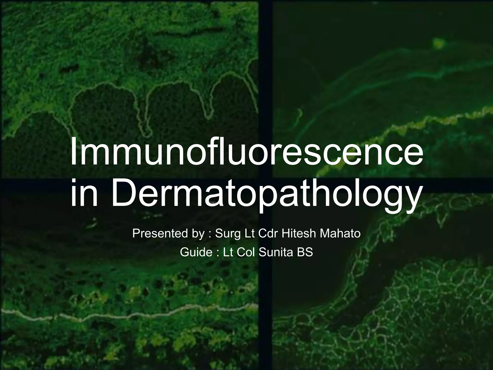
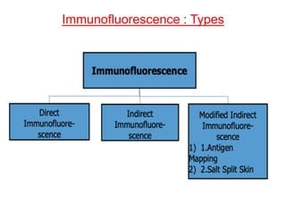
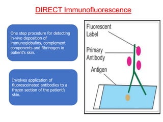
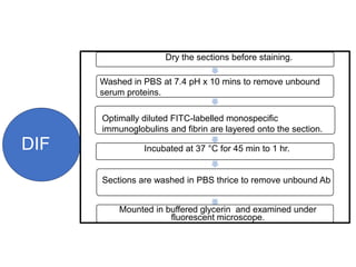
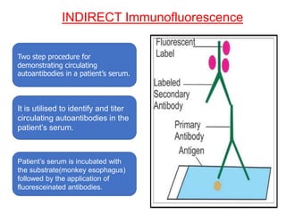
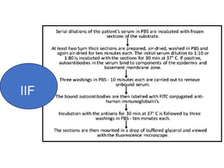
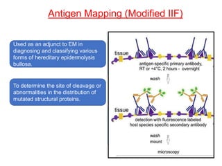
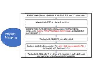
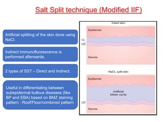
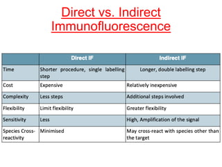
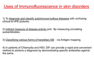
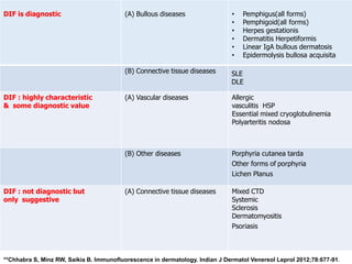
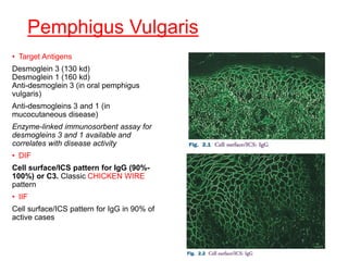
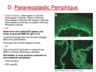
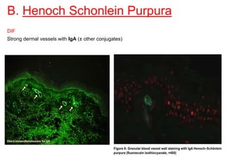
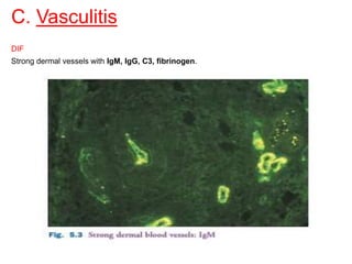
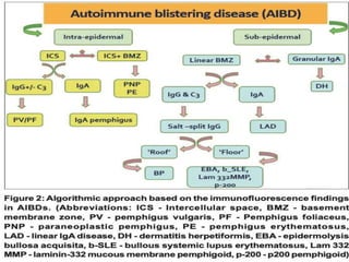
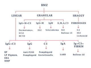
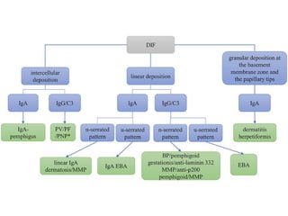
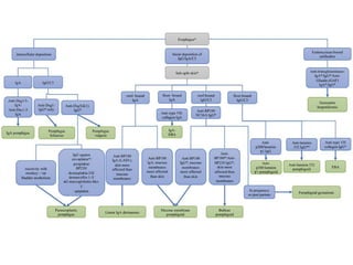
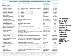
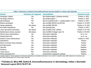
This document provides information about immunofluorescence techniques used in dermatopathology. It begins with the history and components of immunofluorescence microscopy. It then discusses the various applications of immunofluorescence including in skin, muscle, and renal biopsies. Specific techniques for skin biopsies like direct immunofluorescence, indirect immunofluorescence, antigen mapping, and salt split skin technique are explained. Common staining patterns seen in dermatological conditions like pemphigus vulgaris, pemphigus vegetans, pemphigus foliaceus, and paraneoplastic pemphigus are summarized. The document emphasizes the utility of immunofluorescence in diagnosing and classifying autoimmune bullous diseases and connective tissue disorders.
2. Contents
• Immunofluorescence :-
Introduction
Components of an Immunofluorescent microscope
Applications
Techniques
• Classification of various Dermatological disorders according to IF staining pattern.
• Salient IF staining characteristics in various Dermatological disorders.
• Recent advances
• Take home messages
3. Immunofluorescence
• Technique allowing the visualization of a specific protein or antigen in tissue sections or cells
in suspension by binding a specific antibody chemically conjugated with a fluorescent dye.
• Essential to supplement clinical findings and histopathology in the diagnosis of multiple
disorders.
• It permits early diagnosis, treatment, and subsequent monitoring of disease activity in
patients.
4. History
• 1941 - Coons et al, developed the technique of immunofluorescence by
showing the labelling of anti-pneumococcal antibodies with fluorescein in
the pulmonary tissue.
• 1963 – Beginning of immunopathology in dermatology
with the description of the lupus band test (LBT),i.e. deposits of
immunoglobulins and complement at the dermo-epidermal junction.
• 1964 - Beutner and Jordon used the indirect IF technique to demonstrate
antibodies in the sera of Pemphigus patients.
5. Principle of fluorescence
• Property of certain molecules called fluorophores to absorb light at one wave
length (excitation wavelength) and emit light at different wave length
(emission wavelength).
• Selective filters used for this purpose.
1. Energy is absorbed by the atom
which becomes excited.
2. The electron jumps to a higher
energy level.
3. Soon, the electron drops back to
the ground state, emitting a
photon (or a packet of light) –
fluorescence.
6. Immunofluorescence
Microscopy
• The ‘fluorescence microscope’ refers to
any microscope that uses fluorescence
to generate an image.
• Principle : A specific fluorescently
labelled antibody binds to the molecule
of interest and the location of the
antibody is determined by
fluorescence, hence leading to
immunopathological diagnosis.
7. Components :Immunofluorescence
Microscope
Components of a fluorescence microscope are :
1. Light source: Xenon arc lamp or
mercury-vapour lamp; LEDs and lasers
2. Excitation filter
3. Dichroic mirror (Dichroic beam splitter)
4. Emission filter
Epifluorescence: excitation of the fluorophore and
detection of the fluorescence are done through the
same light path (i.e. through the objective)
8. Applications of IF microscopy
1. Renal Biopsy
2. Skin biopsy
3. Muscle biopsy
4. Lung biopsy
5. Autoantibodies in serum and other body fluids
6. Hormones
7. Microbiology
8. Flow cytometry
9. FISH
9. Biopsy techniques : Skin disorders
• A 3- 4 mm punch biopsy is generally
adequate.
• In autoimmune blistering diseases (AIBD)
,an inflammed but unblistered perilesional
area is the ideal specimen.
• In collagenoses, the biospy should be
done in the active lesion in evolution
(avoid recent lesions, with less than 60
days);
• In vasculitis, preference should be given
to recent lesions
10.
11. Recommended sites of biopsy
(i) Bullous diseases- Unblistered perilesional area of fresh lesion.
Extreme precaution in hereditary EB patients due to fragile skin.
(ii) Dermatitis herpetiformis- Normal appearing perilesional skin.
(iii) Lupus erythematosus- Both lesional and apparently normal skin from
sun exposed areas in SLE.
In DLE only from lesional skin.
(iv) Vasculitis- Very fresh lesions.
PAN- deep dermis.
(v) Porphyria– Dorsum of hand and normal skin.
(vi) Lichen Planus- Inflamed mucosa &/ skin.
**Chhabra S, Minz RW, Saikia B. Immunofluorescence in dermatology. Indian J Dermatol Venereol Leprol 2012;78:677-
91.
12. Transportation of biopsy specimen
Michel’s Medium (MM ) : Best and preferred.
Ammonium sulfate, N ethylmaleimide,Potassium citrate buffer, Magnesium
sulfate, distilled water
Can preserve specimen upto 6 months.
pH 7- 7.2
Store at 4ºC
Saline soaked gauze/Saline can be used for transport over short intervals
upto 24 hrs in case Michel’s medium is unavailable.
14. The most important factors are :
1. Preservation of substrate antigen
2. Antibody conjugate
3. Fluorescence microscopy system
4. Staining and incubation
Immunofluorescence Technique
15. 1. SUBSTRATE ANTIGEN(DIF)
• Skin Biopsy - either quick-frozen or placed in Michel’s transport medium
for later quick freezing.
• Quick freezing by isopentane liquid nitrogen is the most widely used
method.
• Unfixed cryostat section (4-5µm) used.(Section thickness is important
because of it’s effect on non-specific staining)
• These are mounted on a slide previouly coated with gelatin/Poly L-lysine
and dried.
• Frozen tissue blocks can be stored in air tight plastic bag in low
temperature cabinets at -70˚C or lower.
16. Handling of sera for Indirect test
• About 3 ml of clotted blood used.
• Hereditory EB- 2-5 ml EDTA blood for mutation analysis.
• All sera should be refrigerated until tests are performed.
• Repeated freezing and thawing should be avoided, since this causes a
rapid loss of antibody activity.
• Positive and negative control sera must be frozen in aliquots of a size
adequate for single experiment.
• EDTA blood should not be frozen.
17. 2. ANTIBODY CONJUGATE
• Two types :-
Monospecific reagents used for direct staining of biopsies.
Human anti-whole-IgG conjugates used for the IIF test of sera.
• Specificity and high degree of reactivity : Most important prerequisites of antibodies
used in IF studies.
(Mentioned by the manufacturer ; confirmation is the responsibility of the user by use of
positive and negative controls.)
• Most commercial conjugates - lyophilized form.
• Require reconstitution with distilled water/ diluents.
• The undiluted stock should be divided into volumes of 0.10–0.50 ml and stored frozen
(−20 °C) until ready to be diluted.
• The diluted conjugates should be stored at 4 °C and not refrozen.
18. Antibodies regularly used for IF
• IgG
• IgM
• IgA
• C3
• Fibrinogen
• C5b-9
• C1q
• Kappa
• Lambda
**ThermoFisher/ Dako/ Pathnsitu :- Most common labs manufacturing
the above
21. DIRECT Immunofluorescence
One step procedure for detecting
in-vivo deposition of
immunoglobulins, complement
components and fibrinogen in
patient’s skin.
Involves application of
fluoresceinated antibodies to a
frozen section of the patient’s
skin.
22. DIF
Dry the sections before staining.
Sections are washed in PBS thrice to remove unbound Ab
Mounted in buffered glycerin and examined under
fluorescent microscope.
Washed in PBS at 7.4 pH x 10 mins to remove unbound
serum proteins.
Optimally diluted FITC-labelled monospecific
immunoglobulins and fibrin are layered onto the section.
Incubated at 37 °C for 45 min to 1 hr.
23. INDIRECT Immunofluorescence
Two step procedure for
demonstrating circulating
autoantibodies in a patient’s serum.
It is utilised to identify and titer
circulating autoantibodies in the
patient’s serum.
Patient’s serum is incubated with
the substrate(monkey esophagus)
followed by the application of
fluoresceinated antibodies.
25. Antigen Mapping (Modified IIF)
Used as an adjunct to EM in
diagnosing and classifying various
forms of hereditary epidermolysis
bullosa.
To determine the site of cleavage or
abnormalities in the distribution of
mutated structural proteins.
26. Antigen
Mapping
Patient’s skin (4 micron) section & NHS/salt split skin on glass slide
Washed with PBS X 15 min & fan dried
Sections treated with panel of primary Ab against known BMZ
components (type IV, XVII& VII collagen; laminin 322) & incubated at
room temp X 1 hr
Washed with PBS X 15 min & fan dried
Sections treated with secondary Ab ( anti – IgG mouse specific Abs )
conjugated with fluorescein dye
Washed with PBS after 1 hr , dried and mounted in buffred glycerol
and examined under fluorescent microscope
27.
28. Salt Split technique (Modified IIF)
Artificial splitting of the skin done using
NaCl.
Indirect Immunofluorescence is
performed afterwards.
2 types of SST – Direct and Indirect.
Useful in differentiating between
subepidermal bullous diseases (like
BP and EBA) based on BMZ staining
pattern : Roof/Floor/combined pattern
29. Direct SST - Patient’s skin &
Indirect SST - NHS/on glass slide
IIF with patient’s serum is carried out
Artificially splitting of the skin at the level of
lamina lucida by incubating it in 1 M solution
of sodium chloride for 24 h
SALT SPLIT SKIN
TECHNIQU
E (SST)
Cryocut sections are prepared
Salt Split
Skin
Technique
31. Uses of Immunofluorescence in skin disorders
1) To diagnose and classify autoimmune bullous diseases with confusing
clinical & HPE pictures.
2) Indirect measure of disease activity and : By measuring circulating
autoantibodies.
3) Classifying various forms of hereditary EB : via Antigen mapping.
4) In patients of Chlamydia and HSV, DIF can provide a rapid and convenient
method to achieve a diagnosis by demonstrating specific antibodies against
the same.
35. Approach to Bullous skin disorders
• Clinical history & classical features
• Age of onset, family history & drug history
• Nature of bullae- flaccid /tense
• Bulla spread sign & Nikolsky sign : to look for acantholysis
• Tzanck smear : acantholytic cells ; eosinophils; neutrophils
• Histopathology : level of split & type of cellular infiltrate
• Immunofluorescence : both direct & indirect for autoimmune bullous
disorders
39. Reporting IF findings in a skin biopsy
• A skin biopsy should be reported under the following heads when seen under a
fluorescent microscope :-
• (i). Type of immunoreactant: IgG, IgA, IgM, C3, fibrin.
• (ii) Location of immune deposits: Intercellular spaces (ICS) in
epidermis/epidermal nuclear staining (ENS) or in vivo ANA/basement membrane
zone (BMZ)/subepidermal blood vessels/hair shaft/cytoid (civatte or colloid) bodies.
• (iii) Extent of staining: Focal or diffuse.
• (iv) Intensity of staining: Semi-quantitative grading of strength of fluorescence: +
to ++++.
• (v) Pattern of immune complex deposits: Granular/linear/shaggy. The granular
pattern is further divided into coarse granules, speckles, threads, and fibrils.
The description of all these staining characteristics leads to immunopathological
diagnosis.
42. Staining patterns in IF of skin
1. Cell surface/Intercellular space pattern
2. Linear BMZ staining pattern.
3. Granular BMZ staining pattern
4. Shaggy BMZ staining pattern
5. Vascular staining pattern and others
• Various conditions in dermatology can be grouped according to the
above staining patterns on IF
44. A. Pemphigus Vulgaris
Clinical Features
• Most common subtype of pemphigus.
• Flaccid bullae rupture, leaving
denuded areas.
• Oral involvement is often the initial
manifestation, in approximately 60% of
patients.
• Positive Nikolsky sign
• Skin manifestations involve scalp,
chest, back, intertriginous areas
Pathology : Pathogenic IgG antibodies to
intra-epidermal cell adhesion molecules
(desmoglein).
Suprabasal blisters.
‘Row of tombstones’ appearance of
basal keratinocytes
46. Pemphigus Vulgaris
• Target Antigens
Desmoglein 3 (130 kd)
Desmoglein 1 (160 kd)
Anti-desmoglein 3 (in oral pemphigus
vulgaris)
Anti-desmogleins 3 and 1 (in
mucocutaneous disease)
Enzyme-linked immunosorbent assay for
desmogleins 3 and 1 available and
correlates with disease activity
• DIF
Cell surface/ICS pattern for IgG (90%-
100%) or C3. Classic CHICKEN WIRE
pattern
• IIF
Cell surface/ICS pattern for IgG in 90% of
active cases
47. B. Pemphigus Vegetans
• DIF
• IIF
• Target Antigens
All findings identical to Pemphigus vulgaris
48. C. Pemphigus Foliaceous
• DIF : Identical to P.vulgaris.
• IIF
Identical to P.vulgaris.
Guinea pig lip or Oesophagus can also be used (in addition to monkey
oesophagus)
• Target Antigens : Desmoglein 1 (160 kd)
49. D. Paraneoplastic Pemphigus
• Target Antigens : Desmoglein 3 (130 kd),
Desmoglein 1(160 kd), Plectin (>500 kd),
Desmoplakin I (250 kd), BP antigen I (230 kd),
Desmoplakin II (210 kd), Envoplakin (210 kd),
Periplakin (190 kd)
• DIF :
Weak focal cell surface/ICS pattern and
linear or granular BMZ for IgG or C3.
Lichenoid changes also may be seen (shaggy
BMZ and cytoid bodies).
Increased rate of false-negative results.
• IIF
Cell surface/ICS (IgG) with or without linear
BMZ on monkey esophagus substrate.
Rat bladder is most sensitive substrate for
paraneoplastic pemphigus.
- 75% sensitive
- 83% specific
50. E. Pemphigus Erythematosus
• C/f : Involves seborrheic areas of
face and trunk, mimicking lupus
erythematosus.
• Target Antigens : Desmoglein 1(160
kd)
• DIF : Cell surface/ICS pattern (IgG
or C3) in >75% cases and granular
deposits of (IgM, C3) at the
DEJ.(Positive Lupus Band TEST)
• IIF Cell surface/ICS pattern: IgG ■
ANA
51. F. IgA Pemphigus
• C/f :
Pruritic vesicles and pustules in an
annular pattern.
Predilection for intertriginous areas.
Rare mucous membrane involvement.
Associated disorders :-
- IgA monoclonal gammopathy
- Crohn disease/gluten-sensitive
enteropathy
• Target Antigens : Desmocollin 1 (subset
of patients target Desmogleins 3 and 1)
• DIF : Cell surface/ICS pattern for IgA.
• IIF : Positive in 50% of patients.
53. • Bullous pemphigoid is the most common
immunobullous disease affecting the elderly.
• Early signs:
• Dermatitis-like: dry or exudative discoid
eczema
• Urticaria-like: erythematous urticated
plaques
• Nonspecific: patchy erythema and/or
dryness
• Tense bullae.
• Nikolsky sign negative.
• Sub-epidermal acantholytic blisters
• Autoantibodies against BPAG2, a component of
the hemidesmosome
A. Bullous pemphigoid
55. HPE
• Unilocular subepidermal blister
• Cell rich type – blisters develop
on erythematous skin.
eosinophils predominant cell in
blister cavity and in dermis
• Cell poor type – blisters develop
on normal skin.
scant perivascular lymphocytic
infiltrate with few eosinophils
56. A. Bullous Pemphigoid
• Target Antigens
BP230 (BPAG1)
BP180 (BPAG2)
-NC16A (immunodominant region of
BP180)
• DIF
Linear basement membrane zone
(BMZ): IgG (90%)
Linear BMZ: C3 (>90%, nearly all)
• IIF
Linear BMZ: IgG on monkey
oesophagus substrate (circulating
IgG antibody in 75% of cases)
57. Salt-split skin (SSS): epidermal pattern(deposition seen on epidermal side)
Occasionally combined epidermal-dermal pattern
58. (B).Pemphigoid Gestationis
• Target Antigens
BP230 (BPAG1)
BP180 (BPAG2) (most important)
-NC16A (immunodominant region of BP180)
• DIF
Linear BMZ: C3 (100% of cases,
diagnostic)
Linear BMZ: IgG (approximately 25% of
cases)
• IIF
Linear BMZ: IgG (<25% of cases, does not
correlate with disease activity)
HG factor: 50% of cases
• Salt-split skin (SSS): epidermal pattern
59. (C).Lichen Planis Pemphigoides
• Target Antigens
BP230 (BPAG1)
BP180 (BPAG2) (most important)
-NC16A (immunodominant region of
BP180)
• DIF
Linear BMZ: IgG and C3 with
changes of lichen planus (i.e., cytoid
bodies with IgM, IgA, C3, and shaggy
BMZ with fibrinogen)
• IIF
Linear BMZ: IgG antibodies in 50% of
patients
• Salt-split skin (SSS): epidermal
pattern
60. (D).Mucous membrane Pemphigoid
• Target Antigens
BP230 (BPAG1)
BP180 (BPAG2) (C-terminus
and some NC16A)
β4 integrin subunit
Laminin-5
Laminin-6
Type VII collagen (290 kd)
• DIF
Linear BMZ: IgG, C3 , IgA
• IIF
Linear BMZ: IgG antibodies in
15-20% of patients.
• Salt-split skin (SSS):
epidermal/dermal/combine
d pattern
61. (E).Bullous Lupus Erythematosus
• Target Antigens
Type VII collagen (290 kd)
-NC1 domain
• DIF
Linear BMZ(>50% cases): IgG
and C3; IgM and IgA also (if
perilesional biopsy of bullae)
Granular BMZ(>25% cases):
IgG, IgM, C3 (if lesional biopsy of
malar rash or other cutaneous
involvement of lupus)
• IIF
Antinuclear antibodies (ANA)
BMZ antibodies not detected on
monkey esophagus but may be
detected on SSS.
• Salt-split skin (SSS): dermal
pattern
62. (F).Epidermolysis Bullosa Acquisita
• Target Antigens
Type VII collagen (290 kd)
-NC1 domain
• DIF
Linear BMZ: IgG (100%) and C3;
occasionally IgA (66%) or IgM
(50%)
• IIF
Linear BMZ: IgG (in 50%
patients)
• Salt-split skin (SSS): dermal
pattern
64. A. Dermatitis Herpetiformis
Clinical Features
• Erythematous papules or vesicles symmetrically distributed over extensor surfaces
of the elbows, knees, buttocks, back, and scalp
• Vesicles may be grouped in a herpetiform configuration
• Intensely pruritic
• Often associated with multiple erosions caused by scratching
• One part of a spectrum of gluten-sensitive disorders that includes celiac disease
• An indirect consequence of a gluten-sensitive enteropathy
HPE:
Early lesions: Accumulation of
neutrophils (microabscesses) at
the tips of dermal papillae.
Older lesions: Subepidermal
vesiculation
65. A. Dermatitis Herpetiformis
• Target Antigens
Tissue transglutaminase in gluten-sensitive
diseases.
Epidermal transglutaminase in skin lesions of
dermatitis herpetiformis.
Circulating IgA antibody testing to tissue
transglutaminase by ELISA is recommended(to
identify the presence of a gluten-sensitive enteropathy
and to monitor response to a gluten-free diet).
• DIF
Granular BMZ pattern for IgA, with stippling of
dermal papillae (100%)
Occasionally C3 (50%); IgG and IgM less often.
• IIF
IgA class endomysial antibody staining
demonstrated in 76% of persons receiving a normal
gluten-containing diet.
Endomysial antibody testing is recommended to
identify a gluten-sensitive enteropathy and to monitor
response to a gluten-free diet.
66. B. Systemic Lupus Erythematosus
• Clinical Features
• Malar “butterfly” rash
• Exacerbated by ultraviolet light
• Waxes and wanes with underlying systemic lupus
• Erythematosus disease activity
• Discoid rash may occur at some point in the disease
• Photosensitivity
• Painless oral ulcers
• Nonerosive arthritis involving two or more peripheral joints
• Serositis
• Central nervous system involvement
• Nephritis
• Anemia, leukopenia, lymphopenia, thrombocytopenia
67. Classification criteria of SLE (2019)
**2019 European League Against Rheumatism/American College of Rheumatology Classification Criteria for Systemic Lupus Erythematosus
68. B.1. Systemic Lupus Erythematosus
• Immunologic disorder
ANA in abnormal titer.
Anti–double-stranded DNA in abnormal titer.
Presence of anti-Sm antibody.
IgG or IgM anticardiolipin antibodies.
• DIF
Granular BMZ pattern for IgG, IgM, IgA, C3
(sun-exposed involved skin >90%; sun-
exposed nonlesional skin 50%; non–sun-
exposed nonlesional skin 30%)
Speckled epidermal nuclei pattern for IgG
in 10% to 15%
High yield with SLE–specific skin lesions:
malar rash, erythematous edematous
plaques, and active disease
• IIF : ANA
69.
70. B.2. Discoid Lupus Erythematosus
• Clinical Features
■ Discoid rash typically presents as sharply demarcated,
erythematous, indurated plaques with hyperkeratosis,
atrophy, telangiectasia, and follicular plugging
■ Hypopigmentation or hyperpigmentation may be
prominent
■ Most frequently involves the face, scalp, ears, V area of
the neck, and extensor aspects of the arms
■ Scalp involvement may lead to scarring alopecia
• Immunologic disorder
■ Antibodies to single-stranded DNA may be present
■ ANA present in low titers in 30% to 40% of patients
■ Ro/SS-A and La/SS-B autoantibodies are rare
■ Antibodies to double-stranded DNA are uncommon
• DIF
■ Granular BMZ pattern for IgG and IgM (involved skin
>90%)
■ May have shaggy, thick BMZ with fibrinogen
■ Cytoid bodies with IgM and IgA
• IIF : None (ANA rarely)
71. B.3. Subacute Cutaneous Lupus
Erythematosus
• Immunologic disorder
■ Autoantibodies to Ro/SS-A
ribonucleoprotein present in 70% to 90%
strongly support the diagnosis
■ Autoantibodies to La/SS-B present in
30% to 50%
■ ANA present in 60% to 80%
DIF
■ Granular BMZ pattern for IgG, IgM, C3
(involved skin >90%)
■ Epidermal/keratinocyte
intracytoplasmic particulate deposition
with IgG
■ Cytoid bodies with IgM and IgA
• IIF : ANA
72. C. Mixed Connective Tissue Disease
• Immunologic disorder
High titers of antibody to the extractable nuclear antigen.
• DIF
■ Granular BMZ pattern rare (15%)
■ Speckled epidermal nuclei for IgG in 46% to 100%
• IIF : ANA
73. D. Systemic Scleroderma
• Clinical Features
■ Skin tightening extends from fingers to upper extremities, trunk, face, and, finally, lower
extremities
■ Raynaud phenomenon
■ Nail-fold capillary changes (giant or sausage-shaped loops)
■ Edema of the hands and fingers
■ Flexion contractures and sclerodactyly with waxy, shiny, atrophic skin
■ Ulcers on fingertips and over knuckles
■ Masklike, expressionless face, with loss of normal facial lines
■ Small, sharp nose, thinning of lips and hair
■ Microstomia with radial furrowing around the mouth
■ Matlike telangiestasias on face and upper trunk.
■ Esophageal dysfunction in more than 90%
■ Pulmonary fibrosis
■ Myocardial fibrosis in 50% to 70%
■ Renal involvement with hypertension
74. D. Systemic Scleroderma
• Immunologic disorder
Anticentromere antibodies only in
12% to 25% of patients (present in
50%-96% of patients with CREST
syndrome)
Scl-70 autoantibodies (30%)
• DIF
Granular BMZ pattern for IgM (sun-
exposed 60%)
Speckled epidermal nuclei pattern in
20%
Shaggy BMZ with fibrinogen
• IIF : ANA
75. D. Dermatomyositis
• Clinical Features
• Erythematous, violaceous papules over the dorsal aspect of the interphalangeal or
metacarpophalangeal joints (termed Gottron papules)
• Symmetric, confluent, violaceous erythema over the interphalangeal or metacarpophalangeal
joints, olecranon process, medial malleoli, and patella (termed Gottron sign)
• Periorbital, violaceous (heliotrope) erythema and edema
• Periungual telangiectasia with or without cuticular hemorrhage and dystrophic cuticles
• Violaceous erythema over dorsal aspect of arms, posterior aspect of shoulder and neck (shawl
sign)
• Poikiloderma atrophicans
• Pruritus can be severe
• Cutaneous calcification is more common in juvenile form (40%-50%)
76. D. Dermatomyositis
• Immunologic disorder
• Increased ANA levels on human
tumor cell substrates (60%-80%)
• Anti-Jo-1 (20% of cases of classic
dermatomyositis and 40% of
cases of polymyositis)
• DIF
• Granular BMZ pattern for IgM,
IgG, and C3 (low intensity)
• Cytoid bodies for IgM and IgA,
shaggy BMZ with fibrinogen.
• IIF : ANA
78. D. Lichenoid Tissue Reactions
• Clinical Features
• Lichen planus
Erythematous to violaceous, flat-topped, polygonal papules with fine, whitish reticulations
termed Wickham striae
Distributed symmetrically over flexural areas of extremities.
Pruritus usually present.
Hypertrophic lichen planus extremely pruritic.
Presence of Koebner phenomenon.
White, reticulated pattern occurs with oral involvement .
Nail pterygium or complete loss of nail plate.
• Lichenoid drug reactions : Beta-adrenergic blockers, Penicillamine, ACE inhibitors
• Lichenoid photodermatoses : photodistributed lichenoid reaction. Caused by
carbamazepine, chlorpromazine, ethambutol, quinine, tetracyclines, thiazide diuretics, and
furosemide.
79. D. Lichenoid Tissue Reactions
• DIF
Shaggy BMZ pattern for fibrinogen
Cytoid bodies for IgM and IgA, occasionally IgG, C3, and fibrinogen
• IIF : ANA for Lupus erythematosus
88. **Chhabra S,
Minz RW,
Saikia B.
Immunofluore
scence in
dermatology.
Indian J
Dermatol
Venereol
Leprol
2012;78:677-
91.
89. **Chhabra S, Minz RW, Saikia B. Immunofluorescence in dermatology. Indian J Dermatol
Venereol Leprol 2012;78:677-91.
90. Potential pitfalls/Limitations of IF
1) Expensive and labour intensive requirements like :-
• Advanced laboratories that are proficient in the performance and interpretation of these
tests.
• A thoroughly trained team comprising of a pathologist + technologist.
• A specialized lab having facilities of cryostat for cutting frozen sections.
• Deep freezers(−20° C/−80 °C) to store these sections until staining.
• Fluorescence microscope to report the DIF findings.
2) Skin biopsy is fragile and needs to be transported in Michel’s fluid only.
3) Photo bleaching : The fluorescence staining quenches rapidly on exposure to light,
more so under the UV light of the fluorescence microscope, resulting in the necessity of
fast reporting and documentation of findings using a digital camera.
91. Potential pitfalls/Limitations
4) No long-term storage period is ideal for reporting of DIF-stained skin biopsy slides.
5) Autofluorescence : Biological autofluorescence in mammalian cells, flavin
coenzymes (NAD, FMN, NADH). Washing with 0.1% sodium borohydride in
phosphate-buffered saline prior to antibody incubation can reduce this. (Always use a
control to differentiate autofluorescence from specific staining)
6) Treated cases may show no IF findings in certain disorders.
7) Skin biopsies gets fixed on exposure to formalin vapours so extreme care is
required in handling the specimen. Dried and/or fixed biopsies are reported unfit for
DIF.
92. Recent advances
AUTOMATED DIF
Why automation??
• DIF staining is labour-intensive.
• Manual handling ->inconsistent
staining results and unspecific
background staining.
• Automated technology for
DIF staining : EUROTide
incubation technique + biochip
based system EUROPath and
applying an automated
procedure for DIF staining.
**Lemcke S, Sokolowski S, Rieckhoff N, Buschtez M et al. Automated direct immunofluorescence analyses of skin biopsies. J Cutan Pathol 2015.
93. Advantages of automated DIF
1) Saves reagent, more accurate.
2) More intense specific IF staining. Less background staining.
3) Costs for antibodies can be reduced almost 2.5 times when using
EUROTide/EUROPath.
4) Upside down incubation prevents any unsolved material to attach to the
tissue and interfere with the reading of the slides.
5) Using macrochips M14, multiple stainings (upto 5) of one patient can be
compiled on a single EUROPath slide thus :-
• accelerating diagnostic assessment
• limiting the risk of confusing patient samples
• sparing storage space.
94.
95. Take home messages
• Immunofluorescence is not a substitute for histopathology but is in fact
complementary to it.
• Always see DIF slides along with H&E slides & correlate with history + clinical
features.
• The values of positive or negative immunofluorescence findings are dependent on
the experience and skill of the laboratory staff and also on the knowledge of the
observer who reports them.
• Always use control to differentiate specific staining from background staining.
• Close cooperation with the clinician is essential, who in turn should select
representative and fresh lesions for biopsy and provide a good history.
96. References
• Rosai and Ackermann’s Surgical Pathology
• Lever’s Histopathology of Skin
• Bancroft Theory and Practice of Histological Techniques
• Amer N. Kalaaji, Atlas of Immunofluorescence in Dermatology: Patterns and
Target Antigens, Mayo Clinic Scientific Press, 2006.
• Chhabra S, Minz RW, Saikia B. Immunofluorescence in dermatology. Indian J
Dermatol Venereol Leprol 2012;78:677-91.
• Lemcke S, Sokolowski S, Rieckhoff N, Buschtez M et al. Automated direct
immunofluorescence analyses of skin biopsies. J Cutan Pathol 2015.