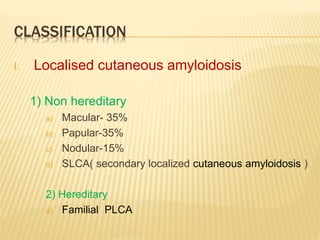
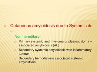
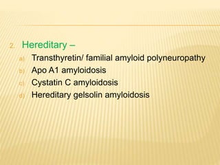
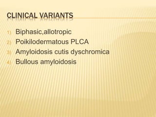
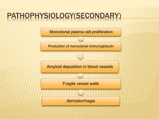
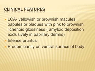
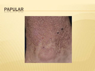
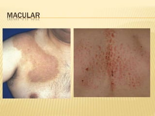
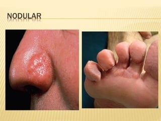
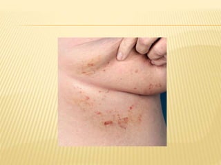
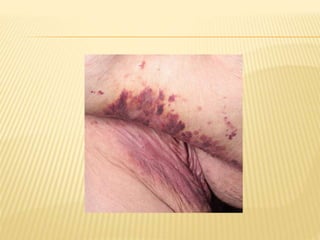
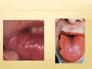
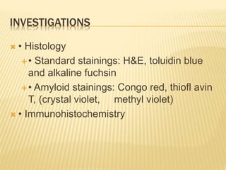
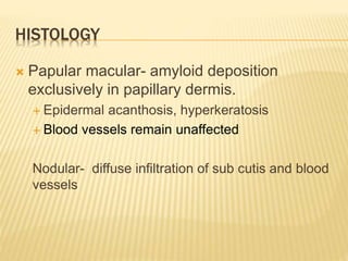
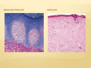
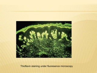
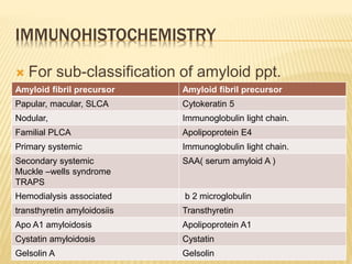
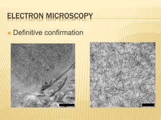
DR. SABHA TALIB NEAZEE
CUTANEOUS AMYLOIDOSIS
DEFINITION
 AMYLOID= STARCH LIKE (LATIN)
 AMYLOIDOSIS IS A CONDITION IN WHICH
THERE IS EXTRACELLULAR DEPOSITION
OF ANY OF GROUP OF AUTOLOGOUS
PROTEINS LEADING TO CHANGES IN
TISSUE ARCHITECTURE AND FUNCTION
 7.5-10 nm wide, linear, non branching
tubular fibrils loosely arranged in meshwork.
 Alpha and beta pleated sheet form.
 Distinction between primary and secondary
– Tt with KmnO4 prior to staining with congo
red.
 Primary amyloid retains its affinity for the dye
and green birefringence under polarized light
, whereas secondary loses both
characteristics
ETIOLOGIC FACTORS
 Racial susceptibility
 Chronic rubbing and friction
 Role of actinic radiation
 Familial cases with AD mode of inheritance
 Increased and reduced expression of various
pro or anti- apoptotic factors respectively
CLASSIFICATION
I. Localised cutaneous amyloidosis
1) Non hereditary
a) Macular- 35%
b) Papular-35%
c) Nodular-15%
d) SLCA( secondary localized cutaneous amyloidosis )
2) Hereditary
a) Familial PLCA
II. Cutaneous amyloidosis due to Systemic ds
–
1. Non hereditary-
a) Primary systemic and myeloma or plasmocytoma –
associated amyloidosis (AL)
b) Secondary systemic amyloidosis with inflammatory
tumour
c) Secondary hemodialysis associated ststemic
amyloidosis
2. Hereditary –
a) Transthyretin/ familial amyloid polyneuropathy
b) Apo A1 amyloidosis
c) Cystatin C amyloidosis
d) Hereditary gelsolin amyloidosis
3) Hereditary systemic ds with secondary
cutaneous amyloidosis
a) Muckle wells syndrome
b) TNF receptor 1 associated periodic fever
syndrome
CLINICAL VARIANTS
1) Biphasic,allotropic
2) Poikilodermatous PLCA
3) Amyloidosis cutis dyschromica
4) Bullous amyloidosis
PATHOPHYSIOLOGY (PRIMARY)
Friction
Friction
Apoptosis of basal keratinocytes
Release of cytokeratins
Covered with autoantibodies
Phagocytosed by macrophages
Degraded by enzymes to amyloid k
PATHOPHYSIOLOGY(SECONDARY)
Monoclonal plasma cell proliferation
Production of monoclonal immunoglobulin
Amyloid deposition in blood vessels
Fragile vessel walls
dermatorrhagia
CLINICAL FEATURES
 LCA- yellowish or brownish macules,
papules or plaques with pink to brownish
lichenoid glossiness ( amyloid deposition
exclusively in papillary dermis)
 Intense pruritus
 Predominantly on ventral surface of body
PAPULAR
MACULAR
NODULAR
 Secondary to systemic ds- petechiae,
ecchymosis and non healing ulcers.( amyloid
deposition in deeper skin layers and blood
vessels )
 Purpura –above nipple line.
 Bleeding in periorbital and intertriginous
areas
 Scleroderma amyloidosum Gottron-
scleroderma like changes by extensive
infiltration of dermis.
 Meretoja syndrome- massive cutis laxa,
extensive patechiae and haemorrhage,
hypotrichosis or alopecia seen in gelsolin
amyloidosis.
 TRAPS- prolong fever episodes, abdominal
pain, myalgias, migratory cutaneous
eryhtemas.
RACCOON EYES
INVESTIGATIONS
 • Histology
• Standard stainings: H&E, toluidin blue
and alkaline fuchsin
• Amyloid stainings: Congo red, thiofl avin
T, (crystal violet, methyl violet)
 • Immunohistochemistry
 • Electron microscopy
• From skin sample: for all kinds of
amyloidoses affecting the skin
• Abdominal fat or rectum biopsy: for all
suspected systemic amyloidoses and
nodular primary localized cutaneous
HISTOLOGY
 Papular macular- amyloid deposition
exclusively in papillary dermis.
 Epidermal acanthosis, hyperkeratosis
 Blood vessels remain unaffected
Nodular- diffuse infiltration of sub cutis and blood
vessels
 Staining properties-
 Congo red-green birefringence under polarized
light = DICHROMISM
 Methyl violet, cresyl violet- metachromatic
staining
 PAS staining
 Van geison`s stain- yellow brown color
 Pagoda red- more specific
 RIT scarlet No.5 – more specific
MACULAR/PAPULAR NODULAR
Thioflavin staining under fluoresence microscopy
IMMUNOHISTOCHEMISTRY
 For sub-classification of amyloid ppt.
Amyloid fibril precursor Amyloid fibril precursor
Papular, macular, SLCA Cytokeratin 5
Nodular, Immunoglobulin light chain.
Familial PLCA Apolipoprotein E4
Primary systemic Immunoglobulin light chain.
Secondary systemic
Muckle –wells syndrome
TRAPS
SAA( serum amyloid A )
Hemodialysis associated b 2 microglobulin
transthyretin amyloidosiis Transthyretin
Apo A1 amyloidosis Apolipoprotein A1
Cystatin amyloidosis Cystatin
Gelsolin A Gelsolin
ELECTRON MICROSCOPY
 Definitive confirmation
MANAGEMENT

Cutaneous amyloidosis

  • 1.
    DR. SABHA TALIBNEAZEE CUTANEOUS AMYLOIDOSIS
  • 2.
    DEFINITION  AMYLOID= STARCHLIKE (LATIN)  AMYLOIDOSIS IS A CONDITION IN WHICH THERE IS EXTRACELLULAR DEPOSITION OF ANY OF GROUP OF AUTOLOGOUS PROTEINS LEADING TO CHANGES IN TISSUE ARCHITECTURE AND FUNCTION
  • 3.
     7.5-10 nmwide, linear, non branching tubular fibrils loosely arranged in meshwork.  Alpha and beta pleated sheet form.  Distinction between primary and secondary – Tt with KmnO4 prior to staining with congo red.  Primary amyloid retains its affinity for the dye and green birefringence under polarized light , whereas secondary loses both characteristics
  • 4.
    ETIOLOGIC FACTORS  Racialsusceptibility  Chronic rubbing and friction  Role of actinic radiation  Familial cases with AD mode of inheritance  Increased and reduced expression of various pro or anti- apoptotic factors respectively
  • 5.
    CLASSIFICATION I. Localised cutaneousamyloidosis 1) Non hereditary a) Macular- 35% b) Papular-35% c) Nodular-15% d) SLCA( secondary localized cutaneous amyloidosis ) 2) Hereditary a) Familial PLCA
  • 6.
    II. Cutaneous amyloidosisdue to Systemic ds – 1. Non hereditary- a) Primary systemic and myeloma or plasmocytoma – associated amyloidosis (AL) b) Secondary systemic amyloidosis with inflammatory tumour c) Secondary hemodialysis associated ststemic amyloidosis
  • 7.
    2. Hereditary – a)Transthyretin/ familial amyloid polyneuropathy b) Apo A1 amyloidosis c) Cystatin C amyloidosis d) Hereditary gelsolin amyloidosis
  • 8.
    3) Hereditary systemicds with secondary cutaneous amyloidosis a) Muckle wells syndrome b) TNF receptor 1 associated periodic fever syndrome
  • 9.
    CLINICAL VARIANTS 1) Biphasic,allotropic 2)Poikilodermatous PLCA 3) Amyloidosis cutis dyschromica 4) Bullous amyloidosis
  • 10.
    PATHOPHYSIOLOGY (PRIMARY) Friction Friction Apoptosis ofbasal keratinocytes Release of cytokeratins Covered with autoantibodies Phagocytosed by macrophages Degraded by enzymes to amyloid k
  • 11.
    PATHOPHYSIOLOGY(SECONDARY) Monoclonal plasma cellproliferation Production of monoclonal immunoglobulin Amyloid deposition in blood vessels Fragile vessel walls dermatorrhagia
  • 12.
    CLINICAL FEATURES  LCA-yellowish or brownish macules, papules or plaques with pink to brownish lichenoid glossiness ( amyloid deposition exclusively in papillary dermis)  Intense pruritus  Predominantly on ventral surface of body
  • 13.
  • 14.
  • 15.
  • 16.
     Secondary tosystemic ds- petechiae, ecchymosis and non healing ulcers.( amyloid deposition in deeper skin layers and blood vessels )  Purpura –above nipple line.  Bleeding in periorbital and intertriginous areas  Scleroderma amyloidosum Gottron- scleroderma like changes by extensive infiltration of dermis.
  • 17.
     Meretoja syndrome-massive cutis laxa, extensive patechiae and haemorrhage, hypotrichosis or alopecia seen in gelsolin amyloidosis.  TRAPS- prolong fever episodes, abdominal pain, myalgias, migratory cutaneous eryhtemas.
  • 18.
  • 22.
    INVESTIGATIONS  • Histology •Standard stainings: H&E, toluidin blue and alkaline fuchsin • Amyloid stainings: Congo red, thiofl avin T, (crystal violet, methyl violet)  • Immunohistochemistry
  • 23.
     • Electronmicroscopy • From skin sample: for all kinds of amyloidoses affecting the skin • Abdominal fat or rectum biopsy: for all suspected systemic amyloidoses and nodular primary localized cutaneous
  • 24.
    HISTOLOGY  Papular macular-amyloid deposition exclusively in papillary dermis.  Epidermal acanthosis, hyperkeratosis  Blood vessels remain unaffected Nodular- diffuse infiltration of sub cutis and blood vessels
  • 25.
     Staining properties- Congo red-green birefringence under polarized light = DICHROMISM  Methyl violet, cresyl violet- metachromatic staining  PAS staining  Van geison`s stain- yellow brown color  Pagoda red- more specific  RIT scarlet No.5 – more specific
  • 26.
  • 27.
    Thioflavin staining underfluoresence microscopy
  • 28.
    IMMUNOHISTOCHEMISTRY  For sub-classificationof amyloid ppt. Amyloid fibril precursor Amyloid fibril precursor Papular, macular, SLCA Cytokeratin 5 Nodular, Immunoglobulin light chain. Familial PLCA Apolipoprotein E4 Primary systemic Immunoglobulin light chain. Secondary systemic Muckle –wells syndrome TRAPS SAA( serum amyloid A ) Hemodialysis associated b 2 microglobulin transthyretin amyloidosiis Transthyretin Apo A1 amyloidosis Apolipoprotein A1 Cystatin amyloidosis Cystatin Gelsolin A Gelsolin
  • 29.
  • 30.