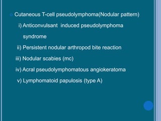
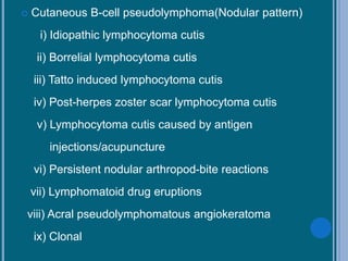
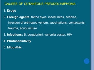
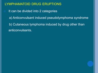
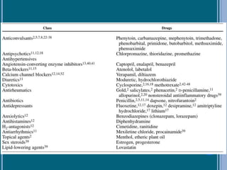
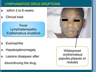
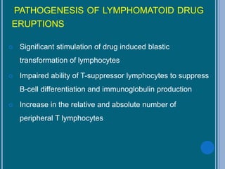
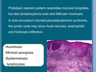
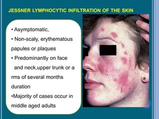
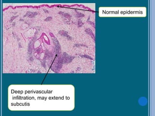
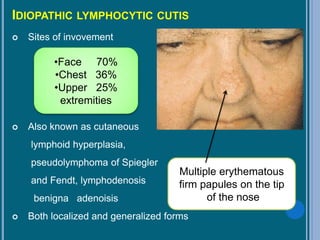
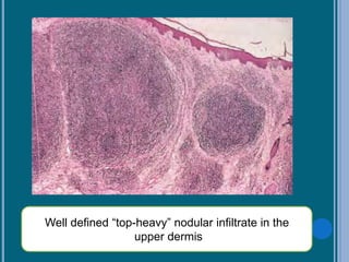
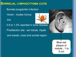
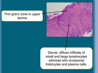
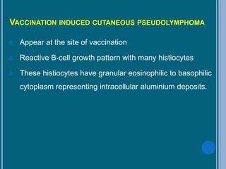
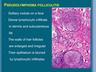
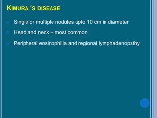
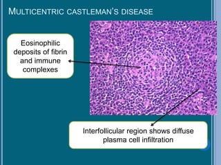
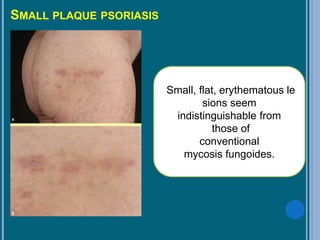
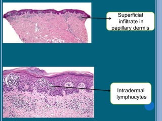
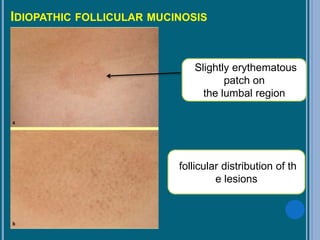
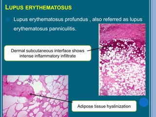
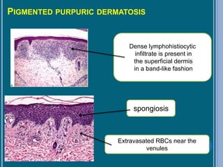
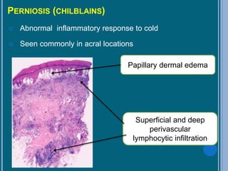
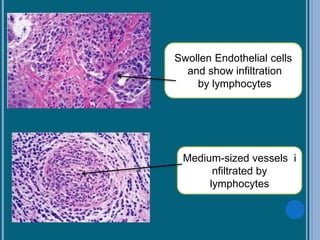
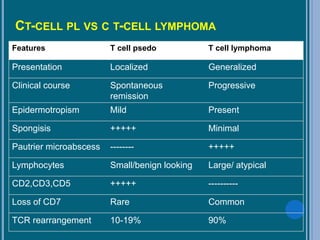
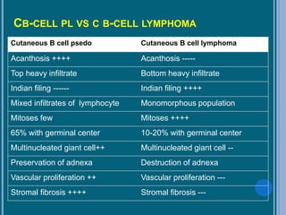
1. Cutaneous T-cell pseudolymphomas
A) Primarily with stripe-like infiltration (the majority of cases)
Lymphomatoid drug eruption (most cases);
Lymphomatoid contact dermatitis;
Actinic reticuloid;
Nodular scabies (individual cases);
Idiopathic forms;
Clonal cutaneous T-cell pseudolymphomas.
B) Primarily with nodular infiltration (a small percentage
of the cases)
Drug-induced – mainly by anti-convulsive drugs
Persistent nodules after insect bites;
Nodular scabies (the majority of cases).
2. Cutaneous B-cell pseudolymphomas (with nodular infiltration)
Cutaneous lymphocytoma from Borrelia burgdorferi;
Cutaneous lymphocytoma after antigens injection;
Cutaneous lymphocytoma resulting from tattoo;
Cutaneous lymphocytoma after Herpes zoster;
Idiopathic forms;
Clonal cutaneous B-cell pseudolymphomas
NEOPLASMS AND PROLIFERATIONS OF FOLLICULAR LINEAGE
NEOPLASMS AND PROLIFERATIONS WITH SEBACEOUS DIFFERENTIATION
NEOPLASMS AND PROLIFERATIONS WITH APOCRINE DIFFERENTIATION
NEOPLASMS AND PROLIFERATIONS WITH ECCRINE DIFFERENTIATION
A comprehensive review of Cutaneous Lymphomas - both B-Cell and T-Cell with latest treatment strategies. Target audience are oncologists, dermatologists, oncology physicians, dermatology and oncology fellows
various cutaneous lymphomas though having low incidence but need to be diagnosed accurately. they can be mimiced by many non neoplastic conditions of skin. so discussing both T and B cell lymphomas
subcorneal
intraepidermal
subepidermal
pemphigus
nikolski's sign
pemphigus foliaceous
pemphigus vulgaris
pseudonikolski's sign
revision notes for dermatology based on lecture notes and high yield topic
based on previous year question
Lichenoid Dermatoses, Characteristics of Lichenoid Dermatoses, What are the Major Lichenoid Dermatoses, Lichen planus (LP), Introduction of LP, Epidemiology of LP, Etiology of LP, Pathogenesis of LP, Clinical Features & Clinical variants of LP, Histopathology of LP, Immunohistochemistry of LP, Differential Diagnosis of LP, Treatment of LP
Medically Important Histoplasma species .pptxNawangSherpa6
The Presentation here is about Medically important Histoplasma species. How does it infect the Human host? What are it's clinical manifestations and How can we diagnose for their infection and potential application for other studies.
Seminar of U.V. Spectroscopy by SAMIR PANDASAMIR PANDA
Spectroscopy is a branch of science dealing the study of interaction of electromagnetic radiation with matter.
Ultraviolet-visible spectroscopy refers to absorption spectroscopy or reflect spectroscopy in the UV-VIS spectral region.
Ultraviolet-visible spectroscopy is an analytical method that can measure the amount of light received by the analyte.
Professional air quality monitoring systems provide immediate, on-site data for analysis, compliance, and decision-making.
Monitor common gases, weather parameters, particulates.
Earliest Galaxies in the JADES Origins Field: Luminosity Function and Cosmic ...Sérgio Sacani
We characterize the earliest galaxy population in the JADES Origins Field (JOF), the deepest
imaging field observed with JWST. We make use of the ancillary Hubble optical images (5 filters
spanning 0.4−0.9µm) and novel JWST images with 14 filters spanning 0.8−5µm, including 7 mediumband filters, and reaching total exposure times of up to 46 hours per filter. We combine all our data
at > 2.3µm to construct an ultradeep image, reaching as deep as ≈ 31.4 AB mag in the stack and
30.3-31.0 AB mag (5σ, r = 0.1” circular aperture) in individual filters. We measure photometric
redshifts and use robust selection criteria to identify a sample of eight galaxy candidates at redshifts
z = 11.5 − 15. These objects show compact half-light radii of R1/2 ∼ 50 − 200pc, stellar masses of
M⋆ ∼ 107−108M⊙, and star-formation rates of SFR ∼ 0.1−1 M⊙ yr−1
. Our search finds no candidates
at 15 < z < 20, placing upper limits at these redshifts. We develop a forward modeling approach to
infer the properties of the evolving luminosity function without binning in redshift or luminosity that
marginalizes over the photometric redshift uncertainty of our candidate galaxies and incorporates the
impact of non-detections. We find a z = 12 luminosity function in good agreement with prior results,
and that the luminosity function normalization and UV luminosity density decline by a factor of ∼ 2.5
from z = 12 to z = 14. We discuss the possible implications of our results in the context of theoretical
models for evolution of the dark matter halo mass function.
Richard's aventures in two entangled wonderlandsRichard Gill
Since the loophole-free Bell experiments of 2020 and the Nobel prizes in physics of 2022, critics of Bell's work have retreated to the fortress of super-determinism. Now, super-determinism is a derogatory word - it just means "determinism". Palmer, Hance and Hossenfelder argue that quantum mechanics and determinism are not incompatible, using a sophisticated mathematical construction based on a subtle thinning of allowed states and measurements in quantum mechanics, such that what is left appears to make Bell's argument fail, without altering the empirical predictions of quantum mechanics. I think however that it is a smoke screen, and the slogan "lost in math" comes to my mind. I will discuss some other recent disproofs of Bell's theorem using the language of causality based on causal graphs. Causal thinking is also central to law and justice. I will mention surprising connections to my work on serial killer nurse cases, in particular the Dutch case of Lucia de Berk and the current UK case of Lucy Letby.
Cancer cell metabolism: special Reference to Lactate PathwayAADYARAJPANDEY1
Normal Cell Metabolism:
Cellular respiration describes the series of steps that cells use to break down sugar and other chemicals to get the energy we need to function.
Energy is stored in the bonds of glucose and when glucose is broken down, much of that energy is released.
Cell utilize energy in the form of ATP.
The first step of respiration is called glycolysis. In a series of steps, glycolysis breaks glucose into two smaller molecules - a chemical called pyruvate. A small amount of ATP is formed during this process.
Most healthy cells continue the breakdown in a second process, called the Kreb's cycle. The Kreb's cycle allows cells to “burn” the pyruvates made in glycolysis to get more ATP.
The last step in the breakdown of glucose is called oxidative phosphorylation (Ox-Phos).
It takes place in specialized cell structures called mitochondria. This process produces a large amount of ATP. Importantly, cells need oxygen to complete oxidative phosphorylation.
If a cell completes only glycolysis, only 2 molecules of ATP are made per glucose. However, if the cell completes the entire respiration process (glycolysis - Kreb's - oxidative phosphorylation), about 36 molecules of ATP are created, giving it much more energy to use.
IN CANCER CELL:
Unlike healthy cells that "burn" the entire molecule of sugar to capture a large amount of energy as ATP, cancer cells are wasteful.
Cancer cells only partially break down sugar molecules. They overuse the first step of respiration, glycolysis. They frequently do not complete the second step, oxidative phosphorylation.
This results in only 2 molecules of ATP per each glucose molecule instead of the 36 or so ATPs healthy cells gain. As a result, cancer cells need to use a lot more sugar molecules to get enough energy to survive.
introduction to WARBERG PHENOMENA:
WARBURG EFFECT Usually, cancer cells are highly glycolytic (glucose addiction) and take up more glucose than do normal cells from outside.
Otto Heinrich Warburg (; 8 October 1883 – 1 August 1970) In 1931 was awarded the Nobel Prize in Physiology for his "discovery of the nature and mode of action of the respiratory enzyme.
WARNBURG EFFECT : cancer cells under aerobic (well-oxygenated) conditions to metabolize glucose to lactate (aerobic glycolysis) is known as the Warburg effect. Warburg made the observation that tumor slices consume glucose and secrete lactate at a higher rate than normal tissues.
A brief information about the SCOP protein database used in bioinformatics.
The Structural Classification of Proteins (SCOP) database is a comprehensive and authoritative resource for the structural and evolutionary relationships of proteins. It provides a detailed and curated classification of protein structures, grouping them into families, superfamilies, and folds based on their structural and sequence similarities.
(May 29th, 2024) Advancements in Intravital Microscopy- Insights for Preclini...Scintica Instrumentation
Intravital microscopy (IVM) is a powerful tool utilized to study cellular behavior over time and space in vivo. Much of our understanding of cell biology has been accomplished using various in vitro and ex vivo methods; however, these studies do not necessarily reflect the natural dynamics of biological processes. Unlike traditional cell culture or fixed tissue imaging, IVM allows for the ultra-fast high-resolution imaging of cellular processes over time and space and were studied in its natural environment. Real-time visualization of biological processes in the context of an intact organism helps maintain physiological relevance and provide insights into the progression of disease, response to treatments or developmental processes.
In this webinar we give an overview of advanced applications of the IVM system in preclinical research. IVIM technology is a provider of all-in-one intravital microscopy systems and solutions optimized for in vivo imaging of live animal models at sub-micron resolution. The system’s unique features and user-friendly software enables researchers to probe fast dynamic biological processes such as immune cell tracking, cell-cell interaction as well as vascularization and tumor metastasis with exceptional detail. This webinar will also give an overview of IVM being utilized in drug development, offering a view into the intricate interaction between drugs/nanoparticles and tissues in vivo and allows for the evaluation of therapeutic intervention in a variety of tissues and organs. This interdisciplinary collaboration continues to drive the advancements of novel therapeutic strategies.
Multi-source connectivity as the driver of solar wind variability in the heli...Sérgio Sacani
The ambient solar wind that flls the heliosphere originates from multiple
sources in the solar corona and is highly structured. It is often described
as high-speed, relatively homogeneous, plasma streams from coronal
holes and slow-speed, highly variable, streams whose source regions are
under debate. A key goal of ESA/NASA’s Solar Orbiter mission is to identify
solar wind sources and understand what drives the complexity seen in the
heliosphere. By combining magnetic feld modelling and spectroscopic
techniques with high-resolution observations and measurements, we show
that the solar wind variability detected in situ by Solar Orbiter in March
2022 is driven by spatio-temporal changes in the magnetic connectivity to
multiple sources in the solar atmosphere. The magnetic feld footpoints
connected to the spacecraft moved from the boundaries of a coronal hole
to one active region (12961) and then across to another region (12957). This
is refected in the in situ measurements, which show the transition from fast
to highly Alfvénic then to slow solar wind that is disrupted by the arrival of
a coronal mass ejection. Our results describe solar wind variability at 0.5 au
but are applicable to near-Earth observatories.
2. HISTORICAL ASPECTS
Cutaneous pseudolymphoma(CPL) was first described
under the term sarcomatosis cutis by Kaposi in 1891.
In 1923, Bilerstein coined term lymphocytoma cutis
Term lymphadenosis benigna cutis was introduced by
bafverstedt in 1943
In 1967, Lever introduced term pseudolymphoma of
Spiegler and Fendt
Subsequently,Caro and Helwig in 1969 introduced term
cutaneous lymphoid hyperplasia
3. DEFINITION
A process that simulates lymphoma, primarily
histologically but sometimes clinically, which at the time
of diagnosis appears to have a benign biologic behaviour
and does not satisfy criteria for malignant lymphoma.
4. This is a heterogeneous group of dermatoses with clinical
manifestations varying from tumor-like nodes to flat cell
infiltrates.
It does not refer a specific disease
It does not imply anything about the cause
But , it implies a process of accumulation of lymphocytes in
the skin in response to variety of known and unknown stimuli.
5. CLASSIFICATION OF CUTANEOUS PSEUDOLYMPHOMA
Cutaneous T-cell pseudolymphoma
i) Band like pattern
ii) Nodular pattern
Cutaneous B cell pseudolymphoma
i) Nodular pattern
9. CAUSES OF CUTANEOUS PSEUDOLYMPHOMA
1. Drugs
2. Foreign agents: tattoo dyes, insect bites, scabies,
injection of arthropod venom, vaccinations, contactants,
trauma, acupuncture
3. Infections: B. burgdorferi, varicella zoster, HIV
4. Photosensitivity
5. Idiopathic
10. LYMPHAMATOID DRUG ERUPTIONS
It can be divided into 2 categories
a) Anticonvulsant induced pseudolymphoma syndrome
b) Cutaneous lymphoma induced by drug other than
anticonvulsants.
11.
12. LYMPHAMATOID DRUG ERUPTIONS
within 2 to 8 weeks
Clinical triad
Eosinophilia
Hepatosplenomegaly
Lesions disappear after
discontinuing the drug.
Fever
Lymphadenopathy
Erythematous eruptions
Widespread
erythematous
papules,plaques or
nodules
13. PATHOGENESIS OF LYMPHOMATOID DRUG
ERUPTIONS
Significant stimulation of drug induced blastic
transformation of lymphocytes
Impaired ability of T-suppressor lymphocytes to suppress
B-cell differentiation and immunoglobulin production
Increase in the relative and absolute number of
peripheral T lymphocytes
14. Prototypic reaction pattern resembles mycosis fungoides
but also lymphocytoma cutis and follicular mucinosis.
In anti-convulsant induced pseudolymphoma syndrome,
the lymph node may show focal necrosis, eosinophilic
and histiocyte infiltration.
•Acanthosis
•Minimal spongiosis
•Epidermotropic
lymphocytes
15. LYMPHOMATOID CONTACT DERMATITIS
Pruritic , generalized, discrete and confluent, erythematous,
scaly papules, and plaques.
Similar histologic features of Mycosis fungoides.
Etiologic elements: gold, nickel and
paraphenylenediamine
•Superficial lymphocytic
dermatitis
•Spongiosis
•Edema in papillary dermis
16. PERSISTENT NODULAR ARTHROPOD-BITE
REACTIONS AND NODULAR SCABIES.
Multiple pruritic firm erythematous to red-brown papules
and nodules
Elbows, abdomen, genitalia, and axillae.
Pathogenesis thought to be a delayed-type
hypersensitivity reaction
18. ACRAL PSEUDOLYMPHOMATOUS ANGIOKERATOMA
2 to 16 yrs
Unilateral eruption of 1 to 5 mm, red or violaceous,
discrete, irregularly shaped, angiomatous papules with
hyperkeratotic collars present on acral regions.
Variant of the persistent nodular arthropod reactions.
Raised , scaly,
undulating lesion
22. LYMPHAMATOID PAPULOSIS
Criteria to diagnose are:
a) Multiple ulcerative
papulonodules
b) Waxing and waning of lesions
c) Less than 3 cm during 3 months of observation.
d) Absence of lymphadenopathy and systemic involvement
Five subtypes: A,B,C,D,E
23. Type A: most common, wedge-shaped infiltrate
comprising large pleomorphic and anaplastic CD30
lymphocytes.The admixed inflammatory infiltrate consists
of histiocytes, neutrophils, and eosinophils
25. Type C: CD 30+ cells with fewer inflammatory cells
CD 30+ cells > 50%
26. Type D: prominent epidermotropism of atypical small-
and medium-sized CD8 and CD30 pleomorphic
lymphocytes.
CD 8+
27. Type E: Angioinvasive type, thrombi, vascular invasion
and cause necrosis and ulceration.
28. JESSNER LYMPHOCYTIC INFILTRATION OF THE SKIN
• Asymptomatic,
• Non-scaly, erythematous
papules or plaques
• Predominantly on face
and neck,upper trunk or a
rms of several months
duration
•Majority of cases occur in
middle aged adults
30. IDIOPATHIC LYMPHOCYTIC CUTIS
Sites of invovement
Also known as cutaneous
lymphoid hyperplasia,
pseudolymphoma of Spiegler
and Fendt, lymphodenosis
benigna adenoisis
Both localized and generalized forms
•Face 70%
•Chest 36%
•Upper 25%
extremities
Multiple erythematous
firm papules on the tip
of the nose
32. BORRELIAL LYMPHOCYTOMA CUTIS
Borrelia burgdorferi infection
Vector : Ixodes ricinus
tick
0.6 to 1.3% reported in lyme disease
Predilection site : ear lobule, nipple
and areola, nose and scrotal region
Blue-red
plaque or
nodule , 1 to
5 cm
33. Thin grenz zone in upper
dermis
Dense, diffuse infiltrate of
small and large lymphocytes
admixed with occasional
histiocytes and plasma cells.
34. VACCINATION INDUCED CUTANEOUS PSEUDOLYMPHOMA
Appear at the site of vaccination
Reactive B-cell growth pattern with many histiocytes
These histiocytes have granular eosinophilic to basophilic
cytoplasm representing intracellular aluminium deposits.
35. PSEUDOLYMPHOMA FOLLICULITIS
Solitary nodule on a face
Dense lymphocytic infiltrate
in dermis and subcutaneous
fat
The walls of hair follicles
are enlarged and irregular
Their epithelium is blurred
by lymphocytic infiltrates
36. KIMURA ‘S DISEASE
Single or multiple nodules upto 10 cm in diameter
Head and neck – most common
Peripheral eosinophilia and regional lymphadenopathy
37. Abundant number of e
osinophils in
germinal centre
Germinal c
entre
formation i
n deeper d
ermis
38. CASTLEMAN’S DISEASE
Also referred to as angiofollicular lymph node hyperplasia
First described by Benjamin Castleman in 1956
Types of Castleman’s disease
Unicentric and Multicentric
Hyaline vascular , Plasmacytic and Mixed cellularity variety
based on histopathology
HIV associated
39. UNICENTRIC CASTLEMAN’S DISEASE
Marked vascular
proliferation with
hyalinization
broad mantle zone consisting o
f a concentric layering of
lymphocytes resulting in an
onion-skin appearance
45. LUPUS ERYTHEMATOSUS
Lupus erythematosus profundus , also referred as lupus
erythematosus panniculitis.
Dermal subcutaneous interface shows
intense inflammatory infiltrate
Adipose tissue hyalinization
46. PIGMENTED PURPURIC DERMATOSIS
Dense lymphohistiocytic
infiltrate is present in
the superficial dermis
in a band-like fashion
spongiosis
Extravasated RBCs near the
venules
47. PERNIOSIS (CHILBLAINS)
Abnormal inflammatory response to cold
Seen commonly in acral locations
Papillary dermal edema
Superficial and deep
perivascular
lymphocytic infiltration
49. HISTOLOGIC PATTERNS OF CUTANEOUS PSEUDOLYMHOMA
Histologic diagnosis of CPL depends on two
considerations
(1) the architectural pattern of the infiltrate and
(2) the composition and cytologic condition of the cells
that comprise the infiltrate.
Patterns of the infiltrate in CPL: a bandlike pattern and a
nodular pattern
50. HISTOLOGIC PATTERNS OF CUTANEOUS T-CELL
PSEUDOLYMHOMA
Mostly band like pattern similar to mycosis fungoides.
Blurring of the dermoepidermal junction.
variable acanthosis and minimal spongiosis.
Epidermotropism of lymphocytes, with occasional
Pautrier microabscess like collections.
51. HISTOLOGIC FEATURES OF CUTANEOUS B-CELL
PSEUDOLYMPHOMA
Nodular or diffuse infiltrate of lymphocytes
Infiltrates involve the papillary dermis
Germinal centers are divided into 2 types
a) small cell nodular form- typical germinal centre and
lacks cellular pleomorphism.
b) large cell nodular form- large pleomorphic
lymphocytes and frequent mitotic figures.
52. IMMUNOHISTOCHEMICAL STUDIES
(1) overexpression or deletion of certain markers in
certain populations of lymphocytes,
(2) the presence of so-called immature markers or
determinants,
(3) the presence of antigens expressed solely by
malignant lymphoid cells
53.
54. IMMUNOHISTOCHEMICAL FEATURES OF CUTANEOUS T-
CELL PSEUDOLYMPHOMA
Most are CD4+ with the exception of actinic reticuloid and
HIV related cutaneous PL which are CD8+.
Lymphomatoid papulosis show CD30+.
In lymphomatoid papulosis, large atypical lymphocytes
are CD30+.
Loss of pan-T-cell markers (CD2, CD3, CD5) described
in CTCL has not been reported in CTPL.
loss of CD7, a common finding in CTCL, is rare in CTPL
55. IMMUNOHISTOCHEMICAL FEATURES OF CUTANEOUS B-
CELL PSEUDOLYMPHOMA
Expression of polyclonal light chains i.e. mixture of kappa
and lambda in pseudolymphoma in contrast to lymphoma
in which one light chain predominates.
MT2/CD45RA and Anti-bcl-2 protein monoclonal
antibodies are also useful markers in distinguishing
primary cutaneous follicular lymphomas from CBPL with
germinal centers.
56. CLONALITY
“Polyclonality signifies benign, monoclonality signifies
malignant”- however is not absolute.
Monoclonality has been demonstrated in some benign or
reactive conditions, such as Lymphomatoid papulosis,
pityriasis lichenoides et varioliformis acuta, and
cutaneous lymphoid hyperplasia.
57. The finding of clonal T- or B-cell populations in
Cutaneous pseudolymphoma suggests that gene
rearrangement analysis cannot be used as an absolute
criterion in the differentiation between CPL and
cutaneous lymphoma.
Presence of clonality must be interpreted in the context of
the clinicopathologic and immunohistochemical features
of cutaneous lymphoproliferative processes
58. CT-CELL PL VS C T-CELL LYMPHOMA
Features T cell psedo T cell lymphoma
Presentation Localized Generalized
Clinical course Spontaneous
remission
Progressive
Epidermotropism Mild Present
Spongisis +++++ Minimal
Pautrier microabscess -------- +++++
Lymphocytes Small/benign looking Large/ atypical
CD2,CD3,CD5 +++++ ----------
Loss of CD7 Rare Common
TCR rearrangement 10-19% 90%
59. CB-CELL PL VS C B-CELL LYMPHOMA
Cutaneous B cell psedo Cutaneous B cell lymphoma
Acanthosis ++++ Acanthosis -----
Top heavy infiltrate Bottom heavy infiltrate
Indian filing ------ Indian filing ++++
Mixed infiltrates of lymphocyte Monomorphous population
Mitoses few Mitoses ++++
65% with germinal center 10-20% with germinal center
Multinucleated giant cell++ Multinucleated giant cell --
Preservation of adnexa Destruction of adnexa
Vascular proliferation ++ Vascular proliferation ---
Stromal fibrosis ++++ Stromal fibrosis ---
60. MODERN CLASSIFICATION OF CUTANEOUS
PSEUDOLYMPHOMA
1. Rijlaarsdam and Willemze’s classification
2. Burg and Braun-Falco classification
61. RIJLAARSDAM AND WILLEMZE’S CLASSIFICATION
1. Cutaneous T-cell pseudolymphoma
a) Primarily with stripe-like infiltration
- Lymphomatoid drug eruptions
- Lymphomatoid contact dermatitis
- Actinic reticuloid
- Nodular scabies
- Idiopathic forms
- Clonal
62. b) Primarily with nodular infiltration
- Drug induced
- Persistent nodules after insect bites
- Nodular scabies
2. Cutaneous B-cell pseudolymphoma
- Cutaneous lymphocytoma from Borrelia burgdorferi
- Cutaneous lymphocytoma after antigen injection
- Cutaneous lymphocytoma resulting from tatoo
- Cutaneous lymphocytoma after Herpes Zoster
- Idiopathic
- Clonal
63. BURG&BRAUN-FALCO; KERL&SMOLE CLASSIFICATION
A) Infiltration from non-lymphoid cell
- Neuroblastoma ,Merkel cell carcinoma
B) Neoplasm rich in lymphocytes
- Cutaneous lymphadenoma (variant of
trichoblastoma)
C) Stroma reaction in epithelial displasia and
malignant neoplasms of the soft tissues
64. D) Diseases which are not directly related to the skin
- Rosai-Dorfmann’s disease, Castleman’s disease,
Kikuchi’s disease
E) Classical dermatological diseases resembling
cutaneous lymphoma
- atypical lymphocyte lobular , panniculitis, lymphomatoid
dermatitis, lymphomatoid folliculitis.
F) Specific cutaneous pseudolymphoma units
- angiolymphoid hyperplasia with eosiniphilia ,Kimura’s
disease, APACHE
65. RECENT ADVANCES
Genotypic analysis has made a major contribution in the
last decade.
Now many pseudolymphomas are considered cutaneous
lymphomas:
a) regressing atypical histiocytosis
b) granulomatous slack skin disease
c) pagetoid reticulosis
Cutaneous
T-cell
lymphoma
66. CLINICAL COURSE AND MANAGEMENT
1. Anti-convulsant induced pseudolymphoma
- Usually regress after 3-4 weeks of withdrawal
- lymphoma has been reported following a period of many
years of drug therapy.
- Phenytoin
2. Other lymphomatoid drug eruption
- complete resolution within 1 to 8 weeks after
discontinuation of the causative drug.
67. 3. Lymphomatoid contact dermatitis
- Topical corticosteroids
- Avoidance of the responsible allergens
4. Nodular scabies
- Antiscabetic therapy is often ineffective
- Spontaneous resolution occurs frequently
- Intralesional corticosteroids are beneficial
68. 5. Actinic reticuloid
- A several-month course of azathioprine leads to a
remission in about two thirds of patients
- Cases of actinic reticuloid progressing to lymphoma have
been reported
- Various combinations of photochemotherapy, UVB
phototherapy, systemic corticosteroids, azathioprine, and
cyclosporine may be beneficial
69. 6. Lymphomatoid papulosis
- disappear without treatment in 3 to 6 weeks
- lymphomas develop in 10% to 20% of patients
- Malignant evolution cannot be predicted by clinical or
histologic features, T-cell receptor gene rearrangement, or
DNA flow cytometry
- Continued observation is essential
Mycosis fungoides 38%
Hodgkin’s lymphoma 24%
CD30+ anaplastic large cell lymphoma 32%
70. 5. Borrelial lymphocytoma
- penicillin 1 gm orally three times daily or
- doxycycline 100 mg orally twice daily for 2 weeks.
- Although, B. burgdorferi-associated Cutaneous B-cell
Lymphoma has been reported.
71. REFERENCES
1. Ploysangum T, Breneman DL, Mutasim DF. Cutaneous
pseudolymphomas. J Am Acad Dermatol. June
1998;38(6):877-98.
2. Bergman R. Pseudolymphoma and cutaneous
lymphoma:Facts and controversies. Clinics of
Dermatology. 2010;28:568-74.
3. Cerroni L. Lymphoproliferative lesions of skin. J Clin
Pathol 2006;59:813–26.
72. 4. Kiyohara T et.al. Linear acral pseudolymphomatous
angiokeratoma of children (APACHE): Further evidence
that APACHE is a cutaneous pseudolymphoma. J am
acad dermatol. Feb 2003;48:15-7.
5. Shtilionova S, Drumeva P, Balabanova M, Krasnaliev.
JofIMAB. 2010;16(3):100-1.
6. Kutlubay Z, Pehlivan O, Engin B. Cutaneous
peudolymphomas. J Turk Acad Dermatol. 2012;6:1-7.
7 Rosai J. Rosai and Ackerman’s Surgical Pathology. 10th
Edition. New York: Mosby; 2011.
73. 8. Elder DE. Lever’s Histopathology of Skin. 11th Edition:
Lippincott Williams and Wilkins;2014.
9. Sternberg SS, Mills SE, Carter D. Sternberg’s diagnostic
surgical pathology. 5th Edition. Philadelphia:Wolters
Kluwer Health/Lippincott Williams & Wilkins;2010.
10. Slater D. Cutaneous pseudolymphoma. Underwood J,
Pignatelli M. Recent Advances in Histopathology 22.
London:Royal Society of Medicine Press Ltd;2007.