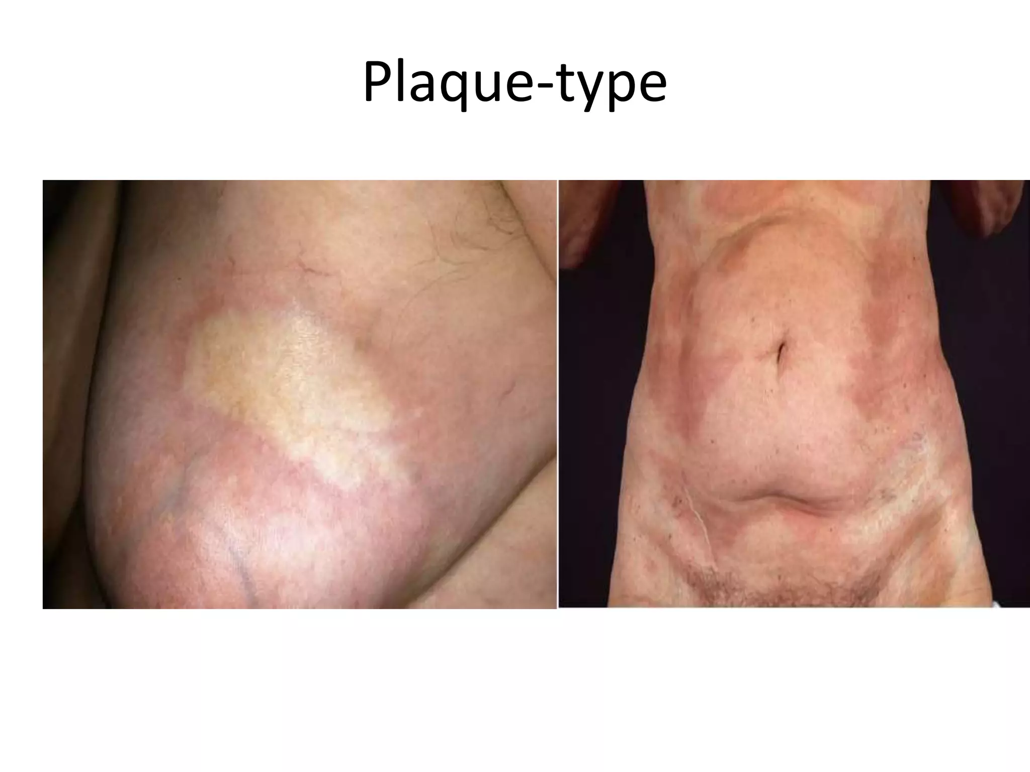
Localized scleroderma (LS), also called morphea, is a rare autoimmune disease that primarily affects the skin, causing hardening and fibrosis. It comes in several subtypes depending on the extent, location, and depth of skin involvement. The most common subtypes are plaque morphea, linear morphea, and generalized morphea. Linear morphea is most common in children and can cause serious complications by restricting growth and movement if not properly treated. While LS only affects the skin, it can lead to significant scarring, contractures, and physical disability depending on the specific subtype and location of lesions.
INTRODUCTION OF PSORIASIS, EPIDEMIOLOGY OF PSORIASIS, CLINICAL FEATURES OF PSORIASIS, PROGNOSIS OF PSORIASIS, HISTOPATHOLOGY OF PSORIASIS, TRIGGERING FACTORS OF PSORIASIS, PATHOGENESIS OF PSORIASIS
Hereditary disorder of keratinization characterized by expanding atrophic anular patch(es) surrounded by prominent keratotic ridge called the cornoid lamella
Pigmentation disorders of skin dermatology revision notesTONY SCARIA
dermatology revision notes for neet pg preparation based on lecture notes with high yield topic & last minute revision notes based on previous year questions
Cutaneous manifestations of hiv infectiontashagarwal
Dermatological problems occur in more than 90% of patients with human immunodeficiency virus (HIV) infection. In some patients, skin is the first organ affected. Skin diseases have proved to be sensitive and useful measures by which HIV progression can be monitored.
Erythroderma is defined as the scaling erythematous dermatitis involving 90% or more of the cutaneous surface.
Also known as exfoliative dermatitis
Idiopathic exfoliative dermatitis – also known as the “red man syndrome”, is characterized by marked palmoplantar keratoderma, dermatopathic lymphadenopathy,increased IgE.
Increased skin perfusion leads to
Temperature dysregulation >
Resulting in skin loss and hypothermia >
High output state >
Cardiac failure
BMR raises to compensate for heat loss
Increased dehydration due to transpiration (similar to burns)
All lead to negative nitrogen balance and characterized by edema, hypoalbuminemia, loss of muscle mass.
Actinic keratoses: Erythematous scaly lesions on sun-damaged skin & considered “precancerous” lesions that have the potential to progress into invasive SCC.
Bowen’s disease: SCC in situ It has the potential to progress to invasive SCC.
Leukoplakia: Leukoplakia refers to a white patch or plaque on the oral mucosa that cannot be wiped off and cannot be characterized clinically or pathologically as any other disease.
this ppt is about malignant tumours of connective tissue origin. classifications, clinical features, radiological features and histological features of all tumors are discussed with pictures.
The non-Hodgkin lymphomas include a diverse and complex group of malignancies of lymphoreticular histogenesis and differentiation.
In most instances, they initially arise within lymph nodes and tend to grow as solid masses.
The non-Hodgkin lymphomas most commonly originate from cells of the B-lymphocyte series, with an estimated 85% of European and American lymphoid neoplasms having this derivation.
Tumors with a T-lymphocyte derivation are less common, whereas true histiocyte-derived lymphomas are even rarer.
Genetic abnormalities like nonrandom chromosomal and molecular rearrangements play an important role in the pathogenesis of many lymphomas and correlate with histology and immunophenotype.
Most lymphomas do not have a familial pattern; however, coexistence of multiple breast cancers, ovarian cancer, sarcomas, and lymphomas in a family may suggest an inherited abnormality in tumor suppressor genes.
Environmental factors also seem to play a role in the development of NHL. Certain chemicals have been linked to the development of NHL include a variety of pesticides and herbicides (e.g. organophosphates, chlorophenols), solvents and organic chemicals (e.g. benzene, carbon tetrachloride), and wood preservatives.
Thus certain workers like pesticide applicators, workers in the petroleum, rubber, plastics, and synthetic industries have a slightly increased risk of NHL.
Patients who receive cancer chemotherapy and/or radiation therapy are at increased risk of developing NHL.
Several viruses have been implicated in the pathogenesis of NHL, including the Epstein-Barr virus in Burkitt’s lymphoma (especially in endemic areas of Africa), sinonasal lymphoma in Asia and South America, and lymphomas in immunocompromised patients; HTLV-1 Human T-lymphotropic Virus in adult T-cell lymphoma/leukemia; and human herpesvirus 8 (HHV 8) in body cavity-based lymphomas in patients with HIV infection.
Immunodeficiency states that seem to predispose to NHL include congenital immunodeficiency states (e.g. ataxia telangiectasia, Wiskott–Aldrich syndrome, common variable hypogammaglobulinemia, severe combined immunodeficiency (SCID) as well as acquired immunodeficiency states (e.g. HIV infection, iatrogenic immunosuppression for solid organ or bone marrow transplant recipients).
Connective-tissue disorders, including Sjögren syndrome, rheumatoid arthritis, chronic lymphocytic thyroiditis, and systemic lupus erythematosus (SLE) are also associated with increased risk of NHL.
The microscopic appearance of the lesional cells was used in the past to classify the tumors as either lymphocytic or histiocytic.
With the development of modern immunologic techniques, however, it is now known that many of the lesions that had been classified as histiocytic were in fact neoplasms composed of transformed B lymphocytes. In the early 1980s, a group of American pathologists devised a classification scheme, known as the Working Formulation for Clinical Use.
Flu Vaccine Alert in Bangalore Karnatakaaddon Scans
As flu season approaches, health officials in Bangalore, Karnataka, are urging residents to get their flu vaccinations. The seasonal flu, while common, can lead to severe health complications, particularly for vulnerable populations such as young children, the elderly, and those with underlying health conditions.
Dr. Vidisha Kumari, a leading epidemiologist in Bangalore, emphasizes the importance of getting vaccinated. "The flu vaccine is our best defense against the influenza virus. It not only protects individuals but also helps prevent the spread of the virus in our communities," he says.
This year, the flu season is expected to coincide with a potential increase in other respiratory illnesses. The Karnataka Health Department has launched an awareness campaign highlighting the significance of flu vaccinations. They have set up multiple vaccination centers across Bangalore, making it convenient for residents to receive their shots.
To encourage widespread vaccination, the government is also collaborating with local schools, workplaces, and community centers to facilitate vaccination drives. Special attention is being given to ensuring that the vaccine is accessible to all, including marginalized communities who may have limited access to healthcare.
Residents are reminded that the flu vaccine is safe and effective. Common side effects are mild and may include soreness at the injection site, mild fever, or muscle aches. These side effects are generally short-lived and far less severe than the flu itself.
Healthcare providers are also stressing the importance of continuing COVID-19 precautions. Wearing masks, practicing good hand hygiene, and maintaining social distancing are still crucial, especially in crowded places.
Protect yourself and your loved ones by getting vaccinated. Together, we can help keep Bangalore healthy and safe this flu season. For more information on vaccination centers and schedules, residents can visit the Karnataka Health Department’s official website or follow their social media pages.
Stay informed, stay safe, and get your flu shot today!
NVBDCP.pptx Nation vector borne disease control programSapna Thakur
NVBDCP was launched in 2003-2004 . Vector-Borne Disease: Disease that results from an infection transmitted to humans and other animals by blood-feeding arthropods, such as mosquitoes, ticks, and fleas. Examples of vector-borne diseases include Dengue fever, West Nile Virus, Lyme disease, and malaria.
Tom Selleck Health: A Comprehensive Look at the Iconic Actor’s Wellness Journeygreendigital
Tom Selleck, an enduring figure in Hollywood. has captivated audiences for decades with his rugged charm, iconic moustache. and memorable roles in television and film. From his breakout role as Thomas Magnum in Magnum P.I. to his current portrayal of Frank Reagan in Blue Bloods. Selleck's career has spanned over 50 years. But beyond his professional achievements. fans have often been curious about Tom Selleck Health. especially as he has aged in the public eye.
Follow us on: Pinterest
Introduction
Many have been interested in Tom Selleck health. not only because of his enduring presence on screen but also because of the challenges. and lifestyle choices he has faced and made over the years. This article delves into the various aspects of Tom Selleck health. exploring his fitness regimen, diet, mental health. and the challenges he has encountered as he ages. We'll look at how he maintains his well-being. the health issues he has faced, and his approach to ageing .
Early Life and Career
Childhood and Athletic Beginnings
Tom Selleck was born on January 29, 1945, in Detroit, Michigan, and grew up in Sherman Oaks, California. From an early age, he was involved in sports, particularly basketball. which played a significant role in his physical development. His athletic pursuits continued into college. where he attended the University of Southern California (USC) on a basketball scholarship. This early involvement in sports laid a strong foundation for his physical health and disciplined lifestyle.
Transition to Acting
Selleck's transition from an athlete to an actor came with its physical demands. His first significant role in "Magnum P.I." required him to perform various stunts and maintain a fit appearance. This role, which he played from 1980 to 1988. necessitated a rigorous fitness routine to meet the show's demands. setting the stage for his long-term commitment to health and wellness.
Fitness Regimen
Workout Routine
Tom Selleck health and fitness regimen has evolved. adapting to his changing roles and age. During his "Magnum, P.I." days. Selleck's workouts were intense and focused on building and maintaining muscle mass. His routine included weightlifting, cardiovascular exercises. and specific training for the stunts he performed on the show.
Selleck adjusted his fitness routine as he aged to suit his body's needs. Today, his workouts focus on maintaining flexibility, strength, and cardiovascular health. He incorporates low-impact exercises such as swimming, walking, and light weightlifting. This balanced approach helps him stay fit without putting undue strain on his joints and muscles.
Importance of Flexibility and Mobility
In recent years, Selleck has emphasized the importance of flexibility and mobility in his fitness regimen. Understanding the natural decline in muscle mass and joint flexibility with age. he includes stretching and yoga in his routine. These practices help prevent injuries, improve posture, and maintain mobilit
TEST BANK for Operations Management, 14th Edition by William J. Stevenson, Ve...kevinkariuki227
TEST BANK for Operations Management, 14th Edition by William J. Stevenson, Verified Chapters 1 - 19, Complete Newest Version.pdf
The prostate is an exocrine gland of the male mammalian reproductive system
It is a walnut-sized gland that forms part of the male reproductive system and is located in front of the rectum and just below the urinary bladder
Function is to store and secrete a clear, slightly alkaline fluid that constitutes 10-30% of the volume of the seminal fluid that along with the spermatozoa, constitutes semen
A healthy human prostate measures (4cm-vertical, by 3cm-horizontal, 2cm ant-post ).
It surrounds the urethra just below the urinary bladder. It has anterior, median, posterior and two lateral lobes
It’s work is regulated by androgens which are responsible for male sex characteristics
Generalised disease of the prostate due to hormonal derangement which leads to non malignant enlargement of the gland (increase in the number of epithelial cells and stromal tissue)to cause compression of the urethra leading to symptoms (LUTS
- Video recording of this lecture in English language: https://youtu.be/lK81BzxMqdo
- Video recording of this lecture in Arabic language: https://youtu.be/Ve4P0COk9OI
- Link to download the book free: https://nephrotube.blogspot.com/p/nephrotube-nephrology-books.html
- Link to NephroTube website: www.NephroTube.com
- Link to NephroTube social media accounts: https://nephrotube.blogspot.com/p/join-nephrotube-on-social-media.html
Knee anatomy and clinical tests 2024.pdfvimalpl1234
This includes all relevant anatomy and clinical tests compiled from standard textbooks, Campbell,netter etc..It is comprehensive and best suited for orthopaedicians and orthopaedic residents.
HOT NEW PRODUCT! BIG SALES FAST SHIPPING NOW FROM CHINA!! EU KU DB BK substit...GL Anaacs
Contact us if you are interested:
Email / Skype : kefaya1771@gmail.com
Threema: PXHY5PDH
New BATCH Ku !!! MUCH IN DEMAND FAST SALE EVERY BATCH HAPPY GOOD EFFECT BIG BATCH !
Contact me on Threema or skype to start big business!!
Hot-sale products:
NEW HOT EUTYLONE WHITE CRYSTAL!!
5cl-adba precursor (semi finished )
5cl-adba raw materials
ADBB precursor (semi finished )
ADBB raw materials
APVP powder
5fadb/4f-adb
Jwh018 / Jwh210
Eutylone crystal
Protonitazene (hydrochloride) CAS: 119276-01-6
Flubrotizolam CAS: 57801-95-3
Metonitazene CAS: 14680-51-4
Payment terms: Western Union,MoneyGram,Bitcoin or USDT.
Deliver Time: Usually 7-15days
Shipping method: FedEx, TNT, DHL,UPS etc.Our deliveries are 100% safe, fast, reliable and discreet.
Samples will be sent for your evaluation!If you are interested in, please contact me, let's talk details.
We specializes in exporting high quality Research chemical, medical intermediate, Pharmaceutical chemicals and so on. Products are exported to USA, Canada, France, Korea, Japan,Russia, Southeast Asia and other countries.
New Directions in Targeted Therapeutic Approaches for Older Adults With Mantl...i3 Health
i3 Health is pleased to make the speaker slides from this activity available for use as a non-accredited self-study or teaching resource.
This slide deck presented by Dr. Kami Maddocks, Professor-Clinical in the Division of Hematology and
Associate Division Director for Ambulatory Operations
The Ohio State University Comprehensive Cancer Center, will provide insight into new directions in targeted therapeutic approaches for older adults with mantle cell lymphoma.
STATEMENT OF NEED
Mantle cell lymphoma (MCL) is a rare, aggressive B-cell non-Hodgkin lymphoma (NHL) accounting for 5% to 7% of all lymphomas. Its prognosis ranges from indolent disease that does not require treatment for years to very aggressive disease, which is associated with poor survival (Silkenstedt et al, 2021). Typically, MCL is diagnosed at advanced stage and in older patients who cannot tolerate intensive therapy (NCCN, 2022). Although recent advances have slightly increased remission rates, recurrence and relapse remain very common, leading to a median overall survival between 3 and 6 years (LLS, 2021). Though there are several effective options, progress is still needed towards establishing an accepted frontline approach for MCL (Castellino et al, 2022). Treatment selection and management of MCL are complicated by the heterogeneity of prognosis, advanced age and comorbidities of patients, and lack of an established standard approach for treatment, making it vital that clinicians be familiar with the latest research and advances in this area. In this activity chaired by Michael Wang, MD, Professor in the Department of Lymphoma & Myeloma at MD Anderson Cancer Center, expert faculty will discuss prognostic factors informing treatment, the promising results of recent trials in new therapeutic approaches, and the implications of treatment resistance in therapeutic selection for MCL.
Target Audience
Hematology/oncology fellows, attending faculty, and other health care professionals involved in the treatment of patients with mantle cell lymphoma (MCL).
Learning Objectives
1.) Identify clinical and biological prognostic factors that can guide treatment decision making for older adults with MCL
2.) Evaluate emerging data on targeted therapeutic approaches for treatment-naive and relapsed/refractory MCL and their applicability to older adults
3.) Assess mechanisms of resistance to targeted therapies for MCL and their implications for treatment selection
Lung Cancer: Artificial Intelligence, Synergetics, Complex System Analysis, S...Oleg Kshivets
RESULTS: Overall life span (LS) was 2252.1±1742.5 days and cumulative 5-year survival (5YS) reached 73.2%, 10 years – 64.8%, 20 years – 42.5%. 513 LCP lived more than 5 years (LS=3124.6±1525.6 days), 148 LCP – more than 10 years (LS=5054.4±1504.1 days).199 LCP died because of LC (LS=562.7±374.5 days). 5YS of LCP after bi/lobectomies was significantly superior in comparison with LCP after pneumonectomies (78.1% vs.63.7%, P=0.00001 by log-rank test). AT significantly improved 5YS (66.3% vs. 34.8%) (P=0.00000 by log-rank test) only for LCP with N1-2. Cox modeling displayed that 5YS of LCP significantly depended on: phase transition (PT) early-invasive LC in terms of synergetics, PT N0—N12, cell ratio factors (ratio between cancer cells- CC and blood cells subpopulations), G1-3, histology, glucose, AT, blood cell circuit, prothrombin index, heparin tolerance, recalcification time (P=0.000-0.038). Neural networks, genetic algorithm selection and bootstrap simulation revealed relationships between 5YS and PT early-invasive LC (rank=1), PT N0—N12 (rank=2), thrombocytes/CC (3), erythrocytes/CC (4), eosinophils/CC (5), healthy cells/CC (6), lymphocytes/CC (7), segmented neutrophils/CC (8), stick neutrophils/CC (9), monocytes/CC (10); leucocytes/CC (11). Correct prediction of 5YS was 100% by neural networks computing (area under ROC curve=1.0; error=0.0).
CONCLUSIONS: 5YS of LCP after radical procedures significantly depended on: 1) PT early-invasive cancer; 2) PT N0--N12; 3) cell ratio factors; 4) blood cell circuit; 5) biochemical factors; 6) hemostasis system; 7) AT; 8) LC characteristics; 9) LC cell dynamics; 10) surgery type: lobectomy/pneumonectomy; 11) anthropometric data. Optimal diagnosis and treatment strategies for LC are: 1) screening and early detection of LC; 2) availability of experienced thoracic surgeons because of complexity of radical procedures; 3) aggressive en block surgery and adequate lymph node dissection for completeness; 4) precise prediction; 5) adjuvant chemoimmunoradiotherapy for LCP with unfavorable prognosis.
2. Introduction
• Localized scleroderma (LS), also called morphea,comprises
a spectrum of sclerotic autoimmune diseases that primarily
affect the skin,primarily of the dermis and subcutaneous
fat, which ultimately leads to a scar-like sclerosis.
• Depending on the respective subtypes, LS might also
involve adjacent tissues such as the fat, fascia, muscle, and
bone. In contrast to systemic sclerosis, LS does not affect
internal organs such as the lungs, heart,kidneys, or
gastrointestinal tract. Although LS and systemic sclerosis
(SSc) share similar pathogenetic pathways including the
release of distinct proinflammatory cytokines and
dysregulation of the connective tissue metabolism, both
diseases rarely coexist, and transition from LS to SSc does
not occur.
3. Epidemiology
• LS is a rare disease that seems to be most frequent in whites, but
may affect all races.
• The prevalence (total number of affected patients per
million)increases with age. It is approximately 500 per million at age
18 and 2200 at age 80 years1.The disease is more prevalent in
women than in men (2.6 to 1).
• The disease may manifest at all ages, but the peak age of incidence
essentially differs in the respective subtypes of LS. The most
frequent subtype of LS,morphea, usually appears in adults between
40 and 50 years of age, whereas linear subtypes primarily present
in childhood between 2 and 14 years of age.
• Several reports of familiar clustering and increased rates of other
autoimmune diseases (e.g., Hashimoto’s thyroiditis, alopecia
areata, vitiligo, and type 1 diabetes) in patients with LS are
suggestive for a genetic component. However, in contrast to SSc,
susceptibility genes for LS are still unknown.
4. Pathogenesis
• The hallmark feature of LS is overproduction of collagen and
increased extracellular matrix deposition.Its exact initiation still
remains unknown. It has been hypothesized that certain stimuli,
e.g.,infections, trauma, radiation, or drugs might cause
microvascular injuries that subsequently result in a release of
various adhesion molecules. Upregulation of some of these
adhesion molecules such as vascular cell adhesion molecule-1 and
intercellular adhesion molecule-1 might induce T cell activation that
in turn activate the release of key player profibrotic cytokines such
as transforming growth factor-beta (TGF-b),platelet-derived growth
factor, connective tissue growth factor, and interleukin 4, 6, and 8.
• This profibrotic pathway additionally includes a spectrum of
chemokines that have demonstrated to significantly contribute to
skin sclerosis.At the end, similar to SSc, activation of all of these
proinflammatory and profibrotic signals lead to excessive collagen
production and decrease of matrix metalloproteinases (MMP)
responsible for collagen degradation.
5. Pathogenesis
• Although much is known about the early inflammatory
phase and the molecular mechanisms involved in the
fibroblastic reaction of LS, little is known about the initial
triggers of the disease. Among infectious agents, Borrelia
organisms have been extensively studied on both sides of
the Atlantic. Although high rates of Borrelia infections,in
part using highly sensitive new detection techniques such
as focus-floating microscopy,have been reported in LS
patients from Europe,a variety of studies from the United
States failed to demonstrate an association.
• Among the few drugs that have been reported to induce LS,
most evidence exists for bleomycin, D-penicillamine,
vitamin K1, and L-5 hydroxytryptophane plus carbidopa.
• Few reports exist on radiation-induced LS, which primarily
occurs in women with breast carcinoma.
6. Classification and clinical
characteristics of LS subtypes
• A German group of experts recently suggested a classification that
considers the extent and depth of fibrosis and refers to the treatment of
the respective subtypes.
7. Plaque-type
• The classical plaque type, is by far the most frequent
subtype of LS, especially in adults. In the early active
phase, morphea usually presents with oval-shaped
lesions surrounded by an erythematous border (the so-
called “lilac ring”). In the later stage of the disease,
morphea lesions become hard and sclerotic in the
center, with a whitish or ivory color. Older lesions may
also become atrophic, hypo-, or hyperpigmented and,
depending on the location of fibrosis, may also lead to
hair loss and loss of the skin appendages.
• Morphea is frequently located on the trunk,especially
the submammary region, and the transitional area
between the hip and inguinal regions.
9. Guttate
morphea
Guttate morphea is a rare subtype
of morphea that presents with
multiple yellowish or whitish,
small sclerotic lesions with a shiny
surface .
Guttate morphea is predominantly
located on the trunk. Early
inflammatory lesions may simply
present as erythematous maculae.
Clinically and histopathologically,
guttate morphea might be difficult
to distinguish from extragenital
lichen sclerosus.
10. Atrophoderma
of Pasini and
Pierini
Atrophoderma of Pasini and
Pierini is
possibly an early abortive
type of morphea. The recently
described term “superficial
morphea” seems to be
synonymous with
atrophoderma of Pasini and
Pierini.
The clinical presentation of
this subtype of LS, which
frequently manifests in
childhood, is characterized by
symmetrical, single or
multiple, sharply demarcated,
hyperpigmented,
nonindurated patches that
are located on the trunk or
extremities.
11. Nodular or
keloid morphea
Here, the
inflammation of
the dermis leads to
thick, keloid-like
nodules
or streaks, clinically
indistinguishable
from indurated
keloids.
12. Bullous morphea
• In some patients, especially those where sclerosis of the skin
is associated with diffuse, rapidly progressive edema,
lymphoceles may result from stasis of lymphatic fluid and may
appear as bullae.
• This is rarely a feature of plaque-type morphea. It seems to be
more frequent in generalized, disabling morphea or
sclerodermoid GVHD. Bullous morphea has to be separated
from mechanical blisters that may occur in the central scar
and are secondary to impaired mechanical stability of the
dermal–epidermal junction.
13. Generalized type
• Generalized LS is a more severe variant of LS.According to
Laxer and Zulian, generalized LS is present in case of four or
more indurated plaques of more than 3 cm in diameter,
involving two or more of seven anatomic sites (head-neck,
each extremity, anterior trunk, and posterior trunk).
• The most commonly affected sites are the trunk,thighs, and
lumbosacral region. The plaques are often distributed
symmetrically and can coalesce into larger lesions.
• A unique and very rare variant of the generalized type of LS
is “disabling pansclerotic morphea.”Disabling pansclerotic
morphea, predominantly occurring in childhood, may lead
to extensive involvement of the skin, fat tissue, fascia,
muscle,and bone, with only a limited tendency to regres
sion of the fibrotic changes. Disabling pansclerotic morphea
often results in severe contractures and poorly healing,
large ulcerations, and skin necroses.
14.
15. Eosinophilic fasciitis
• Eosinophilic fasciitis (also called Shulman syndrome) is
nowadays considered by many experts to be a special
subtype of generalized LS.
• A mechanical trauma often precedes the first
manifestation of the disease. Clinically, eosinophilic fasciitis
exclusively affects the extremities and presents with a rapid
onset of symmetrical swelling of the skin. In the later stage
of disease, lesions become more indurated and fibrotic,
leading to the typical “peau d’orange”-like appearance.
• elevated sedimentation rate, and hypergammaglobulinemia
remain critical elements of the syndrome.
• The diagnosis of eosinophilic fasciitis is suspected in a
patient presenting with characteristic skin changes and
consistent laboratory findings. It is confirmed with full-
thickness biopsy or characteristic MRI findings
16.
17. Linear Morphea
• Linear LS is by far most common subtype of LS in
childhood. Linear LS is characterized by longitudinally
arranged linear, band-like lesions that are
predominantly located on the extremities.Evidence
indicates that linear LS may follow the lines of
Blaschko. In mild disease, the lesions may heal with
residual hyperpigmentation.
• However, depending on the extent of the fibrotic
process, linear LS may lead to severe growth
retardation,muscle atrophy, flexion contractures,
myositis and myalgia, arthritis and arthralgia, and
psychological disability.
19. “en coup de sabre” and Parry–Romberg
Syndrome
• The best known linear subtype of the disease is LS “en coup de sabre”. It is
located on the frontoparietal region of the head, usually ranging
paramedian from the eyebrows into the hair-bearing scalp where it might
cause scarring alopecia. Involvement of the underlying central nervous
system (e.g., seizures, migraine, and headache) and abnormal
ophthalmological findings (e.g., uveitis) are common.
• Several authors have speculated that progressive facial hemiatrophy (also
called Parry–Romberg syndrome) and LS “en coup de sabre” are variants
of the same condition. Progressive facial hemiatrophy is clinically
characterized by a primary atrophy of the subcutaneous tissue,muscle,
and bone. Skin fibrosis is rare in this particular subtype. Progressive facial
hemiatrophy often occurs in childhood or adolescence and may result in
severe facial asymmetry. Occurrence of simultaneous linear LS “en coup
de sabre” and progressive facial hemiatrophy is quite frequent,with a
reported coincidence of up to 40%. In the classification proposed in this
presentation,progressive facial hemiatrophy is listed under the linear
subtypes of LS, although with exclusive involvement of extracutaneous
structures, it may also be classified as a “deep subtype” of LS.
20.
21. Deep morphea
• The deep type of LS, called deep morphea, is by
far the most rare variant (<5% of patients).
• In deep morphea, the fibrotic process mainly
affects the deeper layers of the connective tissue,
i.e., fat tissue, fascia, and underlying muscle.
• Deep morphea lesions are typically arranged
symmetrically and predominantly located on the
extremities.
• Deep morphea may initiate during childhood and
may sometimes manifest without any clinical
signs of inflammation
23. Differential diagnoses
• LS may present with a broad spectrum of subtypes and
clinical stages of disease. Accordingly, a variety of different
differential diagnoses should be considered . In daily
routine, the physicians’ pivotal challenge is to differentiate
LS from SSc. Importantly, typical facial (e.g.,
teleangiectases,beak-shaped nose, and microstomia) and
vascular (e.g., Raynaud’s phenomenon, pitting scars, and
digital ulcers) features of SSc as well as highly specific
serum antibodies (e.g., anticentromere antibodies and anti-
Scl-70 antibodies) are absent in LS.
• The most relevant differential diagnoses for limited LS
(morphea) are extragenital lichen sclerosus and
acrodermatitis chronic atrophicans, for generalized LS
chronic graft versus host disease,SSc, and nephrogenic
systemic fibrosis, and for linear LS lupus erythematosus
profundus and other types of panniculitis.
24.
25.
26.
27.
28. Serologic features
• High incidence of autoimmune phenomena has been reported in LS
patients. Serum antinuclear antibodies, most of them with a homogenous
pattern, have been detected in 20% to 80% of LS patients.
• Active childhood LS might be associated with antihistone antibodies,
hypergammaglobulinemia , and eosinophilia.
• In patients with linear LS of the extremities with concomitant joint
involvement, increased levels of rheumatoid factor may be present, and
do sometimes correlate with the clinical degree of arthritis activity.
• Numerous cytokines (e.g., tumor necrosis factor-a, interleukin 2, 4, 6, 8,
and 13) and soluble factors (e.g., CD23 or CD30) have been reported in LS
patients, and some of them seem to correlate with the respective subtype
and disease activity.However, none of them have so far been proven to be
consistently useful for assessing disease activity or clinical course. The
same applies to procollagen III peptide.
• Therefore, at present, treatment success in LS can purely be measured by
clinical features.
29. Radiologic examination
• Patients with LS “en coup de sabre” and progressive facial
hemiatrophy often suffer from neurological symptoms (e.g.,
migraine, headaches, and epilepsy).
• In these cases, cranial magnetic resonance imaging (MRI) should be
considered to detect potential involvement of the central nervous
system because subcortical calcifications and brain atrophy are
common. Despite such abnormalities of the central nervous system,
many patients are asymptomatic.
• In addition, MRI and computed tomography studies might be
helpful for surgical planning (e.g., in LS “en coup de sabre”type) and
to detect muscle or bone involvement,for instance in linear LS of
the extremities. Bone scintigraphy should be considered in cases
with linear LS of the extremities that might have concomitant
arthritis.
31. Calcipotriol Topical therapy corticosteroid
• Although no well-performed studies exist on the use of topical
corticosteroids, they are the mainstay of topical treatment in LS.
Therapy with moderate to high-potent corticosteroids should be
performed in the active phase of disease, and their application
should be restricted to a total of 3 months.
• Intralesional steroid therapy might be performed in LS “en coup de
sabre,” with injections into the active margin.
• So far, two uncontrolled studies exist on the use of topical
calcipotriol in LS, in one of which calcipotriol 0.005% was
administered along with low dose UVA1 phototherapy . In both
studies,administration was performed twice daily. In the
monotherapy study, calcipotriol 0.005% was applied under
occlusion.
• Calcipotriol 0.005% should be considered for active inflammatory
superficial types of LS with a low degree of sclerosis. Treatment
should be performed twice daily for a minimum of 3 months.
32. Imiquimod Topical therapy calcineurin inhibitors
• A recent double-blind,placebo controlled pilot study has
shown that topical tacrolimus significantly improves LS.
Outcome measures in this study were the changes of
surface area, a clinical score for erythema, induration,
dyspigmentation, telangiectasia, and atrophy. Early
inflammatory lesions resolved and late sclerotic lesions
softened,whereas no effects were seen on preexisting skin
atrophy.
• Thus, tacrolimus ointment might be an effective treatment
option for active LS lesions.
• The topical immune response modifier imiquimod has been
reported to significantly improve abnormal pigmentation,
sclerosis, and erythema in LS. The mechanism of imiquimod
action in LS might be explained by induction of interferon-
g, which inhibits TGF-b, thereby exhibiting a broad
antifibrotic Effect.
33. phototherapy
• The first experience of the successful use of UV phototherapy in LS
has been reported in 1994. Since then, much information has been
gained on the entire spectrum of antifibrotic and anti inflammatory
effects of UV phototherapy in skin sclerosis.
• Because longer wavelengths in the UVA range (320–400 nm)
penetrate deeper into the dermis than does UVB (280–320), most
studies have focused on UVA. A number of uncontrolled studies on
psoralen plus UVA (PUVA; a study on bath-PUVA with 17 patients
and a study on cream-PUVA with four patients), broad-band UVA
(three studies from Egypt with 15, 22, and 67 patients),and UVA1-
phototherapy (six open uncontrolled studies with a total of 88
patients) have demonstrated that phototherapy is very effective in
LS.
• Before initiating UV phototherapy in LS, it should be considered
that UV rays only penetrate into the deep dermis. Therefore, UV
phototherapy (in combination with topical treatment, e.g.,topical
corticosteroids or topical vitamin D-analogs) is an effective
treatment option for limited disease restricted to the skin, but not
in LS subtypes with affection of deeper structures (e.g.,fat tissue,
fascia, muscle, or bone). Such subtypes require systemic therapy.
34. Systemic therapy
corticosteroid
• Similar to topical corticosteroids, there is a paucity of data
on systemic corticosteroids, although they are widely used
agents in LS, particularly in linear,generalized, and deep
subtypes. In the only published uncontrolled study on 17
patients with LS (corticosteroid dosage: 0.5–1.0 mg/kg
body weight daily), a marked improvement was noticed in
nearly all of the patients.
• Systemic corticosteroids are safe and effective in active
lesions of LS,and should be considered in patients with
severe disease affecting extracutaneous structures (e.g.,fat
tissue, fascia, muscle, and bone).Moreover, systemic
corticosteroids are the first-line treatment option in
eosinophilic fasciitis.
35. Systemic therapy
MXT
• Among systemic treatment of LS, best evidence exists
for the use of methotrexate (MTX). MTX inhibits
several cytokines that play a central role in sclerotic
skin diseases, such as interleukins 2, 4,and 6.
• Recently, a large randomized placebo-controlled study
on 85 patients with childhood LS treated with oral
corticosteroids and low-dose MTX confirmed the
efficacy of this combination therapy for LS.
• Thus, MTX alone or in combination with corticosteroids
are currently first-line options for LS, especially in
linear, generalized, and deep subtypes.
36. Systemic therapy
• Recently, a small case series of seven MTXresistant LS
patients treated with mycophenolate mofetil (MMF) has
shown improvement of skin sclerosis and inflammation.
• In vitro studies have shown that MMF inhibits the
proliferation of lymphocytes, but also of other cell types,
including smooth muscle cells and fibroblasts, indicating
that it has direct antifibrotic properties in addition to its
well-known immunosuppressive effects.These preliminary
observations make MMF an interesting new candidate for
further clinical studies.
• Numerous other systemics have reported in cases of LS,
including infliximab, bosentan, antimalarial drugs, imatinib,
extracorporeal photopheresis,cyclosporine, and penicillin,
but none of them has so far been confirmed in a larger
number of subjects.