This meta-analysis reviewed 40 studies on supracondylar osteotomy for treating cubitus varus deformity in children. It found that lateral wedge osteotomy was the most common procedure, achieving 84% excellent results in correcting the deformity and restoring carrying angle. On average, procedures corrected 27 degrees of varus deformity and improved range of motion by 20 degrees. Major complications occurred in less than 10% of cases and included residual deformity, nerve injury, infection, and loss of fixation. The study found equivalent results across treatment methods but noted k-wire fixation had a higher risk of complications compared to screws or external fixation. It recommends surgeons choose treatment based on individual case factors and discuss risks and expectations thoroughly with parents.
Posterolateral corner injuries of knee joint Samir Dwidmuthe
Missed posterolateral corner injuries of knee joint is a common cause for failure of ACL and PCL reconstruction only next to malpositioned tunnels.
Isolated PLC injuries are uncommon, making up <2% of all acute knee ligamentous injuries. Covey JBJS 2001
Incidence of PLC injuries associated with concomitant ACL and PCL disruptions are much more common (43% to 80%). Ranawat JAAOS 2008
A recent (MRI) analysis of surgical tibialplateau fractures demonstrated an incidence of PLC injuries in 68% of cases. Gardner JOT 2005
Take home message
PLC injuries to be ruled out in every case of ACL& PCL rupture.
Neurovascular integrity to be checked in every case.
Grade I & II can be managed conservatively.
Grade III Acute- Repair.
Grade III Chronic- Anatomic PLC recon.
Beware of varus knee alignment.
Tendoachilles rupture and its managementRohan Vakta
Achilles tendon is the strongest tendon of body. There are many causes of its rupture. It can be acute or chronic rupture. Management of chronic rupture by semitendinosus tendon is mentioned here.
Aspect particulier en traumatologie pédiatriqueROBERT ELBAUM
L ’enfant n’est pas un petit adulte
Particularités propres à la traumatologie pédiatrique
Connaissance du potentiel de remodelage mais ne pas le surestimer
Connaissance RX des points d ’ossifications
Incision or transection of bone.
Uses:-
to correct deformity.
to change shape of bone.
to redirect load trajectories in a limb so as to influence joint function.
Mandibular fractures
Dr. Ahmed M. Adawy
Professor Emeritus, Dept. Oral & Maxillofacial Surg.
Former Dean, Faculty of Dental Medicine
Al-Azhar University
Fractures of the mandible are a common form of facial injury in adults and occur most frequently in males during the third decade of life. The main causes of mandibular fractures are road traffic accidents, interpersonal violence, falls and sport injuries. Mandibular fractures are classified according to various criteria. The three main factors to consider are the cause of the fracture, the type of fracture and the site of the fracture. Clinical diagnosis as well as radiographic examinations are presented. Treatment modalities are discussed. Moreover, treatment-related complications are given.
Guided Growth for Angular Knee Deformities in Nutritional Rickets ChildrenTamer El-Sobky
Nutritional rickets in children is a global health concern. It manifests in generalized skeletal deformities including angular or coronal plane knee deformities. Guided growth surgery is a recognized treatment option for angular knee deformities in general. However, there is insufficient citations on its use in the treatment of angular knee deformities in children with nutritional rickets. Rachitic lower limb deformities can be complex. They are usually multiostotic, multiapex and multiplane and require extensive corrective osteotomies. However osteotomies are fraught with complications and can be technically demanding. In this presentation we present our experience with the use of surgical guided growth as a minimally invasive treatment option to correct angular knee deformities in children with nutritional rickets.
Arthroscopic Management of Anterior, Posterior, and Multidirectional Shoulder...Peter Millett MD
Arthroscopic treatment of the unstable shoulder has evolved rapidly and significantly in recent years. Better understanding of the pathoanatomy, advancements in technology, and improved surgical techniques have led to dramatic improvements in outcome. An arthroscopic approach includes significant advantages. Arthroscopy provides better identification of concomitant pathology, lower morbidity, less soft tissue dissection, maximal preservation of motion, shorter surgical time, and improved cosmesis. There is less pain, and many patients have an easier functional recovery, with greater returns in motion compared with traditional open techniques. Finally, some of the inherent risks of open procedures, such as postoperative subscapularis rupture, are virtually eliminated. Surgeons can now routinely expect results that are at least comparable, if not better than, those achieved with open techniques. For more shoulder surgery and instability studies, visit Dr. Millett, Orthopedic Surgeon, Vail Colorado http://drmillett.com/shoulder-studies
‘Double X’ Cross Fixationin Paediatric Supracondylar Humerus Fractures: A 20-...clinicsoncology
Over the last 50 years, the developments emerged in the diagnosis and treatment of supracondylar humerus fractures (SHF) have significantly reduced the number of severe complications while certain complications with dreadful evolution, such as elbow stiffness or Volkmann’s syndrome, have completely vanished. During my residency, in 1982, on the suggestion of Prof. Pesamosca, I have performed a surgical intervention for a patient diagnosed with SHF
Within a period from January 2010 to January 2016, there were total of 920 surgically treated patients of Orthopaedy and Traumatology Department, Dubrovnik County Hospital, Croatia, which is a single acute hospital in Dubrovacko-neretvanska County where all patients with proximal femoral fracture are treated within the Orthopaedic-Traumatology Department. The aim of this retrospective study is to compare used implants according to type of the proximal femoral region fracture (femoral neck, pertrochanteric , subtrochanteric) and used osteosynthetic implant depending on the type of fracture. A new surgical techniques were used more commonly. Osteoporotic proximal femoral fractures stayed the major and growing problem in the geriatric traumatology and the traumatollogy in general as well.
- Video recording of this lecture in English language: https://youtu.be/lK81BzxMqdo
- Video recording of this lecture in Arabic language: https://youtu.be/Ve4P0COk9OI
- Link to download the book free: https://nephrotube.blogspot.com/p/nephrotube-nephrology-books.html
- Link to NephroTube website: www.NephroTube.com
- Link to NephroTube social media accounts: https://nephrotube.blogspot.com/p/join-nephrotube-on-social-media.html
Ozempic: Preoperative Management of Patients on GLP-1 Receptor Agonists Saeid Safari
Preoperative Management of Patients on GLP-1 Receptor Agonists like Ozempic and Semiglutide
ASA GUIDELINE
NYSORA Guideline
2 Case Reports of Gastric Ultrasound
ARTIFICIAL INTELLIGENCE IN HEALTHCARE.pdfAnujkumaranit
Artificial intelligence (AI) refers to the simulation of human intelligence processes by machines, especially computer systems. It encompasses tasks such as learning, reasoning, problem-solving, perception, and language understanding. AI technologies are revolutionizing various fields, from healthcare to finance, by enabling machines to perform tasks that typically require human intelligence.
Factory Supply Best Quality Pmk Oil CAS 28578–16–7 PMK Powder in Stockrebeccabio
Factory Supply Best Quality Pmk Oil CAS 28578–16–7 PMK Powder in Stock
Telegram: bmksupplier
signal: +85264872720
threema: TUD4A6YC
You can contact me on Telegram or Threema
Communicate promptly and reply
Free of customs clearance, Double Clearance 100% pass delivery to USA, Canada, Spain, Germany, Netherland, Poland, Italy, Sweden, UK, Czech Republic, Australia, Mexico, Russia, Ukraine, Kazakhstan.Door to door service
Hot Selling Organic intermediates
The prostate is an exocrine gland of the male mammalian reproductive system
It is a walnut-sized gland that forms part of the male reproductive system and is located in front of the rectum and just below the urinary bladder
Function is to store and secrete a clear, slightly alkaline fluid that constitutes 10-30% of the volume of the seminal fluid that along with the spermatozoa, constitutes semen
A healthy human prostate measures (4cm-vertical, by 3cm-horizontal, 2cm ant-post ).
It surrounds the urethra just below the urinary bladder. It has anterior, median, posterior and two lateral lobes
It’s work is regulated by androgens which are responsible for male sex characteristics
Generalised disease of the prostate due to hormonal derangement which leads to non malignant enlargement of the gland (increase in the number of epithelial cells and stromal tissue)to cause compression of the urethra leading to symptoms (LUTS
Tom Selleck Health: A Comprehensive Look at the Iconic Actor’s Wellness Journeygreendigital
Tom Selleck, an enduring figure in Hollywood. has captivated audiences for decades with his rugged charm, iconic moustache. and memorable roles in television and film. From his breakout role as Thomas Magnum in Magnum P.I. to his current portrayal of Frank Reagan in Blue Bloods. Selleck's career has spanned over 50 years. But beyond his professional achievements. fans have often been curious about Tom Selleck Health. especially as he has aged in the public eye.
Follow us on: Pinterest
Introduction
Many have been interested in Tom Selleck health. not only because of his enduring presence on screen but also because of the challenges. and lifestyle choices he has faced and made over the years. This article delves into the various aspects of Tom Selleck health. exploring his fitness regimen, diet, mental health. and the challenges he has encountered as he ages. We'll look at how he maintains his well-being. the health issues he has faced, and his approach to ageing .
Early Life and Career
Childhood and Athletic Beginnings
Tom Selleck was born on January 29, 1945, in Detroit, Michigan, and grew up in Sherman Oaks, California. From an early age, he was involved in sports, particularly basketball. which played a significant role in his physical development. His athletic pursuits continued into college. where he attended the University of Southern California (USC) on a basketball scholarship. This early involvement in sports laid a strong foundation for his physical health and disciplined lifestyle.
Transition to Acting
Selleck's transition from an athlete to an actor came with its physical demands. His first significant role in "Magnum P.I." required him to perform various stunts and maintain a fit appearance. This role, which he played from 1980 to 1988. necessitated a rigorous fitness routine to meet the show's demands. setting the stage for his long-term commitment to health and wellness.
Fitness Regimen
Workout Routine
Tom Selleck health and fitness regimen has evolved. adapting to his changing roles and age. During his "Magnum, P.I." days. Selleck's workouts were intense and focused on building and maintaining muscle mass. His routine included weightlifting, cardiovascular exercises. and specific training for the stunts he performed on the show.
Selleck adjusted his fitness routine as he aged to suit his body's needs. Today, his workouts focus on maintaining flexibility, strength, and cardiovascular health. He incorporates low-impact exercises such as swimming, walking, and light weightlifting. This balanced approach helps him stay fit without putting undue strain on his joints and muscles.
Importance of Flexibility and Mobility
In recent years, Selleck has emphasized the importance of flexibility and mobility in his fitness regimen. Understanding the natural decline in muscle mass and joint flexibility with age. he includes stretching and yoga in his routine. These practices help prevent injuries, improve posture, and maintain mobilit
Couples presenting to the infertility clinic- Do they really have infertility...Sujoy Dasgupta
Dr Sujoy Dasgupta presented the study on "Couples presenting to the infertility clinic- Do they really have infertility? – The unexplored stories of non-consummation" in the 13th Congress of the Asia Pacific Initiative on Reproduction (ASPIRE 2024) at Manila on 24 May, 2024.
Lung Cancer: Artificial Intelligence, Synergetics, Complex System Analysis, S...Oleg Kshivets
RESULTS: Overall life span (LS) was 2252.1±1742.5 days and cumulative 5-year survival (5YS) reached 73.2%, 10 years – 64.8%, 20 years – 42.5%. 513 LCP lived more than 5 years (LS=3124.6±1525.6 days), 148 LCP – more than 10 years (LS=5054.4±1504.1 days).199 LCP died because of LC (LS=562.7±374.5 days). 5YS of LCP after bi/lobectomies was significantly superior in comparison with LCP after pneumonectomies (78.1% vs.63.7%, P=0.00001 by log-rank test). AT significantly improved 5YS (66.3% vs. 34.8%) (P=0.00000 by log-rank test) only for LCP with N1-2. Cox modeling displayed that 5YS of LCP significantly depended on: phase transition (PT) early-invasive LC in terms of synergetics, PT N0—N12, cell ratio factors (ratio between cancer cells- CC and blood cells subpopulations), G1-3, histology, glucose, AT, blood cell circuit, prothrombin index, heparin tolerance, recalcification time (P=0.000-0.038). Neural networks, genetic algorithm selection and bootstrap simulation revealed relationships between 5YS and PT early-invasive LC (rank=1), PT N0—N12 (rank=2), thrombocytes/CC (3), erythrocytes/CC (4), eosinophils/CC (5), healthy cells/CC (6), lymphocytes/CC (7), segmented neutrophils/CC (8), stick neutrophils/CC (9), monocytes/CC (10); leucocytes/CC (11). Correct prediction of 5YS was 100% by neural networks computing (area under ROC curve=1.0; error=0.0).
CONCLUSIONS: 5YS of LCP after radical procedures significantly depended on: 1) PT early-invasive cancer; 2) PT N0--N12; 3) cell ratio factors; 4) blood cell circuit; 5) biochemical factors; 6) hemostasis system; 7) AT; 8) LC characteristics; 9) LC cell dynamics; 10) surgery type: lobectomy/pneumonectomy; 11) anthropometric data. Optimal diagnosis and treatment strategies for LC are: 1) screening and early detection of LC; 2) availability of experienced thoracic surgeons because of complexity of radical procedures; 3) aggressive en block surgery and adequate lymph node dissection for completeness; 4) precise prediction; 5) adjuvant chemoimmunoradiotherapy for LCP with unfavorable prognosis.
Supracondylar osteotomy for treatment of cubitus varus
1. Supracondylar osteotomy for treatment
of cubitus varus in children
A systematic review published in The bone and Joint
Journal, May 2014
Presented by : Harjot Singh Gurudatta
Moderator : DR. GAGAN KHANNA
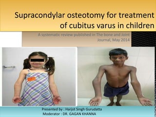
2. INTRODUCTION
Cubitus varus is the most common significant late complication of supracondylar fracture. This
deformity typically represents fracture malunion and rarely results from partial
growth arrest of the medial condylar growth plate. Malunion may be avoided by careful
attention to anatomic reduction and secure fixation at the time of initial management. Cubitus
varus is generally considered a cosmetically acceptable deformity, but increased risk of lateral
condyle fracture, tardy ulnar palsy, posterolateral rotary instability of the elbow, and posterior
shoulder instability has also been reported. Also, increased awareness among parents has
resulted in more children seeking attention
3. WHY OPERATE?
An increased risk of fracture,especially of the
lateral condyle, has been linked with cubitus
varus deformity.
tardy posterolateral rotatory instability
Less flexion and hyperextention
Tardy ulnar nerve palsy also has been associated with cubitus varus and internal rotational malalignment. With a cubitus
varus deformity, the olecranon fossa moves to the ulnar side of the distal humerus, and the triceps shifts a bit ulnarward.
Investigators theorized that this ulnar shift might compress the ulnar nerve against the medial epicondyle, narrowing the
cubital tunnel and resulting in chronic neuropathy. Also, a fibrous band running between the heads of the flexor carpi ulnaris
was thought to cause ulnar nerve compression.
4. TREATMENT OPTIONS
Medial displacement and rotation of the distal fragment have been cited most often, but
experimental studies showed that varus tilting of the distal fragment was the most important
cause of change in the carrying angle. Other suggested causes include varus tilting of the distal
fragment and growth disturbance in the distal humerus, especially overgrowth of the
lateral condyle. Osteonecrosis and delayed growth of the trochlea, with relative overgrowth of
the normal lateral side of the distal humeral epiphysis, is a rare cause of progressive
cubitus varus deformity after supracondylar fracture.
Accordingly, We can either go for
(i) observation with expected remodeling, (ii) hemiepiphysiodesis and growth alteration,
and (iii) corrective osteotomy.
Observation is not recommened in this era.
Hemiepiphysiodesis is also seldom recommended as the distal humerus accounts for
only 20% of Humeral growth and even less in the >6yrs age group.
Three basic types of osteotomies have been described:
a medial opening wedge osteotomy with a bone graft, an
oblique osteotomy with derotation, and a lateral closing
wedge osteotomy. The latter being the most popular.
5.
6.
7.
8. This study is a metaanalysis of various studies conducted for cubitus varus deformity correction.
A total of 330 studies were shortlisted out of which 40 were selected for this analysis based on
no. of cases, age, operative technique and many other factors.
Outcome assessment was done according to the correction achieved, ROM attained,alignment
,complications, function etc.
Major complications included Residual deformity, nerve injury, infection, loss of fixation,
stiffness.
Good correction with <5 deg varus with good function without pain and no complications were
included as excellent result
9. RESULTS
Lateral wedge osteotomy was the main method of treatment in 24 of the studies, with 473
patients resulting in 84% excellent results
Distraction osteogenesis was done in 2 studies, 37 patients with equivalent results.
Dome and complex(multiplanar) osteotomy done in 7 studies, 100 patients with 90% excellent
results.
The most common fixation method used were k wires(46%), ex-fix(12%), Tension band with
screws(11%), screw(8%), plates(4%).
Mean preop carrying angle was 20 deg varus and postop 7 deg valgus, with around 27 deg
correction.
Mean time to union was 8 weeks, and mean ROM improved by 20 deg.
Nerve injury incidences and infection were uncommon mean being 2 % in all groups.
Most common complication was residual varus , Being 6% in all groups
As far as fixation method is concerned , 20% of those with k – wires sustained
complications(infection(3%), residual varus(10%), loss of fixation).
Screws alone and Ex-fix had the lowest complications., But the p value was insignificant.
10. Approach also had no differnce in complications with 2% nerve injuries in all post med or lat
approach
This study found the results to be similar in all study groups in terms of complications, and
effectiveness.
This study recommends the surgeons to chose the method of treatment with care and as per
the norms in their locality, practice and teaching. It is most important to take the parents in
confidence with detailed discussions regarding the possible complications, fixation method,
functional outcome and residual deformity