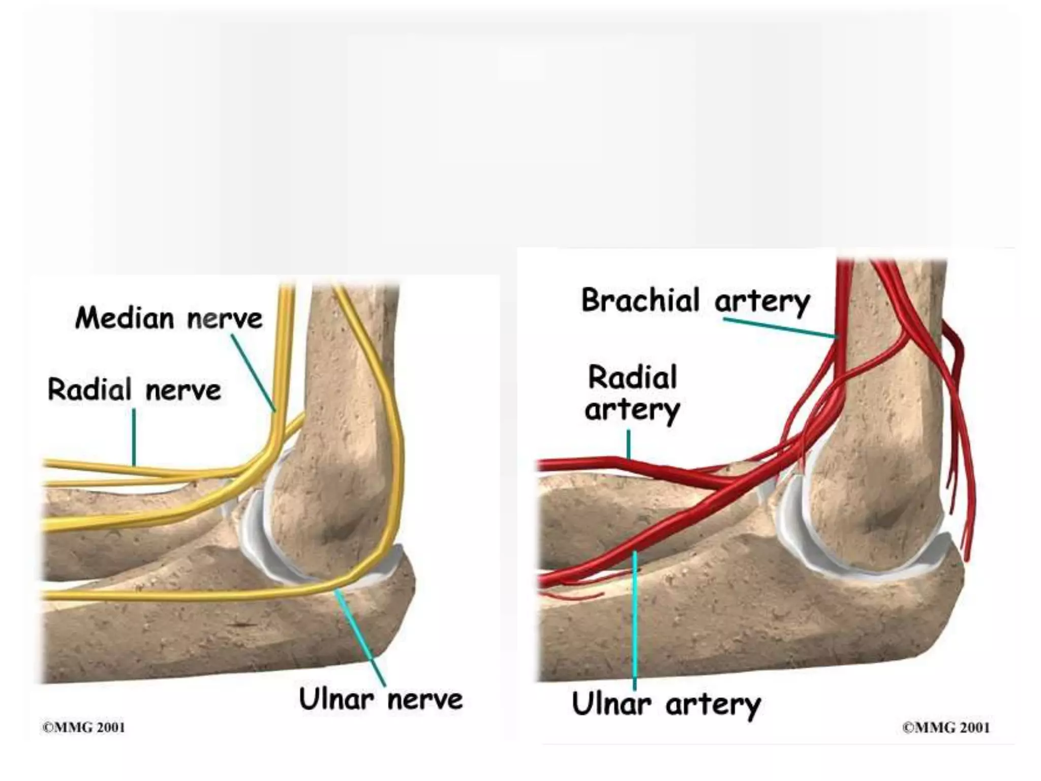
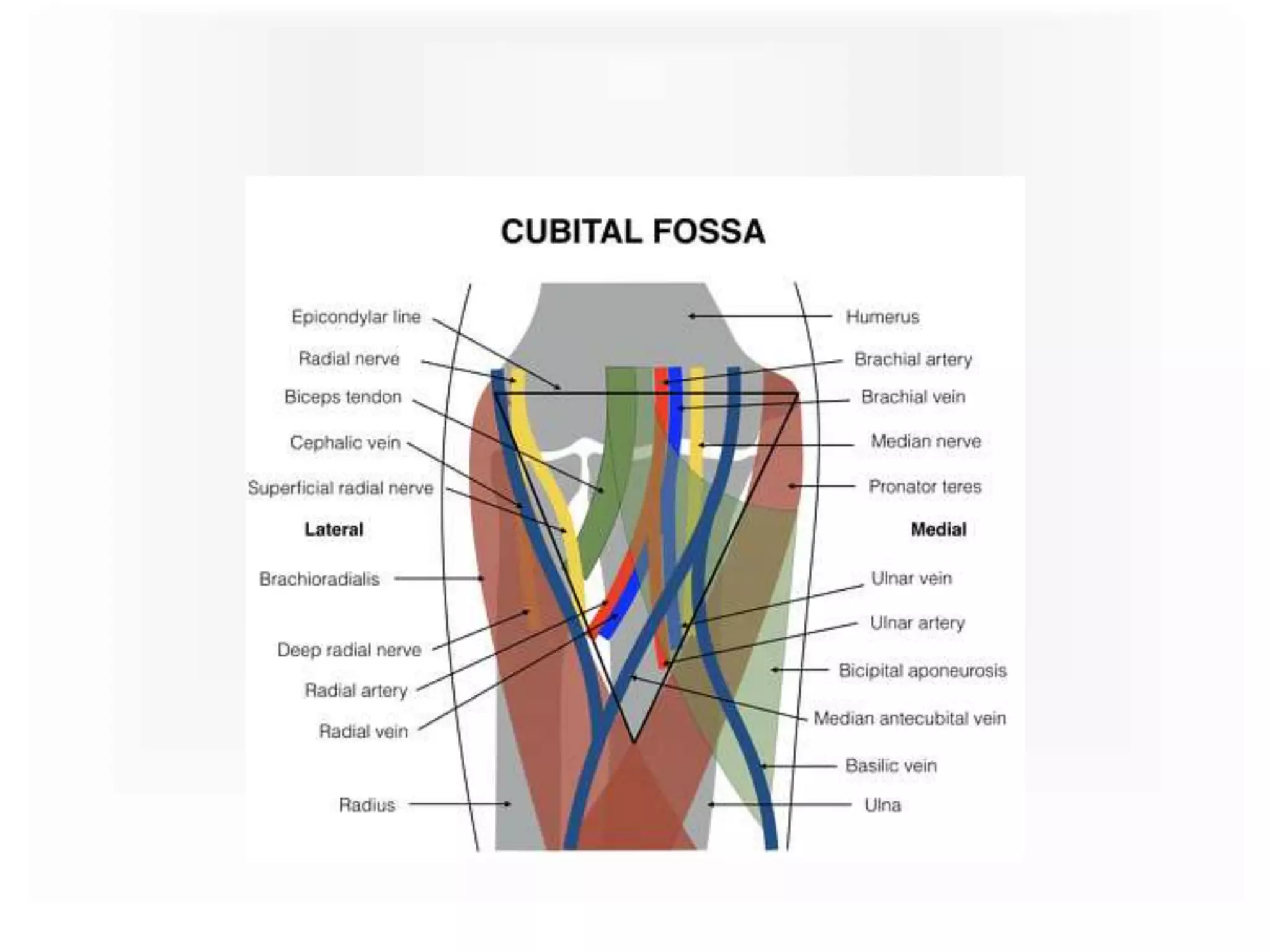
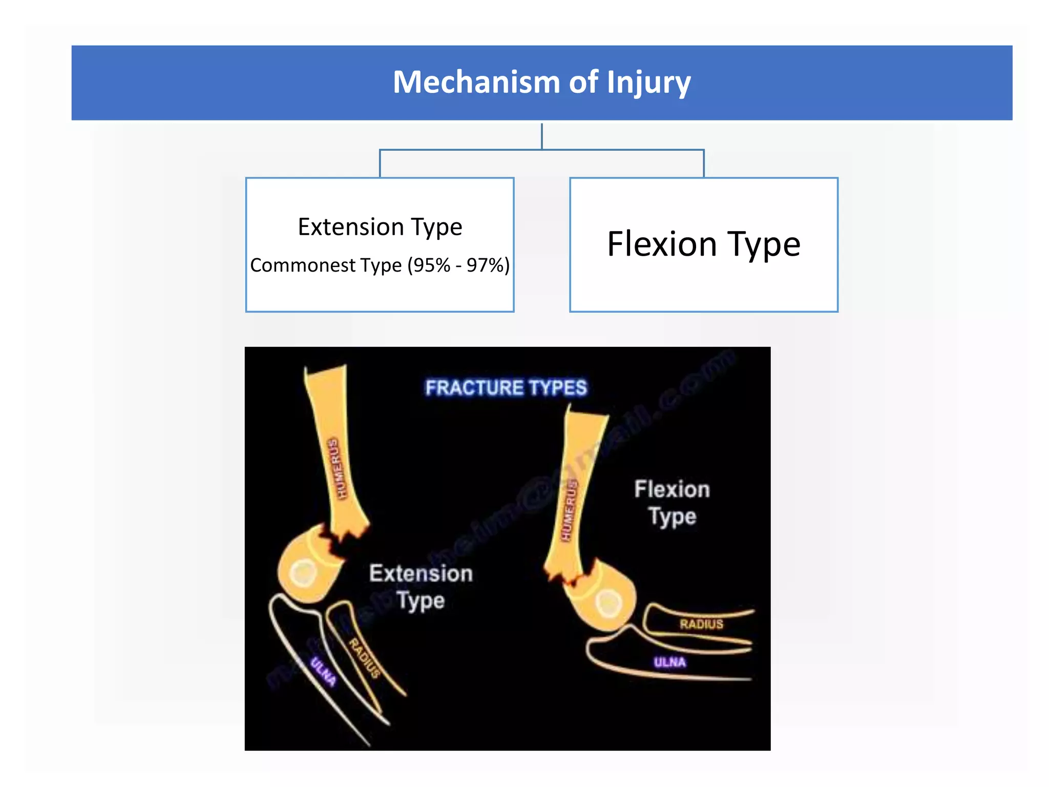
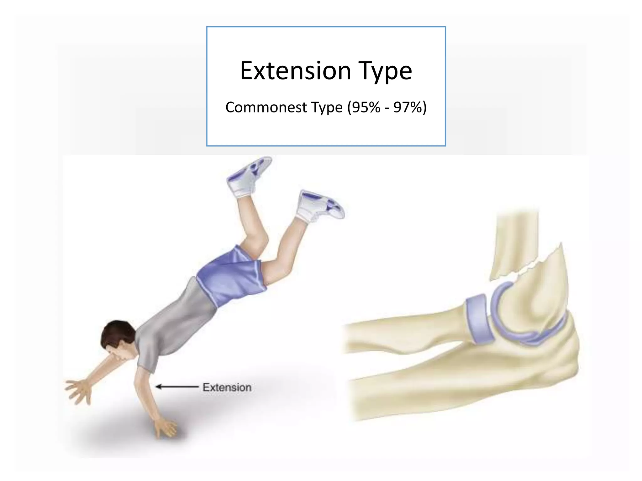
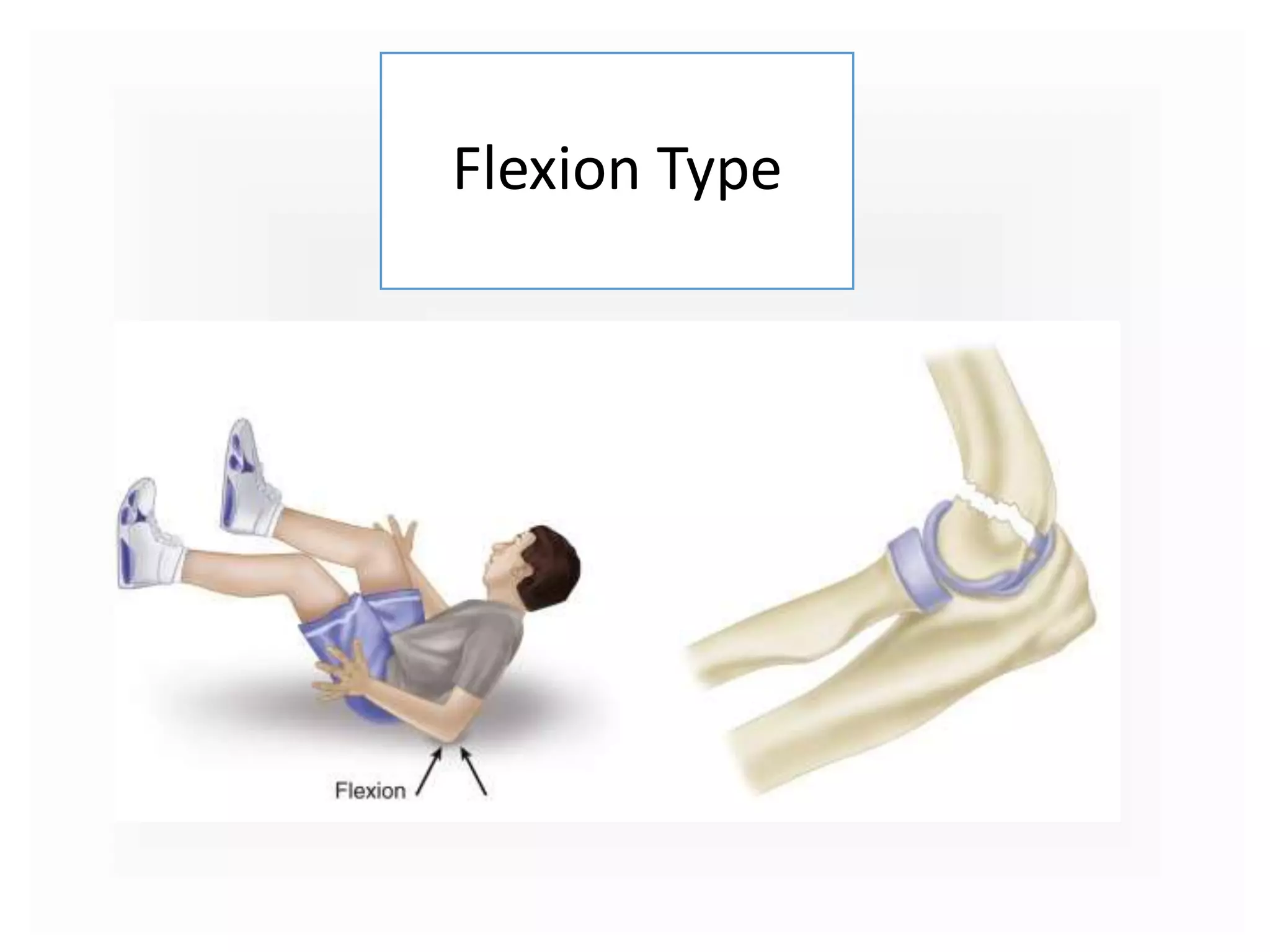
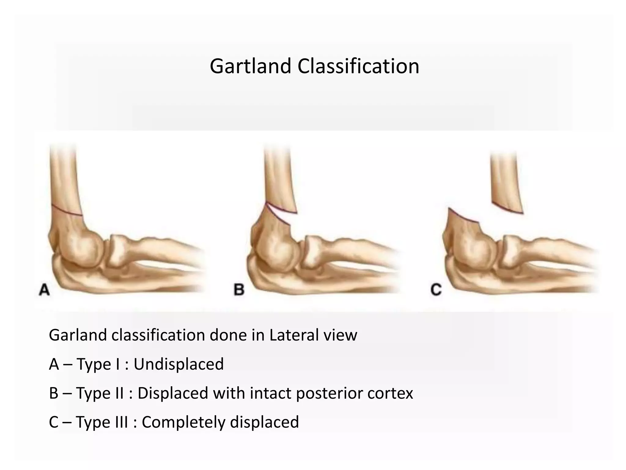
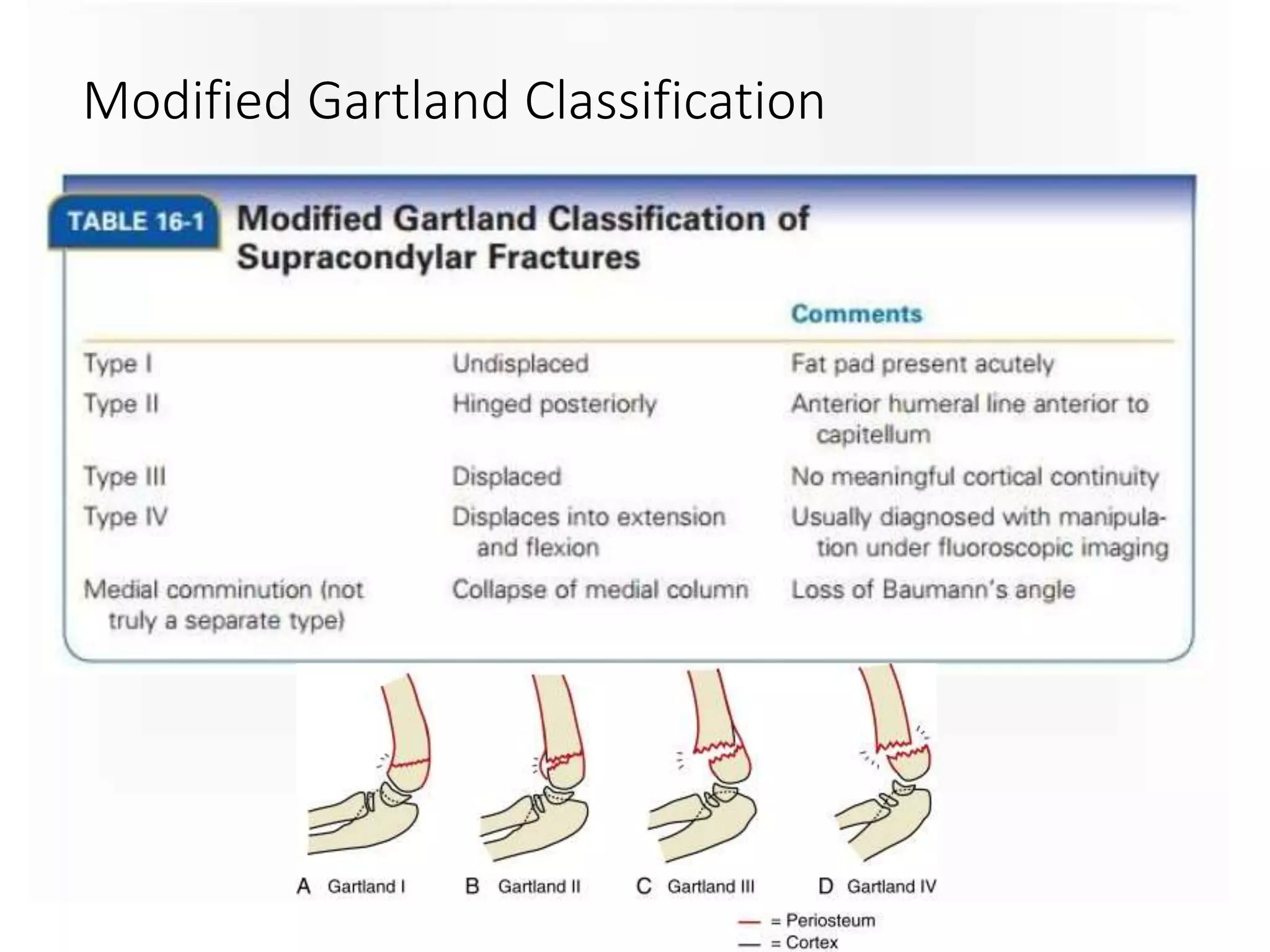
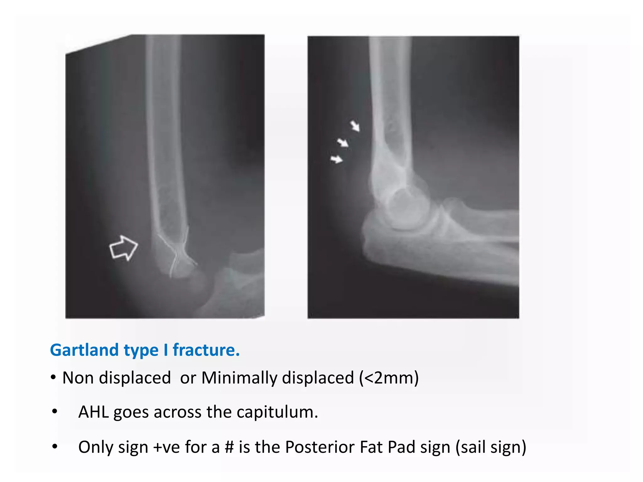
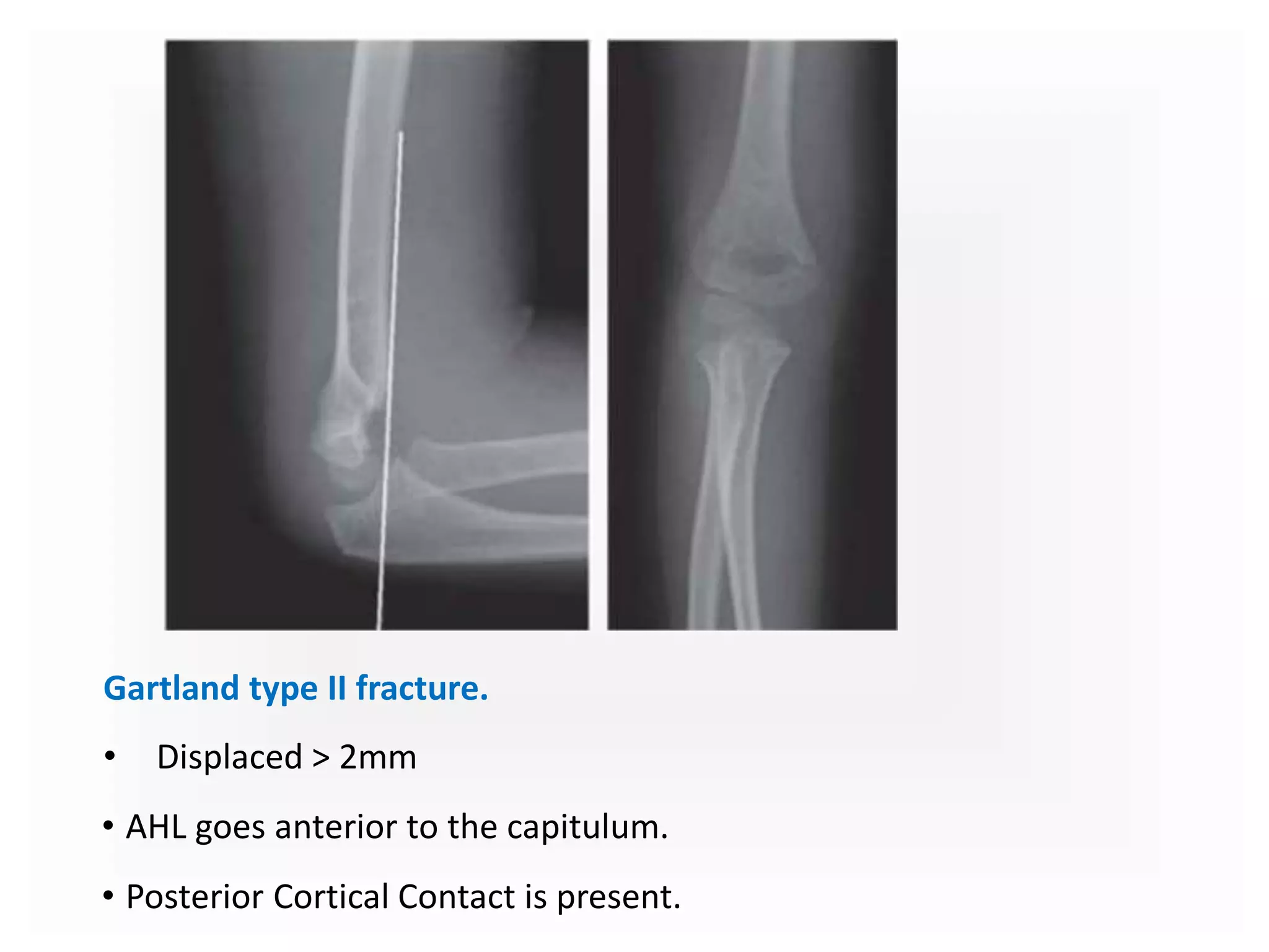
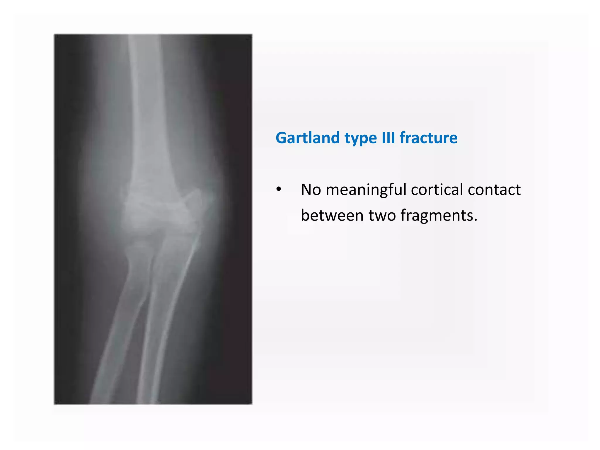
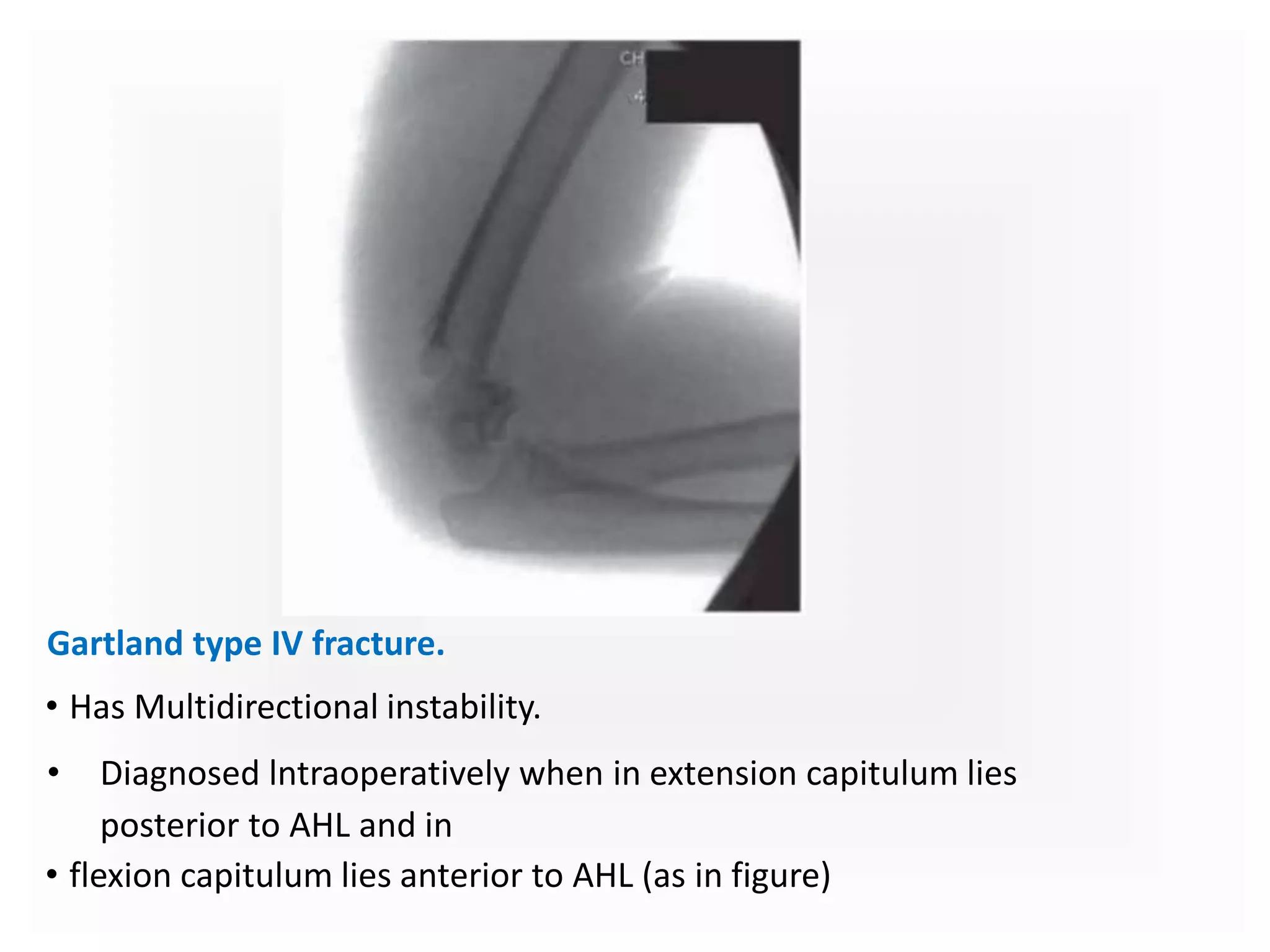
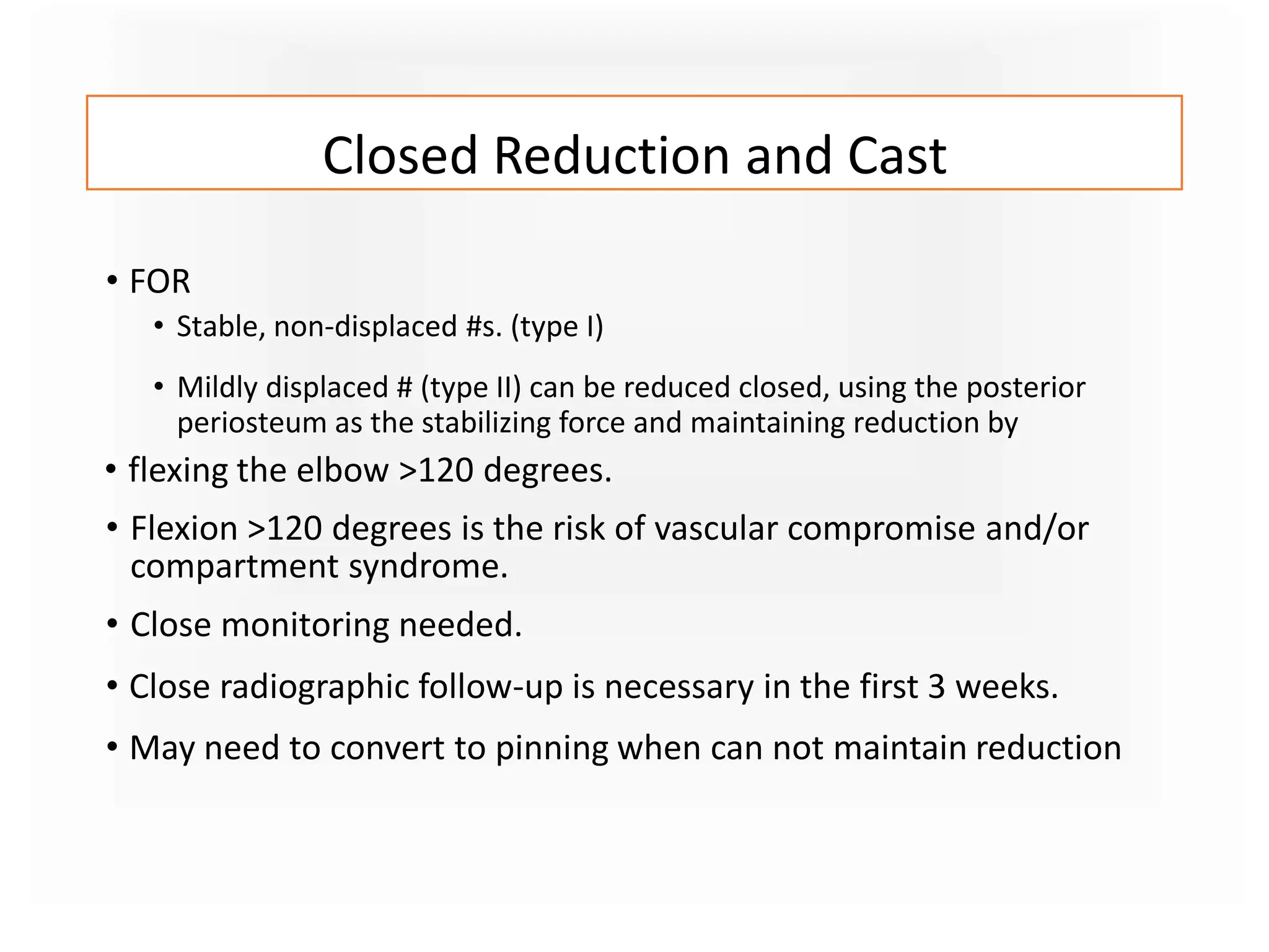
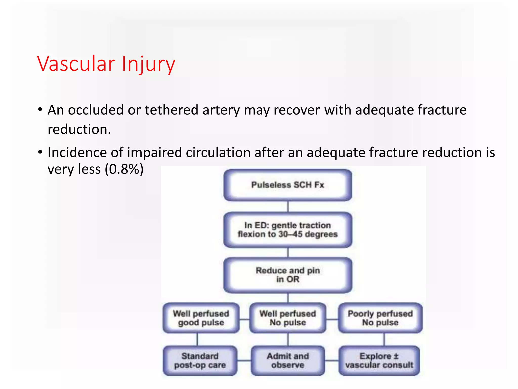
SUPRACONDYLAR HUMERUS
FRACTURE & COMPLICATION
Dr Yash Oza
Introduction
• Fracture of distal humerus
which is proximaI to the both
condyles.
• Often runs through the apices
of coronoid and olecranon
fossae, just above the fossae
or through the metaphysis of
humerus.
• # goes through the thinnest
portion of the bone
Epidemiology
• Most common elbow injury in Children (around 60%}
• Becomes less common with increasing age
• Peak age : Bimodal
• (1) 5 - 7yrs : Boys > Girls
• (2) Around 80yrs female
• Non Dominant Limb > Dominant Limb
• Nerve injuries - 7% (Radial > Median > Ulnar)
• Vascular Injuries - 1%
• Open # - < 1%
Anatomy
• Ossification Centers around the elbow
Mechanism of Injury
Extension Type
Commonest Type (95% - 97%)
Flexion Type
Extension Type
Commonest Type (95% - 97%)
Flexion Type
Patient Evalution
• Patient comes with History of fall
• Symptoms
• pain
• refusal to move the elbow
• Physical examination
• On inspection
• gross deformity
• swelling
• Ecchymosis in antecubital fossa
• motion
• Very limited active elbow motion
• Neurovascular examination is done
• Time and mechanism of injury is noted
Examination
• Vascular Examination
• Warm or Cold periphery
• Red or Blue periphery
• Radial pulse present or absent
• Signs of developing compartment syndrome.
• Maintain high index of suspicion
• Pain on passive flexion and extension of fingers
• Swelling , ecchymosis and anterior skin puckering
• Tenseness of the volar compartment
• Anxiety , Agitation and Increased analgesic requirement (3As)
Examination
• Neurological deficits
• Radial Nerve - Sensation over the dorsal 1st web space , Wrist
and finger extension
• AIN - OK sign
• Median Nerve - Sensation over the index finger , Flexion of
Fingers
• Ulnar Nerve - Sensation over the little finger, lnterrosei function.
• Median nerve injury may mask the pain of compartment syndrome
, Thus close monitoring needed.
• All Documented and informed to relatives
Classification
• During the 1950s, these injuries were called the "misunderstood
fracture" as such injuries often resulted in bony deformity and
Volkmann's contracture.
• In 1959, Gartland described a simple classification scheme to
reemphasize principles underlying treatment of patients with a
supracondylar humerus fracture.
Gartland Classification
Garland classification done in Lateral view
A – Type I : Undisplaced
B – Type II : Displaced with intact posterior cortex
C – Type III : Completely displaced
Modified Gartland Classification
AO Muller Classification of Distal humeral #
• X-Ray Views
•True AP
• Lateral
• Oblique
•Axial (jones view)
•AP of an elbow in 90 degrees of flexion
will give a roughly 45 degree angulated
view of the distal humerus and proximal
radius and ulna
Radiographic Diagnosis
Anterior Humeral Line (AHL) Baumann’s Angle
Anterior Humeral Line Baumann’s Angle
AHL should cross through
capitulum in dead lateral
view
• A rule of thumb is that a
Baumann's angle >10
degrees is OK.
• A decrease in Baumann's
angle compared to the
other side is a sign that a
fracture is in varus
angulation.
Gartland type I fracture.
• Non displaced or Minimally displaced (<2mm)
• AHL goes across the capitulum.
• Only sign +ve for a # is the Posterior Fat Pad sign (sail sign)
Gartland type II fracture.
• Displaced > 2mm
• AHL goes anterior to the capitulum.
• Posterior Cortical Contact is present.
Gartland type III fracture
• No meaningful cortical contact
between two fragments.
Gartland type IV fracture.
• Has Multidirectional instability.
• Diagnosed lntraoperatively when in extension capitulum lies
posterior to AHL and in
• flexion capitulum lies anterior to AHL (as in figure)
• Initially kept splinted with elbow in a comfortable position. (20 - 40
degree of flexion).
• Extreme flexion of extension may increase compartment pressure.
• Avoid tight bandage and splinting.
• Elbow and hand elevated above heart.
Initial Management
Urgency of treatment
•Treatment should be urgent In the presence of,
o poor distal perfusion
o firm compartments
o associated forearm #
o considerable swelling
o antecubital ecchymosis and skin
Methods of treatment
1. Cast only
2. Closed Reduction and Cast
3. Closed Reduction and Percutaneous
Pinning
4. Open Reduction
Closed Reduction and Cast
• FOR
• Stable, non-displaced #s. (type I)
• Mildly displaced # (type II) can be reduced closed, using the posterior
periosteum as the stabilizing force and maintaining reduction by
• flexing the elbow >120 degrees.
• Flexion >120 degrees is the risk of vascular compromise and/or
compartment syndrome.
• Close monitoring needed.
• Close radiographic follow-up is necessary in the first 3 weeks.
• May need to convert to pinning when can not maintain reduction
Technique of reduction
Closed Reduction and Percutaneous Pinning
(CRPP)
• Most common operative treatment.
• Initial attempt at closed reduction is indicated in almost all displaced
supracondylar fractures that are not open fractures.
• Technique:
• C-arm is needed
• The elbow is then flexed while pushing the olecranon anteriorly to correct the
sagittal deformity and reduce the fracture.
• Wires are passed
Open Reduction
• Indicated in cases of failed closed reduction , a loss of pulse or
poorly perfused hand following reduction, and open fractures.
• Problems with Open reductions are –
• elbow stiffness
• myositis ossificans
• ugly scarring
• iatrogenic neurovascular injury.
• In setting of severe soft tissue injury and bone injury, better
results seen with open reduction.
Vascular Injury
• An occluded or tethered artery may recover with adequate fracture
reduction.
• Incidence of impaired circulation after an adequate fracture reduction is
very less (0.8%)
• Pin migration
• most common complication (~2%)
• Pin tract Infection
• occurs in 1-2.4%
• typically superficial and treated with oral antibiotics
• Cubitus valgus
• caused by fracture malunion
• can lead to tardy ulnar nerve palsy
• Cubitus varus (gunstock deformity)
• caused by fracture varus malunion, especially in medial comminution
pattern
• is NOT caused by growth disturbance
• usually a cosmetic issue with little functional limitations, but may be present
Complications
• Nerve palsy from injury
• usually resolve, nerves rarely torn
• extension type fractures
• neuropraxia in 11%
• most commonly AIN
• mechanism = tenting of nerve on fracture, or entrapment in
the fracture site
• flexion type fractures
• neuropraxia in 17%
• most commonly cause ulnar neuropraxia
• Vascular Injury
• radial pulse absent on initial presentation in 7-12%
• pulseless hand after closed reduction and pinning (3-4%)
• decision to explore is based on quality of extremity perfusion
rather than absence of pulse
• Volkmann ischemic contracture
• rare, but dreaded complication
• may result from elbow hyperflexion casting
• increase in deep volar forearm compartment pressures and loss of
radial pulse with elbow flexed >90°
• rarely seen with CRPP and postoperative immobilization in less than
90°
• Postoperative stiffness
• rare after casting or after pinning procedures
• remove pins and allow gentle ROM at 3-4 weeks postop
• resolves by 6 months
• Non-union
• rare
• Myositis Ossificans ( Heterotrophic ossification of muscle)
• Rare. Can be managed conservatively.
• Avascular Necrosis
• Of trochlear
• Fragile blood supply to ossification center (more distal # this
supply is at risk)
• Posterior approach to open reduction has increased risk

Supracondylar humerus fracture & complication for MBBS students

  • 1.
    SUPRACONDYLAR HUMERUS FRACTURE &COMPLICATION Dr Yash Oza
  • 2.
    Introduction • Fracture ofdistal humerus which is proximaI to the both condyles. • Often runs through the apices of coronoid and olecranon fossae, just above the fossae or through the metaphysis of humerus. • # goes through the thinnest portion of the bone
  • 3.
    Epidemiology • Most commonelbow injury in Children (around 60%} • Becomes less common with increasing age • Peak age : Bimodal • (1) 5 - 7yrs : Boys > Girls • (2) Around 80yrs female • Non Dominant Limb > Dominant Limb • Nerve injuries - 7% (Radial > Median > Ulnar) • Vascular Injuries - 1% • Open # - < 1%
  • 4.
  • 7.
    • Ossification Centersaround the elbow
  • 8.
    Mechanism of Injury ExtensionType Commonest Type (95% - 97%) Flexion Type
  • 9.
  • 10.
  • 11.
    Patient Evalution • Patientcomes with History of fall • Symptoms • pain • refusal to move the elbow • Physical examination • On inspection • gross deformity • swelling • Ecchymosis in antecubital fossa • motion • Very limited active elbow motion • Neurovascular examination is done • Time and mechanism of injury is noted
  • 12.
    Examination • Vascular Examination •Warm or Cold periphery • Red or Blue periphery • Radial pulse present or absent • Signs of developing compartment syndrome. • Maintain high index of suspicion • Pain on passive flexion and extension of fingers • Swelling , ecchymosis and anterior skin puckering • Tenseness of the volar compartment • Anxiety , Agitation and Increased analgesic requirement (3As)
  • 13.
    Examination • Neurological deficits •Radial Nerve - Sensation over the dorsal 1st web space , Wrist and finger extension • AIN - OK sign • Median Nerve - Sensation over the index finger , Flexion of Fingers • Ulnar Nerve - Sensation over the little finger, lnterrosei function. • Median nerve injury may mask the pain of compartment syndrome , Thus close monitoring needed. • All Documented and informed to relatives
  • 14.
    Classification • During the1950s, these injuries were called the "misunderstood fracture" as such injuries often resulted in bony deformity and Volkmann's contracture. • In 1959, Gartland described a simple classification scheme to reemphasize principles underlying treatment of patients with a supracondylar humerus fracture.
  • 15.
    Gartland Classification Garland classificationdone in Lateral view A – Type I : Undisplaced B – Type II : Displaced with intact posterior cortex C – Type III : Completely displaced
  • 16.
  • 17.
    AO Muller Classificationof Distal humeral #
  • 18.
    • X-Ray Views •TrueAP • Lateral • Oblique •Axial (jones view) •AP of an elbow in 90 degrees of flexion will give a roughly 45 degree angulated view of the distal humerus and proximal radius and ulna Radiographic Diagnosis
  • 19.
    Anterior Humeral Line(AHL) Baumann’s Angle
  • 20.
    Anterior Humeral LineBaumann’s Angle AHL should cross through capitulum in dead lateral view • A rule of thumb is that a Baumann's angle >10 degrees is OK. • A decrease in Baumann's angle compared to the other side is a sign that a fracture is in varus angulation.
  • 21.
    Gartland type Ifracture. • Non displaced or Minimally displaced (<2mm) • AHL goes across the capitulum. • Only sign +ve for a # is the Posterior Fat Pad sign (sail sign)
  • 22.
    Gartland type IIfracture. • Displaced > 2mm • AHL goes anterior to the capitulum. • Posterior Cortical Contact is present.
  • 23.
    Gartland type IIIfracture • No meaningful cortical contact between two fragments.
  • 24.
    Gartland type IVfracture. • Has Multidirectional instability. • Diagnosed lntraoperatively when in extension capitulum lies posterior to AHL and in • flexion capitulum lies anterior to AHL (as in figure)
  • 25.
    • Initially keptsplinted with elbow in a comfortable position. (20 - 40 degree of flexion). • Extreme flexion of extension may increase compartment pressure. • Avoid tight bandage and splinting. • Elbow and hand elevated above heart. Initial Management
  • 26.
    Urgency of treatment •Treatmentshould be urgent In the presence of, o poor distal perfusion o firm compartments o associated forearm # o considerable swelling o antecubital ecchymosis and skin
  • 27.
    Methods of treatment 1.Cast only 2. Closed Reduction and Cast 3. Closed Reduction and Percutaneous Pinning 4. Open Reduction
  • 28.
    Closed Reduction andCast • FOR • Stable, non-displaced #s. (type I) • Mildly displaced # (type II) can be reduced closed, using the posterior periosteum as the stabilizing force and maintaining reduction by • flexing the elbow >120 degrees. • Flexion >120 degrees is the risk of vascular compromise and/or compartment syndrome. • Close monitoring needed. • Close radiographic follow-up is necessary in the first 3 weeks. • May need to convert to pinning when can not maintain reduction
  • 29.
  • 30.
    Closed Reduction andPercutaneous Pinning (CRPP) • Most common operative treatment. • Initial attempt at closed reduction is indicated in almost all displaced supracondylar fractures that are not open fractures. • Technique: • C-arm is needed • The elbow is then flexed while pushing the olecranon anteriorly to correct the sagittal deformity and reduce the fracture. • Wires are passed
  • 31.
    Open Reduction • Indicatedin cases of failed closed reduction , a loss of pulse or poorly perfused hand following reduction, and open fractures. • Problems with Open reductions are – • elbow stiffness • myositis ossificans • ugly scarring • iatrogenic neurovascular injury. • In setting of severe soft tissue injury and bone injury, better results seen with open reduction.
  • 32.
    Vascular Injury • Anoccluded or tethered artery may recover with adequate fracture reduction. • Incidence of impaired circulation after an adequate fracture reduction is very less (0.8%)
  • 33.
    • Pin migration •most common complication (~2%) • Pin tract Infection • occurs in 1-2.4% • typically superficial and treated with oral antibiotics • Cubitus valgus • caused by fracture malunion • can lead to tardy ulnar nerve palsy • Cubitus varus (gunstock deformity) • caused by fracture varus malunion, especially in medial comminution pattern • is NOT caused by growth disturbance • usually a cosmetic issue with little functional limitations, but may be present Complications
  • 34.
    • Nerve palsyfrom injury • usually resolve, nerves rarely torn • extension type fractures • neuropraxia in 11% • most commonly AIN • mechanism = tenting of nerve on fracture, or entrapment in the fracture site • flexion type fractures • neuropraxia in 17% • most commonly cause ulnar neuropraxia • Vascular Injury • radial pulse absent on initial presentation in 7-12% • pulseless hand after closed reduction and pinning (3-4%) • decision to explore is based on quality of extremity perfusion rather than absence of pulse
  • 35.
    • Volkmann ischemiccontracture • rare, but dreaded complication • may result from elbow hyperflexion casting • increase in deep volar forearm compartment pressures and loss of radial pulse with elbow flexed >90° • rarely seen with CRPP and postoperative immobilization in less than 90° • Postoperative stiffness • rare after casting or after pinning procedures • remove pins and allow gentle ROM at 3-4 weeks postop • resolves by 6 months • Non-union • rare
  • 36.
    • Myositis Ossificans( Heterotrophic ossification of muscle) • Rare. Can be managed conservatively. • Avascular Necrosis • Of trochlear • Fragile blood supply to ossification center (more distal # this supply is at risk) • Posterior approach to open reduction has increased risk