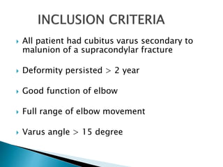
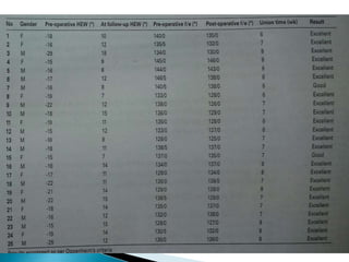
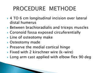
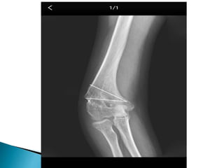
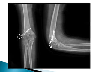
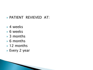
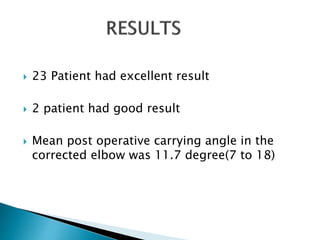
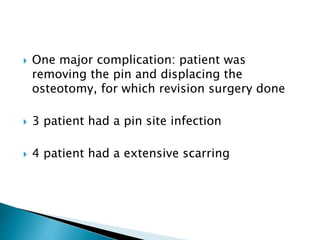
PRESENTER : DR. SANDEEP TRIPATHI
MODERATOR : PROF.SURENDHER KUMAR
 To describe a new technique for
correcting post traumatic cubitus
varus using a lateral closing wedge
isosceles triangular osteotomy
CUBITUS VARUS:/gunstock elbow
deviation of forearm toward the midline of the
body, and carrying angle decreased
(5 to 15 degree normal)
Most common complication of supracondylar
fracture
It produce cosmetic deformity
PATIENT
 Performed in 25 patient(15-male, 10-female)
 Age group: 6 to 12 year
 From may 2010 to april 2013
 All patient had cubitus varus secondary to
malunion of a supracondylar fracture
 Deformity persisted > 2 year
 Good function of elbow
 Full range of elbow movement
 Varus angle > 15 degree
 4 TO 6 cm longitudinal incision over lateral
distal humerus
 Between brachioradialis and triceps muscles
 Coronoid fossa exposed circuferentially
 Line of osteotomy make
 Osteotomy made
 Preserve the medial cortical hinge
 Fixed with 2 kirschner wire (k-wire)
 Long arm cast applied with elbow flex 90 deg
 PATIENT REVIEVED AT:
 4 weeks
 6 weeks
 3 months
 6 months
 12 months
 Every 2 year
 23 Patient had excellent result
 2 patient had good result
 Mean post operative carrying angle in the
corrected elbow was 11.7 degree(7 to 18)
 One major complication: patient was
removing the pin and displacing the
osteotomy, for which revision surgery done
 3 patient had a pin site infection
 4 patient had a extensive scarring
 A lateral isosceles triangular osteotomy and
k-wire fixation is a
 Practical
 Effective
 Reliable
 Safe & simple method
 Excellent cosmesis
 This approach reduced the incidence of post
operative stiffness & ulnar nerve injury
DISCUSSION
CUBITUS VARUS:
(extension+internal rotation+varus angulation
Of distal fragment)
is a complication of supracondylar fracture of
humerus in childhood arising as a result:
Inadequate reduction
Loss of reduction during healing
Growth disturbance due to physeal injury
Limitation of the technique:
 Can not correct severe internal rotation
deformity

Lateral closing isosceles triangular osteotomy for the treatment

  • 1.
    PRESENTER : DR.SANDEEP TRIPATHI MODERATOR : PROF.SURENDHER KUMAR
  • 2.
     To describea new technique for correcting post traumatic cubitus varus using a lateral closing wedge isosceles triangular osteotomy
  • 3.
    CUBITUS VARUS:/gunstock elbow deviationof forearm toward the midline of the body, and carrying angle decreased (5 to 15 degree normal) Most common complication of supracondylar fracture It produce cosmetic deformity
  • 4.
    PATIENT  Performed in25 patient(15-male, 10-female)  Age group: 6 to 12 year  From may 2010 to april 2013
  • 5.
     All patienthad cubitus varus secondary to malunion of a supracondylar fracture  Deformity persisted > 2 year  Good function of elbow  Full range of elbow movement  Varus angle > 15 degree
  • 7.
     4 TO6 cm longitudinal incision over lateral distal humerus  Between brachioradialis and triceps muscles  Coronoid fossa exposed circuferentially  Line of osteotomy make  Osteotomy made  Preserve the medial cortical hinge  Fixed with 2 kirschner wire (k-wire)  Long arm cast applied with elbow flex 90 deg
  • 10.
     PATIENT REVIEVEDAT:  4 weeks  6 weeks  3 months  6 months  12 months  Every 2 year
  • 11.
     23 Patienthad excellent result  2 patient had good result  Mean post operative carrying angle in the corrected elbow was 11.7 degree(7 to 18)
  • 12.
     One majorcomplication: patient was removing the pin and displacing the osteotomy, for which revision surgery done  3 patient had a pin site infection  4 patient had a extensive scarring
  • 13.
     A lateralisosceles triangular osteotomy and k-wire fixation is a  Practical  Effective  Reliable  Safe & simple method  Excellent cosmesis  This approach reduced the incidence of post operative stiffness & ulnar nerve injury
  • 14.
    DISCUSSION CUBITUS VARUS: (extension+internal rotation+varusangulation Of distal fragment) is a complication of supracondylar fracture of humerus in childhood arising as a result: Inadequate reduction Loss of reduction during healing Growth disturbance due to physeal injury
  • 15.
    Limitation of thetechnique:  Can not correct severe internal rotation deformity