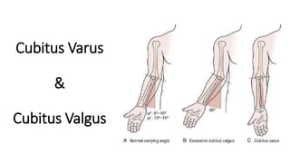
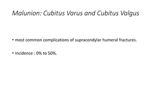
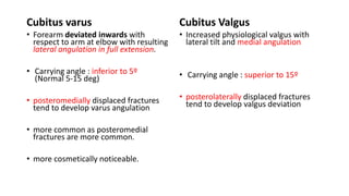
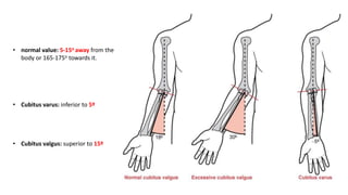
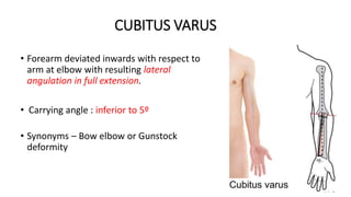
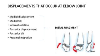
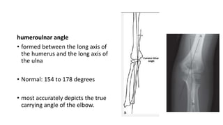
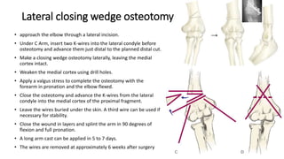
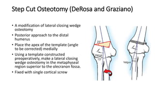
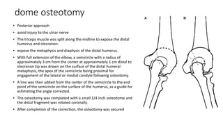
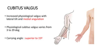
Cubitus Varus
&
Cubitus Valgus
Malunion: Cubitus Varus and Cubitus Valgus
• most common complications of supracondylar humeral fractures.
• incidence : 0% to 50%.
Cubitus varus
• Forearm deviated inwards with
respect to arm at elbow with resulting
lateral angulation in full extension.
• Carrying angle : inferior to 5º
(Normal 5-15 deg)
• posteromedially displaced fractures
tend to develop varus angulation
• more common as posteromedial
fractures are more common.
• more cosmetically noticeable.
Cubitus Valgus
• Increased physiological valgus with
lateral tilt and medial angulation
• Carrying angle : superior to 15º
• posterolaterally displaced fractures
tend to develop valgus deviation
Carrying angle of the arm
(To assess Cubitus varus or valgus)
• the angle created by the medial border of
the fully supinated forearm and medial
border of the humerus, with the elbow
extended
• considerable individual variation.
• comparison should be made with the
contralateral side
• normal value: 5-15o away from the
body or 165-175o towards it.
• Cubitus varus: inferior to 5º
• Cubitus valgus: superior to 15º
• Carrying angle decreases with elbow extension, thus hyperextension
tends increases a cubitus varus deformity
• whereas a flexion contracture can create the appearance of cubitus
valgus
Problems arising from cubitus varus or valgus:
• Cosmetic deformity
• Functional limitation
• Recurrent elbow fracture
• tardy ulnar nerve symptoms
• cubitus valgus and varus are primarily cosmetic deformities
• mild degrees of malunion can be treated by simple reassurance.
• The resultant cubitus varus deformity is a combined
• deformity of varus
• extension
• internal rotation
• Most corrective osteotomies are to correct varus and extension deformity.
• The rotational deformity is well tolerated and best left untreated because
rotation of the distal fragment makes the osteotomy unstable.
CUBITUS VARUS
• Forearm deviated inwards with respect to
arm at elbow with resulting lateral
angulation in full extension.
• Carrying angle : inferior to 5º
• Synonyms – Bow elbow or Gunstock
deformity
Causes
• Post traumatic malunited supracondylar humerus fracture (most common)
• Malunited lateral condyle fracture
• Malunited intercondylar fracture
• Malunited medial condyle fracture
• NEOPLASTIC: secondary to exostosis in distal, lateral humerus
• Congenital : epiphyseal dysplasia
• VASCULAR : Trochlear Osteonecrosis
• INFECTIVE: medial growth plate damage
factors for malunion
• Impacted / comminuted type I supracondylar fractures
• Rotationally unstable type II fractures treated in a cast with
subsequent loss of reduction
• Poorly stabilized or reduced type III fractures or
• delayed neglected fractures
Patient presents with
• Previous history trauma to elbow
• history institutional treatment or local treatment
• Deformity of elbow joint – on extension of elbow
• Gunstock deformity
• Cosmetic problem No functional disability as such
• Ulnar nerve irritation
ON EXAMINATION
• Inspection
• Hyperextension deformity
• Limited flexion
• Medial tilt and lateral angulation at elbow
• Prominence of lateral condyle of humerus
• Gunstock deformity
• Wasting of muscles
“Gun-stock Deformity”
• Looks like a loading stock of old
long barrel guns
• PALPATION:
• Thickening and irregularity of supracondylar ridges
• Medial epicondyle tip higher
DISPLACEMENTS THAT OCCUR AT ELBOW JOINT
• Medial displacement
• Medial tilt
• Internal rotation
• Posterior displacement
• Posterior tilt
• Proximal migration
DISTAL FRAGMENT
Investigations:
• Plain radiograph of elbow AP and lateral view
• Assess the Carrying angle
MEASUREMENTS ON XRAY :- AP VIEW
Baumann’s angle or the shaft-physeal
angle
• between the longitudinal axis of the
humerus and a line through the physis
of the lateral condyle
• Decrease in normal physiological
valgus
• Increase in Baumann’s Angle
(Normal : 64 ̊to 81 ̊)
metaphyseal-diaphyseal angle
• between the long axis of the
humerus and a line connecting
the lateral and medial
epicondyles
• Normal: 72 to 95 degrees
humeroulnar angle
• formed between the long axis of
the humerus and the long axis of
the ulna
• Normal: 154 to 178 degrees
• most accurately depicts the true
carrying angle of the elbow.
LATERAL VIEW
• Normally no overlap between
the lateral condylar epiphysis
and olecranon epiphysis
• If significant tilt of distal
fragment occurs, there is overlap
between the two, which appears
like a crescent → ‘Crescent Sign”
TREATMENT :- MODALITIES
1. Observation with expected remodeling
2. Hemiepiphysiodesis and growth alteration
3. Corrective osteotomy
4. Distraction osteogenesis by Ilizarov technique
• Treatment is primarily “Cosmetic Correction”
Corrective Osteotomy
Pre-requisites
• At least 1 year following fracture (Bone remodeling and tissue
equilibrium)
• Patient demanding surgery
• Calculation of wedge to be removed
→ Normal side Xray → Wedge angle = Varus + Normal physiological Valgus
CORRECTIVE OSTEOTOMY
Options include:
• Medial open wedge osteotomy
• Lateral closing wedge osteotomy also known as French osteotomy .
• Oblique osteotomy with derotation.
• Dome osteotomy .
• Step cut osteotomy
• Arch
• Pentalateral
Osteotomies
Lateral closing wedge osteotomy
• approach the elbow through a lateral incision.
• Under C Arm, insert two K-wires into the lateral condyle before
osteotomy and advance them just distal to the planned distal cut.
• Make a closing wedge osteotomy laterally, leaving the medial
cortex intact.
• Weaken the medial cortex using drill holes.
• Apply a valgus stress to complete the osteotomy with the
forearm in pronation and the elbow flexed.
• Close the osteotomy and advance the K-wires from the lateral
condyle into the medial cortex of the proximal fragment.
• Leave the wires buried under the skin. A third wire can be used if
necessary for stability.
• Close the wound in layers and splint the arm in 90 degrees of
flexion and full pronation.
• A long arm cast can be applied in 5 to 7 days.
• The wires are removed at approximately 6 weeks after surgery
Step Cut Osteotomy (DeRosa and Graziano)
• A modification of lateral closing wedge
osteotomy
• Posterior approach to the distal
humerus
• Place the apex of the template (angle
to be corrected) medially
• Using a template constructed
preoperatively, make a lateral closing
wedge osteotomy in the metaphyseal
region superior to the olecranon fossa.
• Fixed with single cortical screw
dome osteotomy
• Posterior approach
• avoid injury to the ulnar nerve
• The triceps muscle was split along the midline to expose the distal
humerus and olecranon.
• expose the metaphysis and diaphysis of the distal humerus.
• With full extension of the elbow, a semicircle with a radius of
approximately 3 cm from the center at approximately 1 cm distal to
olecranon tip was drawn on the surface of the distal humeral
metaphysis, the apex of the semicircle being proximal for
engagement of the lateral or medial condyle following osteotomy.
• A line was then added from the center of the semicircle to the end
point of the semicircle on the surface of the humerus, as a guide for
estimating the angle corrected.
• The osteotomy was completed with a small 1/4 inch osteotome and
the distal fragment was rotated coronally.
• After completion of the correction, the osteotomy was secured
COMPLICATIONS OF OSTEOTOMY
• Stiffness
• Nerve injury
• Persistent deformity (under correction)
• Recurrent deformity
• Non-union
• Osteomyelitis
• Skin sloughing
CUBITUS VALGUS
• Increased physiological valgus with
lateral tilt and medial angulation
• Physiological cubitus valgus varies from
3 to 29 deg
• Carrying angle : superior to 15º
Causes
• Non-union fracture lateral condyle of humerus
• Malunited supracondylar fracture humerus
• Osteonecrosis of lateral trochlea
• Malunited intercondylar fracture
• Radial head fracture dislocation
• Medial epiphyseal injury and growth stimulation
• History of fracture of lateral condyle of humerus
• Patient presents with external deformity of elbow joint
• Usually asymptomatic till patient develops TARDY ULNAR NERVE
PALSY
Tardy ulnar nerve palsy
• due to gradual stretching of the ulnar nerve during the progression of
valgus deformity of elbow
• Symptoms – tingling and paraesthesia over ulnar nerve distribution
• Can also be seen in cubitus varus (friction neuropathy), medial
condyle fracture, olecranon fracture and Monteggia fracture
dislocation
Investigations
• Plain radiograph AP and lateral view of elbow
• Assess the Carrying angle
• Nerve conduction studies
Treatment
• Deformity correction for cosmetic reasons – Osteotomies, Ilizarov
technique
• Anterior transposition of ulnar nerve for Tardy ulnar nerve palsy
Cubitus valgus varus

Cubitus valgus varus

  • 1.
  • 2.
    Malunion: Cubitus Varusand Cubitus Valgus • most common complications of supracondylar humeral fractures. • incidence : 0% to 50%.
  • 3.
    Cubitus varus • Forearmdeviated inwards with respect to arm at elbow with resulting lateral angulation in full extension. • Carrying angle : inferior to 5º (Normal 5-15 deg) • posteromedially displaced fractures tend to develop varus angulation • more common as posteromedial fractures are more common. • more cosmetically noticeable. Cubitus Valgus • Increased physiological valgus with lateral tilt and medial angulation • Carrying angle : superior to 15º • posterolaterally displaced fractures tend to develop valgus deviation
  • 4.
    Carrying angle ofthe arm (To assess Cubitus varus or valgus) • the angle created by the medial border of the fully supinated forearm and medial border of the humerus, with the elbow extended • considerable individual variation. • comparison should be made with the contralateral side
  • 5.
    • normal value:5-15o away from the body or 165-175o towards it. • Cubitus varus: inferior to 5º • Cubitus valgus: superior to 15º
  • 6.
    • Carrying angledecreases with elbow extension, thus hyperextension tends increases a cubitus varus deformity • whereas a flexion contracture can create the appearance of cubitus valgus
  • 7.
    Problems arising fromcubitus varus or valgus: • Cosmetic deformity • Functional limitation • Recurrent elbow fracture • tardy ulnar nerve symptoms
  • 8.
    • cubitus valgusand varus are primarily cosmetic deformities • mild degrees of malunion can be treated by simple reassurance.
  • 9.
    • The resultantcubitus varus deformity is a combined • deformity of varus • extension • internal rotation • Most corrective osteotomies are to correct varus and extension deformity. • The rotational deformity is well tolerated and best left untreated because rotation of the distal fragment makes the osteotomy unstable.
  • 10.
    CUBITUS VARUS • Forearmdeviated inwards with respect to arm at elbow with resulting lateral angulation in full extension. • Carrying angle : inferior to 5º • Synonyms – Bow elbow or Gunstock deformity
  • 11.
    Causes • Post traumaticmalunited supracondylar humerus fracture (most common) • Malunited lateral condyle fracture • Malunited intercondylar fracture • Malunited medial condyle fracture • NEOPLASTIC: secondary to exostosis in distal, lateral humerus • Congenital : epiphyseal dysplasia • VASCULAR : Trochlear Osteonecrosis • INFECTIVE: medial growth plate damage
  • 12.
    factors for malunion •Impacted / comminuted type I supracondylar fractures • Rotationally unstable type II fractures treated in a cast with subsequent loss of reduction • Poorly stabilized or reduced type III fractures or • delayed neglected fractures
  • 13.
    Patient presents with •Previous history trauma to elbow • history institutional treatment or local treatment • Deformity of elbow joint – on extension of elbow • Gunstock deformity • Cosmetic problem No functional disability as such • Ulnar nerve irritation
  • 14.
    ON EXAMINATION • Inspection •Hyperextension deformity • Limited flexion • Medial tilt and lateral angulation at elbow • Prominence of lateral condyle of humerus • Gunstock deformity • Wasting of muscles
  • 15.
    “Gun-stock Deformity” • Lookslike a loading stock of old long barrel guns
  • 16.
    • PALPATION: • Thickeningand irregularity of supracondylar ridges • Medial epicondyle tip higher
  • 17.
    DISPLACEMENTS THAT OCCURAT ELBOW JOINT • Medial displacement • Medial tilt • Internal rotation • Posterior displacement • Posterior tilt • Proximal migration DISTAL FRAGMENT
  • 18.
    Investigations: • Plain radiographof elbow AP and lateral view • Assess the Carrying angle
  • 19.
    MEASUREMENTS ON XRAY:- AP VIEW Baumann’s angle or the shaft-physeal angle • between the longitudinal axis of the humerus and a line through the physis of the lateral condyle • Decrease in normal physiological valgus • Increase in Baumann’s Angle (Normal : 64 ̊to 81 ̊)
  • 20.
    metaphyseal-diaphyseal angle • betweenthe long axis of the humerus and a line connecting the lateral and medial epicondyles • Normal: 72 to 95 degrees
  • 21.
    humeroulnar angle • formedbetween the long axis of the humerus and the long axis of the ulna • Normal: 154 to 178 degrees • most accurately depicts the true carrying angle of the elbow.
  • 22.
    LATERAL VIEW • Normallyno overlap between the lateral condylar epiphysis and olecranon epiphysis • If significant tilt of distal fragment occurs, there is overlap between the two, which appears like a crescent → ‘Crescent Sign”
  • 23.
    TREATMENT :- MODALITIES 1.Observation with expected remodeling 2. Hemiepiphysiodesis and growth alteration 3. Corrective osteotomy 4. Distraction osteogenesis by Ilizarov technique • Treatment is primarily “Cosmetic Correction”
  • 24.
    Corrective Osteotomy Pre-requisites • Atleast 1 year following fracture (Bone remodeling and tissue equilibrium) • Patient demanding surgery • Calculation of wedge to be removed → Normal side Xray → Wedge angle = Varus + Normal physiological Valgus
  • 25.
    CORRECTIVE OSTEOTOMY Options include: •Medial open wedge osteotomy • Lateral closing wedge osteotomy also known as French osteotomy . • Oblique osteotomy with derotation. • Dome osteotomy . • Step cut osteotomy • Arch • Pentalateral
  • 26.
  • 27.
    Lateral closing wedgeosteotomy • approach the elbow through a lateral incision. • Under C Arm, insert two K-wires into the lateral condyle before osteotomy and advance them just distal to the planned distal cut. • Make a closing wedge osteotomy laterally, leaving the medial cortex intact. • Weaken the medial cortex using drill holes. • Apply a valgus stress to complete the osteotomy with the forearm in pronation and the elbow flexed. • Close the osteotomy and advance the K-wires from the lateral condyle into the medial cortex of the proximal fragment. • Leave the wires buried under the skin. A third wire can be used if necessary for stability. • Close the wound in layers and splint the arm in 90 degrees of flexion and full pronation. • A long arm cast can be applied in 5 to 7 days. • The wires are removed at approximately 6 weeks after surgery
  • 28.
    Step Cut Osteotomy(DeRosa and Graziano) • A modification of lateral closing wedge osteotomy • Posterior approach to the distal humerus • Place the apex of the template (angle to be corrected) medially • Using a template constructed preoperatively, make a lateral closing wedge osteotomy in the metaphyseal region superior to the olecranon fossa. • Fixed with single cortical screw
  • 29.
    dome osteotomy • Posteriorapproach • avoid injury to the ulnar nerve • The triceps muscle was split along the midline to expose the distal humerus and olecranon. • expose the metaphysis and diaphysis of the distal humerus. • With full extension of the elbow, a semicircle with a radius of approximately 3 cm from the center at approximately 1 cm distal to olecranon tip was drawn on the surface of the distal humeral metaphysis, the apex of the semicircle being proximal for engagement of the lateral or medial condyle following osteotomy. • A line was then added from the center of the semicircle to the end point of the semicircle on the surface of the humerus, as a guide for estimating the angle corrected. • The osteotomy was completed with a small 1/4 inch osteotome and the distal fragment was rotated coronally. • After completion of the correction, the osteotomy was secured
  • 30.
    COMPLICATIONS OF OSTEOTOMY •Stiffness • Nerve injury • Persistent deformity (under correction) • Recurrent deformity • Non-union • Osteomyelitis • Skin sloughing
  • 31.
    CUBITUS VALGUS • Increasedphysiological valgus with lateral tilt and medial angulation • Physiological cubitus valgus varies from 3 to 29 deg • Carrying angle : superior to 15º
  • 32.
    Causes • Non-union fracturelateral condyle of humerus • Malunited supracondylar fracture humerus • Osteonecrosis of lateral trochlea • Malunited intercondylar fracture • Radial head fracture dislocation • Medial epiphyseal injury and growth stimulation
  • 33.
    • History offracture of lateral condyle of humerus • Patient presents with external deformity of elbow joint • Usually asymptomatic till patient develops TARDY ULNAR NERVE PALSY
  • 34.
    Tardy ulnar nervepalsy • due to gradual stretching of the ulnar nerve during the progression of valgus deformity of elbow • Symptoms – tingling and paraesthesia over ulnar nerve distribution • Can also be seen in cubitus varus (friction neuropathy), medial condyle fracture, olecranon fracture and Monteggia fracture dislocation
  • 35.
    Investigations • Plain radiographAP and lateral view of elbow • Assess the Carrying angle • Nerve conduction studies
  • 36.
    Treatment • Deformity correctionfor cosmetic reasons – Osteotomies, Ilizarov technique • Anterior transposition of ulnar nerve for Tardy ulnar nerve palsy