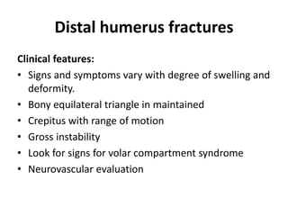
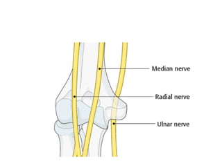
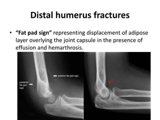
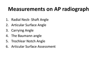
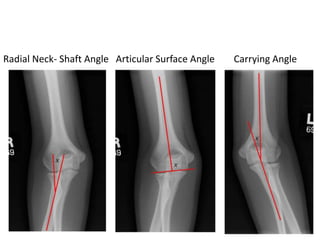
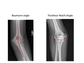
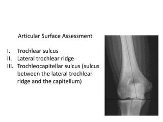
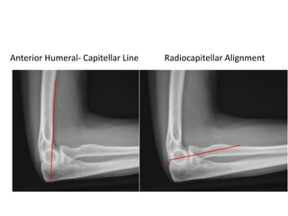
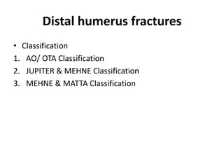
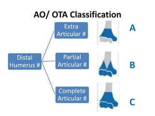
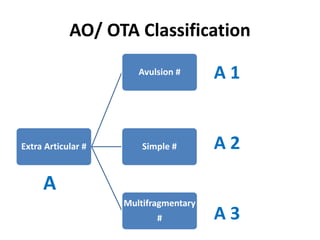
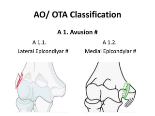
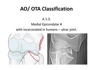
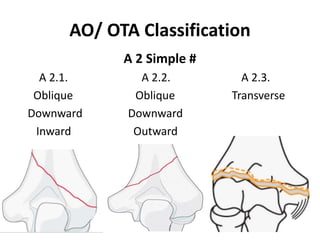
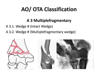
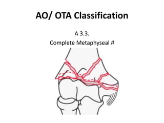
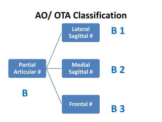
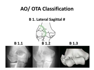
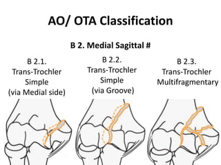
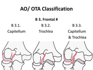
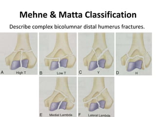
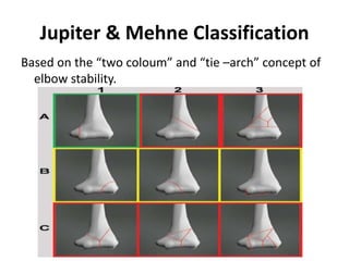
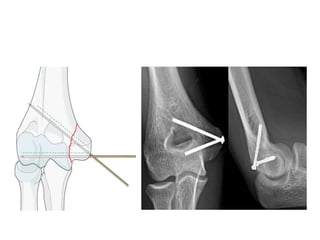
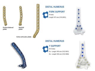
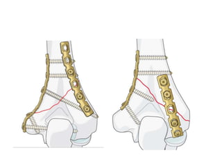
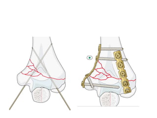
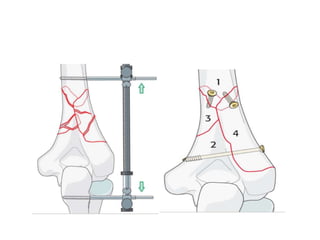
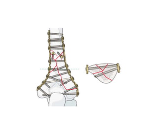
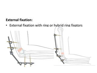
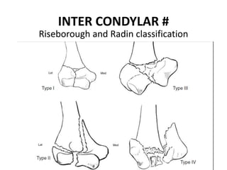
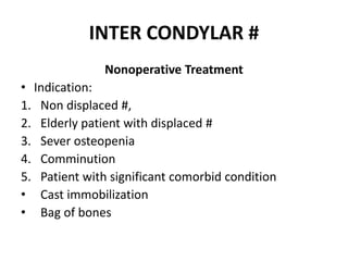
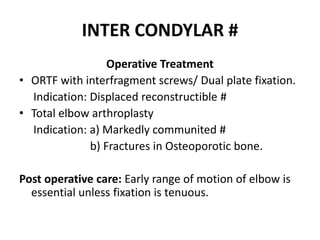
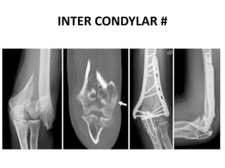
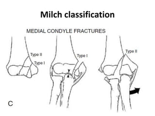
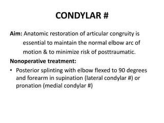
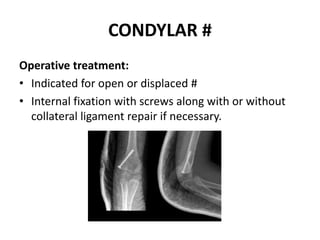
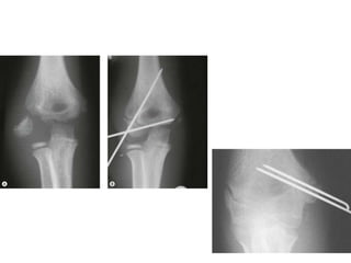
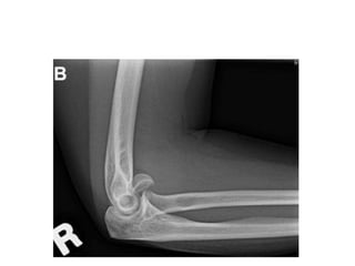
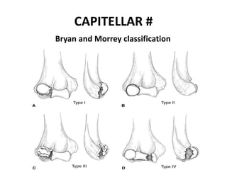
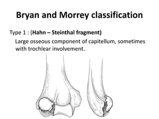
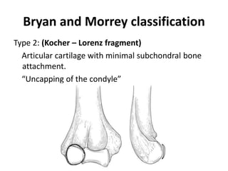
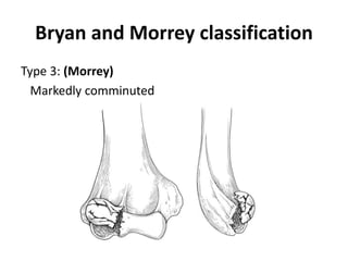
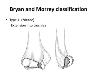
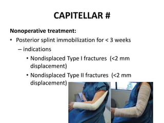
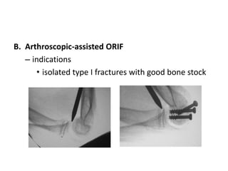
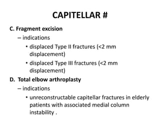
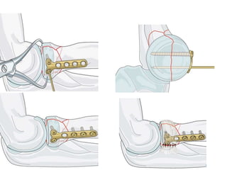
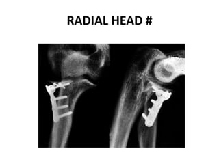
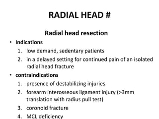
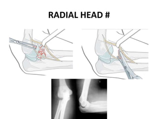
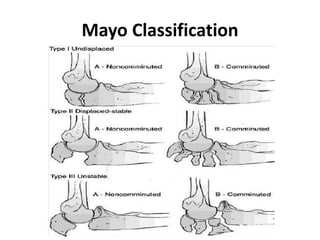
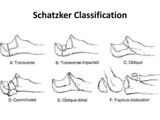
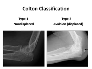
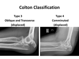
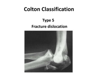
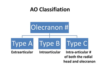
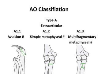
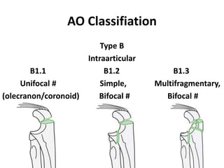
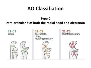
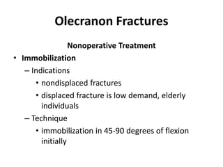
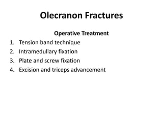
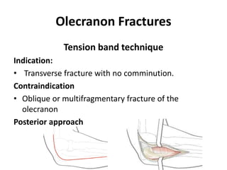
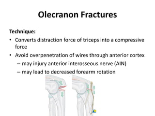
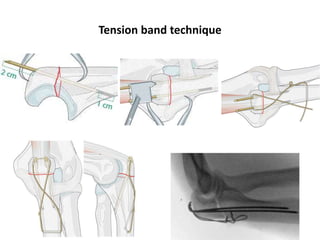
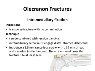
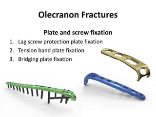
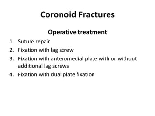
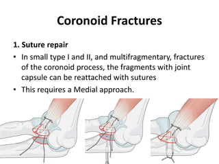
This document discusses various types of fractures of the distal humerus. It begins by describing distal humerus fractures in general, including common mechanisms of injury and clinical features. It then discusses specific fracture types - supracondylar, intercondylar, condylar, trochlear, and capitellar - providing details on classification systems, treatment options, and potential complications for each. Measurement techniques for radiographic evaluation and several classification systems used for distal humerus fractures are also summarized.
Tom Selleck Health: A Comprehensive Look at the Iconic Actor’s Wellness Journeygreendigital
Tom Selleck, an enduring figure in Hollywood. has captivated audiences for decades with his rugged charm, iconic moustache. and memorable roles in television and film. From his breakout role as Thomas Magnum in Magnum P.I. to his current portrayal of Frank Reagan in Blue Bloods. Selleck's career has spanned over 50 years. But beyond his professional achievements. fans have often been curious about Tom Selleck Health. especially as he has aged in the public eye.
Follow us on: Pinterest
Introduction
Many have been interested in Tom Selleck health. not only because of his enduring presence on screen but also because of the challenges. and lifestyle choices he has faced and made over the years. This article delves into the various aspects of Tom Selleck health. exploring his fitness regimen, diet, mental health. and the challenges he has encountered as he ages. We'll look at how he maintains his well-being. the health issues he has faced, and his approach to ageing .
Early Life and Career
Childhood and Athletic Beginnings
Tom Selleck was born on January 29, 1945, in Detroit, Michigan, and grew up in Sherman Oaks, California. From an early age, he was involved in sports, particularly basketball. which played a significant role in his physical development. His athletic pursuits continued into college. where he attended the University of Southern California (USC) on a basketball scholarship. This early involvement in sports laid a strong foundation for his physical health and disciplined lifestyle.
Transition to Acting
Selleck's transition from an athlete to an actor came with its physical demands. His first significant role in "Magnum P.I." required him to perform various stunts and maintain a fit appearance. This role, which he played from 1980 to 1988. necessitated a rigorous fitness routine to meet the show's demands. setting the stage for his long-term commitment to health and wellness.
Fitness Regimen
Workout Routine
Tom Selleck health and fitness regimen has evolved. adapting to his changing roles and age. During his "Magnum, P.I." days. Selleck's workouts were intense and focused on building and maintaining muscle mass. His routine included weightlifting, cardiovascular exercises. and specific training for the stunts he performed on the show.
Selleck adjusted his fitness routine as he aged to suit his body's needs. Today, his workouts focus on maintaining flexibility, strength, and cardiovascular health. He incorporates low-impact exercises such as swimming, walking, and light weightlifting. This balanced approach helps him stay fit without putting undue strain on his joints and muscles.
Importance of Flexibility and Mobility
In recent years, Selleck has emphasized the importance of flexibility and mobility in his fitness regimen. Understanding the natural decline in muscle mass and joint flexibility with age. he includes stretching and yoga in his routine. These practices help prevent injuries, improve posture, and maintain mobilit
Couples presenting to the infertility clinic- Do they really have infertility...Sujoy Dasgupta
Dr Sujoy Dasgupta presented the study on "Couples presenting to the infertility clinic- Do they really have infertility? – The unexplored stories of non-consummation" in the 13th Congress of the Asia Pacific Initiative on Reproduction (ASPIRE 2024) at Manila on 24 May, 2024.
HOT NEW PRODUCT! BIG SALES FAST SHIPPING NOW FROM CHINA!! EU KU DB BK substit...GL Anaacs
Contact us if you are interested:
Email / Skype : kefaya1771@gmail.com
Threema: PXHY5PDH
New BATCH Ku !!! MUCH IN DEMAND FAST SALE EVERY BATCH HAPPY GOOD EFFECT BIG BATCH !
Contact me on Threema or skype to start big business!!
Hot-sale products:
NEW HOT EUTYLONE WHITE CRYSTAL!!
5cl-adba precursor (semi finished )
5cl-adba raw materials
ADBB precursor (semi finished )
ADBB raw materials
APVP powder
5fadb/4f-adb
Jwh018 / Jwh210
Eutylone crystal
Protonitazene (hydrochloride) CAS: 119276-01-6
Flubrotizolam CAS: 57801-95-3
Metonitazene CAS: 14680-51-4
Payment terms: Western Union,MoneyGram,Bitcoin or USDT.
Deliver Time: Usually 7-15days
Shipping method: FedEx, TNT, DHL,UPS etc.Our deliveries are 100% safe, fast, reliable and discreet.
Samples will be sent for your evaluation!If you are interested in, please contact me, let's talk details.
We specializes in exporting high quality Research chemical, medical intermediate, Pharmaceutical chemicals and so on. Products are exported to USA, Canada, France, Korea, Japan,Russia, Southeast Asia and other countries.
Lung Cancer: Artificial Intelligence, Synergetics, Complex System Analysis, S...Oleg Kshivets
RESULTS: Overall life span (LS) was 2252.1±1742.5 days and cumulative 5-year survival (5YS) reached 73.2%, 10 years – 64.8%, 20 years – 42.5%. 513 LCP lived more than 5 years (LS=3124.6±1525.6 days), 148 LCP – more than 10 years (LS=5054.4±1504.1 days).199 LCP died because of LC (LS=562.7±374.5 days). 5YS of LCP after bi/lobectomies was significantly superior in comparison with LCP after pneumonectomies (78.1% vs.63.7%, P=0.00001 by log-rank test). AT significantly improved 5YS (66.3% vs. 34.8%) (P=0.00000 by log-rank test) only for LCP with N1-2. Cox modeling displayed that 5YS of LCP significantly depended on: phase transition (PT) early-invasive LC in terms of synergetics, PT N0—N12, cell ratio factors (ratio between cancer cells- CC and blood cells subpopulations), G1-3, histology, glucose, AT, blood cell circuit, prothrombin index, heparin tolerance, recalcification time (P=0.000-0.038). Neural networks, genetic algorithm selection and bootstrap simulation revealed relationships between 5YS and PT early-invasive LC (rank=1), PT N0—N12 (rank=2), thrombocytes/CC (3), erythrocytes/CC (4), eosinophils/CC (5), healthy cells/CC (6), lymphocytes/CC (7), segmented neutrophils/CC (8), stick neutrophils/CC (9), monocytes/CC (10); leucocytes/CC (11). Correct prediction of 5YS was 100% by neural networks computing (area under ROC curve=1.0; error=0.0).
CONCLUSIONS: 5YS of LCP after radical procedures significantly depended on: 1) PT early-invasive cancer; 2) PT N0--N12; 3) cell ratio factors; 4) blood cell circuit; 5) biochemical factors; 6) hemostasis system; 7) AT; 8) LC characteristics; 9) LC cell dynamics; 10) surgery type: lobectomy/pneumonectomy; 11) anthropometric data. Optimal diagnosis and treatment strategies for LC are: 1) screening and early detection of LC; 2) availability of experienced thoracic surgeons because of complexity of radical procedures; 3) aggressive en block surgery and adequate lymph node dissection for completeness; 4) precise prediction; 5) adjuvant chemoimmunoradiotherapy for LCP with unfavorable prognosis.
Factory Supply Best Quality Pmk Oil CAS 28578–16–7 PMK Powder in Stockrebeccabio
Factory Supply Best Quality Pmk Oil CAS 28578–16–7 PMK Powder in Stock
Telegram: bmksupplier
signal: +85264872720
threema: TUD4A6YC
You can contact me on Telegram or Threema
Communicate promptly and reply
Free of customs clearance, Double Clearance 100% pass delivery to USA, Canada, Spain, Germany, Netherland, Poland, Italy, Sweden, UK, Czech Republic, Australia, Mexico, Russia, Ukraine, Kazakhstan.Door to door service
Hot Selling Organic intermediates
These lecture slides, by Dr Sidra Arshad, offer a quick overview of physiological basis of a normal electrocardiogram.
Learning objectives:
1. Define an electrocardiogram (ECG) and electrocardiography
2. Describe how dipoles generated by the heart produce the waveforms of the ECG
3. Describe the components of a normal electrocardiogram of a typical bipolar leads (limb II)
4. Differentiate between intervals and segments
5. Enlist some common indications for obtaining an ECG
Study Resources:
1. Chapter 11, Guyton and Hall Textbook of Medical Physiology, 14th edition
2. Chapter 9, Human Physiology - From Cells to Systems, Lauralee Sherwood, 9th edition
3. Chapter 29, Ganong’s Review of Medical Physiology, 26th edition
4. Electrocardiogram, StatPearls - https://www.ncbi.nlm.nih.gov/books/NBK549803/
5. ECG in Medical Practice by ABM Abdullah, 4th edition
6. ECG Basics, http://www.nataliescasebook.com/tag/e-c-g-basics
Prix Galien International 2024 Forum ProgramLevi Shapiro
June 20, 2024, Prix Galien International and Jerusalem Ethics Forum in ROME. Detailed agenda including panels:
- ADVANCES IN CARDIOLOGY: A NEW PARADIGM IS COMING
- WOMEN’S HEALTH: FERTILITY PRESERVATION
- WHAT’S NEW IN THE TREATMENT OF INFECTIOUS,
ONCOLOGICAL AND INFLAMMATORY SKIN DISEASES?
- ARTIFICIAL INTELLIGENCE AND ETHICS
- GENE THERAPY
- BEYOND BORDERS: GLOBAL INITIATIVES FOR DEMOCRATIZING LIFE SCIENCE TECHNOLOGIES AND PROMOTING ACCESS TO HEALTHCARE
- ETHICAL CHALLENGES IN LIFE SCIENCES
- Prix Galien International Awards Ceremony
Anti ulcer drugs and their Advance pharmacology ||
Anti-ulcer drugs are medications used to prevent and treat ulcers in the stomach and upper part of the small intestine (duodenal ulcers). These ulcers are often caused by an imbalance between stomach acid and the mucosal lining, which protects the stomach lining.
||Scope: Overview of various classes of anti-ulcer drugs, their mechanisms of action, indications, side effects, and clinical considerations.
TEST BANK for Operations Management, 14th Edition by William J. Stevenson, Ve...kevinkariuki227
TEST BANK for Operations Management, 14th Edition by William J. Stevenson, Verified Chapters 1 - 19, Complete Newest Version.pdf
Report Back from SGO 2024: What’s the Latest in Cervical Cancer?bkling
Are you curious about what’s new in cervical cancer research or unsure what the findings mean? Join Dr. Emily Ko, a gynecologic oncologist at Penn Medicine, to learn about the latest updates from the Society of Gynecologic Oncology (SGO) 2024 Annual Meeting on Women’s Cancer. Dr. Ko will discuss what the research presented at the conference means for you and answer your questions about the new developments.
5. Distal humerus fractures
Mechanism of injury:
• Most low energy injury result from a simple fall in
middle aged & in elderly women in which the elbow
is either struck directly or axially loaded in a fall onto
outstretched hand.
• Motor vehicle and sporting accidents are more
common cause of injury in young individuals.
6. Distal humerus fractures
Clinical features:
• Signs and symptoms vary with degree of swelling and
deformity.
• Bony equilateral triangle in maintained
• Crepitus with range of motion
• Gross instability
• Look for signs for volar compartment syndrome
• Neurovascular evaluation
7.
8. Distal humerus fractures
• “Fat pad sign” representing displacement of adipose
layer overlying the joint capsule in the presence of
effusion and hemarthrosis.
9. Measurements on AP radiograph
1. Radial Neck- Shaft Angle
2. Articular Surface Angle
3. Carrying Angle
4. The Baumann angle
5. Trochlear Notch Angle
6. Articular Surface Assessment
12. Articular Surface Assessment
I. Trochlear sulcus
II. Lateral trochlear ridge
III. Trochleocapitellar sulcus (sulcus
between the lateral trochlear
ridge and the capitellum)
13. Measurements on Lateral Radiograph
1. Anterior Humeral- Capitellar Line.
2. Radiocapitellar Alignment.
3. Anterior Angulation of Articular Surface of Distal
Humerus.
4. Olecranon- coronoid angle.
5. Greater Sigmoid Notch Circle
31. Jupiter & Mehne Classification
Based on the “two coloum” and “tie –arch” concept of
elbow stability.
32. General treatment principles:
• Anatomical articular reduction
• Stable internal fixation of articular surface.
• Restoration of articular axial alignment.
• Early range of elbow motion.
33. Treatment options for the various types of humeral #:
• Epicondylar avulsion # (Type A1) with minimal (<1
cm) displacment can be treated conservatively
• Single column # without comminution (Type B1-B3)
can be treated conservatively at first but will likely
require surgery.
• Comminuted or two column # (Type A2, A3, and C1-
3 ) require surgery.
34. Conservative management:
• Nonsurgical treatment may be recommended for
stable fractures in which the pieces of bone are not
out of place (displaced).
• It may also be recommended for patients who are at
higher risk for surgical complications.
1. Cast or splint stabilization
2. Traction
3. Bag of bones technique
35. • Cast or splint stabilization:
Splints are typically worn for 6 weeks before
supervised motion is started. If the fracture shifts out
of position during this time, you may need surgery to
put the bones back together.
37. Surgical approach:
1. Lateral column fractures addressed with kocher
approach
2. Medial column fractures are apporached with
posterior triceps reflecting or transolecranon
approach.
3. Bicolumn fractures of distal humerus are operated
with olecranon osteotomy, triceps-splitting or Triceps
Reflecting Anconeus Pedicle approach.
38. Internal fixation:
• Use of two orthogonal plates is the most
stable method of treating these fractures.
• 90-90* with medial and posterolateral
position.
• Medial and lateral position.
47. Total elbow arthroplasty
• Semiconstrained hinge design is used to treat
unsalvageable nonunions.
• All the previous implants are removed and
arthroplasty is proceeded.
48.
49. COMPLICATIONS
1. Volkmann ischemic contracture.
2. Loss of elbow range of motion.
a. Loss of motion due to joint stiffness.
b. Loss of extension dueto callus formation in
olecuranon fossa.
c. Loss of flexion due to capsular contracture.
3. Heterotopic bone formation may occur.
50. CLINICAL FEATURES
SC fracture Humerus Post Dislocation
1. Younger children 1. Slightly older
2. Arm is short 2. Forearm is short
3. Bony triangle maintained 3. Triangle is disrupted
4. Swelling is more 4. Swelling is less
5. Crepitus is present 5. Crepitus is absent
6. Olecranonbelow
intercondylar line
6. Olecranonabove
intercondylar line
7. Step sign negative 7. Step sign positive
8. Restricted movements 8. Grossly restricted
9. Radial nerve affected 9. Median & ulnar N injured
51. INTER CONDYLAR #
• This is most common distal humeral #.
• Comminution is common.
• Mechanism of injury:
Force is directed against the posterior aspect of an
elbow flexed >90 degrees, thus driving the ulna into
the trochlea.
53. INTER CONDYLAR #
Nonoperative Treatment
• Indication:
1. Non displaced #,
2. Elderly patient with displaced #
3. Sever osteopenia
4. Comminution
5. Patient with significant comorbid condition
• Cast immobilization
• Bag of bones
54. INTER CONDYLAR #
Operative Treatment
• ORTF with interfragment screws/ Dual plate fixation.
Indication: Displaced reconstructible #
• Total elbow arthroplasty
Indication: a) Markedly communited #
b) Fractures in Osteoporotic bone.
Post operative care: Early range of motion of elbow is
essential unless fixation is tenuous.
56. INTER CONDYLAR #
Complications:
1. Post traumatic arthritis
2. Failure fixation
3. Loss of motion
4. Heterotopic bone
5. Neurological injury (upto 15%)
6. Nonunion (5 to 15%)
7. Infection
57. CONDYLAR #
• Rare in adults and much more common in the
pediatric age group.
• Less than 5% of all distal humerus #
• Lateral # more common than medial.
• Mechanism of injury:
Abduction/ Adduction of forearm with elbow
extension.
58. CONDYLAR # - CLASSIFICATION
1. Milch classification of lateral condylar #
2. Milch classification of medial condylar #
61. CONDYLAR #
Aim: Anatomic restoration of articular congruity is
essential to maintain the normal elbow arc of
motion & to minimize risk of posttraumatic.
Nonoperative treatment:
• Posterior splinting with elbow flexed to 90 degrees
and forearm in supination (lateral condylar #) or
pronation (medial condylar #)
62. CONDYLAR #
Operative treatment:
• Indicated for open or displaced #
• Internal fixation with screws along with or without
collateral ligament repair if necessary.
63.
64. Complications:
• Stiffness
– most common complication
• Nonunion
• AVN
– occurs 1-3 years after fracture
– posterior dissection can result in lateral condyle
osteonecrosis (may also occur in the trochlea)
• Tardy ulnar nerve palsy
– slow, progressive ulnar nerve palsy caused by stretch in
cubitus valgus
– usually late finding, presenting many years after initial
fracture
65. • Malunion
– caused from delay in diagnosis and improper treatment
– 20% cubitus varus in nondisplaced/minimally displaced fractures
• traumatic inflammation leads to lateral overgrowth (see
spurring below)
– 10% cubitus valgus ± tardy ulnar nerve palsy
• because of lateral physeal arrest as fracture is Salter Harris IV
– fishtail deformity
• area between medial ossification center and lateral condyle
ossification center resorbs or fails to develop
• does NOT predispose to arthritis
• may predispose to further fracture
– treatment
• supracondylar osteotomy
66. CAPITELLAR #
• 1% of elbow fractures
• 6% of all distal humerus fractures
• Occur in the coronal plane, parallel to the anterior
humerus.
• Anterior displacement of the articular fragment into
the coronoid or radial fossae may result in a block to
flexion.
67. Mechanism of injury:
• Fall on outstretched arm (typically fall from standing)
• Typically, elbow is in semi-flexed elbow position
• Force is transmitted through radial head to
capitellum. # occur secondary to shear.
68. • Symptoms
– elbow pain, deformity, swelling
• Physical exam
– Inspection and Palpation
• Ecchymosis, swelling
• Diffuse tenderness
– Range of motion & Instability
• May have mechanical block to
flexion/extension and/or rotation
– Neurovascular exam
71. Bryan and Morrey classification
Type 1 : (Hahn – Steinthal fragment)
Large osseous component of capitellum, sometimes
with trochlear involvement.
72. Bryan and Morrey classification
Type 2: (Kocher – Lorenz fragment)
Articular cartilage with minimal subchondral bone
attachment.
“Uncapping of the condyle”
73. Bryan and Morrey classification
Type 3: (Morrey)
Markedly comminuted
74. Bryan and Morrey classification
• Type 4: (McKee)
Extension into trochlea
75. CAPITELLAR #
Nonoperative treatment:
• Posterior splint immobilization for < 3 weeks
– indications
• Nondisplaced Type I fractures (<2 mm
displacement)
• Nondisplaced Type II fractures (<2 mm
displacement)
76. CAPITELLAR #
Operative treatment:
1. Open reduction and internal fixation
2. Arthroscopic-assisted ORIF
3. Fragment excision
4. Total elbow arthroplasty
77. CAPITELLAR #
A. Open reduction and internal fixation
• Indications:
– displaced Type I fractures (<2 mm displacement)
– Type IV fractures
• Technique:
1. ORIF with lateral column approach
2. ORIF with posterior approach with or without
olecranon osteotomy
78. ORIF with lateral column approach
• Indications
– isolated capitellar fractures
– type IV fractures that can have trochlear
involvement
• Approach
– lateral approach recommended for isolated Type I
and Type IV fx
79. • Technique
– Headless screw fixation
– Avoid disruption of the blood supply that comes
from the posterolateral aspect of the elbow
– Do not destabilize LUCL
80. CAPITELLAR #
ORIF with posterior approach with or without
olecranon osteotomy
• Indications
– capitellar fractures with associated
fractures/injuries to distal humuers/olecranon
and/or medial side of the elbow
• Approach
– indicated when more extensive articular work is
needed
– lateral decubitus positioning
83. CAPITELLAR #
C. Fragment excision
– indications
• displaced Type II fractures (<2 mm
displacement)
• displaced Type III fractures (<2 mm
displacement)
D. Total elbow arthroplasty
– indications
• unreconstructable capitellar fractures in elderly
patients with associated medial column
instability .
84. CAPITELLAR #
Complications:
• Elbow contracture/stiffness
(most common)
• Nonunion (1-11% with ORIF)
• Ulnar nerve injury
• Heterotopic ossification
(4% with ORIF)
• AVN of capitellum
• Nonunion of olecranon
osteotomy
• Instability
• Post-traumatic arthritis
• Cubital valgus
• Tardy ulnar nerve palsy
• Infection
85. TROCHLEA #
Laugier’s #
• Extremely rare
• It is associated with elbow dislocation
• Mechanism of injury: Tangential sharing force
resulting from elbow dislocaton.
• Treatment:
Undisplaced # - posterior splinting
Displaced # - ORIF with Kirschner wire or screw
• Complication:
Posttraumatic arthritis; Restricted range of motion
86. RADIAL HEAD #
• 1.5-4% of all fractures
• Most common elbow fractures (33%)
• Radial head fractures can occur in isolation or as part of a
more complex elbow dislocation.
seen in
1. Terrible Triad
(Radial head # dislocation + ULCL + Coronoid process #)
2. Essex-Lopresti injury
(Radial head # dislocation + DRUJ injury + Interosseous
membrane distruption)
87. RADIAL HEAD #
Mechanism of injury:
• Most common is fall onto the outstretched hand.
• Higher energy injuries representing fall from a height
or during sports.
• Elbow in extension + Forearm in pronation, most
force transmitted from wrist to radial head.
89. RADIAL HEAD #
Clinical evaluation
• Typically present with limited elbow and forearm
motion and pain on passive rotation of the forearm.
• Tenderness over radial head as well as elbow
effusion.
• Stability
1. Elbow
2. DRUJ
3. Interosseous membrane
90. RADIAL HEAD #
• Elbow
– lateral pivot shift test (tests LUCL)
– valgus stress test (tests MCL)
• DRUJ
– palpate wrist for tenderness
• Interosseous membrane
– palpate along interosseous membrane for
tenderness
– radius pull test
• >3mm translation concerning for longitudinal
forearm instability (Essex-Lopresti)
91. RADIAL HEAD #
Imaging
1. Radiographs
• recommended views
– AP and lateral elbow
• check for fat pad sign indicating occult minimally
displaced fracture
92. RADIAL HEAD #
• Additional views
– Radiocapitellar view (Greenspan view)
• oblique lateral view of elbow
93. RADIAL HEAD #
2. CT scan
• further delineate fragments in comminuted fractures
• identify associated injuries in complex fracture
dislocations
94. RADIAL HEAD #
Goal of Treatment
1. Pain free
2. Stable arc of motion
3. Correction of any block to forearm rotation
4. Early range of elbow and forearm motion
95. RADIAL HEAD #
Nonoperative Treatment
• Short period of immobilization followed by early
ROM
– indications
• isolated minimally displaced fractures with no
mechanical blocks (Mason Type I)
– outcomes
• elbow stiffness with prolonged immobilization
• good results in 85% to 95% of patients
96. RADIAL HEAD #
Operative Treatment
1. ORIF
2. Fragment excision (partial excision)
3. Radial head arthroplasty
4. Radial head resection
97. RADIAL HEAD #
Approaches to Radial
Head
A. Kocher approach
(Intervalbetween ECU
& Anconeus)
B. Kaplan approach
(Intervalbetween EDC
& ECRB )
98. RADIAL HEAD #
ORIF
• Indications
– Mason Type II with mechanical block
– Mason Type III where ORIF feasible
– presence of other complex ipsilateral elbow
injuries
• Outcomes
• ORIF shown to have worse outcome with 3 or
more fragments compared to ORIF with < 3
fragments
99. RADIAL HEAD #
• Posterolateral placement
Safe zone (nonarticular area) consists of
90-110 degree arc from radial styloid to
Lister's tubercle, with arm in neutral
rotation to avoid impingement of ulna
with forearm rotation.
100. RADIAL HEAD #
Mini fragment 1.5 mm or 2.0 mm screws, Mini fragment 1.5 or 2.0 T-plate
Headless compression screws
(Herbert or similar screws)
104. RADIAL HEAD #
Radial head resection
• Indications
1. low demand, sedentary patients
2. in a delayed setting for continued pain of an isolated
radial head fracture
• contraindications
1. presence of destabilizing injuries
2. forearm interosseous ligament injury (>3mm
translation with radius pull test)
3. coronoid fracture
4. MCL deficiency
106. RADIAL HEAD #
Complications after excision of the radial head
include:
1. Muscle weakness
2. Wrist pain
3. Valgus elbow instability
4. Heterotopic ossification
5. Arthritis
6. Proximal radial migration
7. Decreased strength
8. Cubitus valgus
107. RADIAL HEAD #
Replacement arthroplasty
Indication
• Unreconstructable radial head fracture associated with
an unstable fracture of the coronoid process.
• After radial head excision with evidence of medial
collateral ligament insufficiency or ulnohumeral
instability.
• Unreconstructable radial head fracture associated with
interosseous membrane injury.
• Unreconstructable radial head fracture with acute
distal radioulnar joint injury (Essex-Lopresti injury).
109. Complications
1. Displacement of fracture
2. Posterior interosseous nerve injury (with operative
management)
3. Loss of fixation
4. Loss of forearm rotation
5. Elbow stiffness
6. Radiocapitellar joint arthritis
7. Infection
8. Heterotopic ossification
9. Hardware loosening
10. Complex regional pain syndrome
110. Olecranon Fractures
• Bimodal distribution.
• high energy injuries in young
• secondary to falls in the elderly
• 8% to 10% of all elbow fractures
Mechanismof injury
• Direct blow
• A fall on an outstretched hand with the elbow in flexion
• Sudden and violent triceps muscle contraction can
produce an avulsion fracture of varying size of the
olecranon tip
111. Olecranon Fractures
• Symptoms
– pain well localized to posterior elbow
• Physical exam
– palpable defect
• indicates displaced fracture or severe
comminution
– inability to extend elbow
• indicates discontinuity of triceps (extensor)
mechanism
112. Olecranon Fractures
• Radiographs
– recommended views
• AP/lateral radiographs
–true lateral essential for determination of
fracture pattern
– additional views
• radiocapitellar may be helpful for
–radial head fracture
–capitellar shear fracture
125. Olecranon Fractures
Tension band technique
Indication:
• Transverse fracture with no comminution.
Contraindication
• Oblique or multifragmentary fracture of the
olecranon
Posterior approach
126. Olecranon Fractures
Technique:
• Converts distraction force of triceps into a compressive
force
• Avoid overpenetration of wires through anterior cortex
– may injury anterior interosseous nerve (AIN)
– may lead to decreased forearm rotation
128. Olecranon Fractures
Intramedullary fixation
Indications
• transverse fracture with no comminution
Technique
• can be combined with tension banding
• intramedullary screw must engage distal intramedullary canal
• Introduce a 6.5 mm cancellous screw with a 32 mm thread
and a washer inside the canal. The screw should cross the
fracture site at least 7cm.
129. Olecranon Fractures
Plate and screw fixation
1. Lag screw protection plate fixation
2. Tension band plate fixation
3. Bridging plate fixation
130. Olecranon Fractures
1. Lag screw protection plate
Indication
• Intraarticular oblique fracture of the olecranon
Contraindication
• Multifragmentary #
• Simple transverse #
131. Olecranon Fractures
2. Tension band plate
Indication
• Multifragmentary olecranon
fracture in which articular
surface is intact
• More distal olecranon
fractures
Contraindication
• Multifragmentation of the
articular surface
132. Olecranon Fractures
3. Bridging plate
Indication
• Multifragmentary articular fragmentation of the ulna
Contraindication
• Simple fracture
• Bony buttress in the joint
133. Olecranon Fractures
Excision and triceps advancement
Indications
1. elderly patients with osteoporotic bone
2. fracture must involve <50% of joint surface
3. nonunions
Technique
• Triceps tendon reattached with nonabsorbable
sutures passed through drill holes in proximal ulna
136. Coronoid Fractures
• Coronoid fractures are pathognomonic of an episode
of elbow instability.
• Occur in 10% to 15% of elbow dislocation
Mechanism
– Traumatic shear injury
• Typically occurs as distal humerus is driven
against coronoid with an episode of severe
varus stress or posterior subluxation.
137. Coronoid Fractures
Regan and Morrey Classification
•Type I
Coronoid process tip #
•Type II
Fracture of 50% or less of height
•Type III
Fracture of more than 50% of height
140. Coronoid Fractures
Associated conditions
• Posteromedial rotatory instability
– coronoid anteromedial facet fracture and LCL
disruption. Results from a varus deforming force
• Posterolateral rotatory instability
– coronoid tip fracture, radial head fracture, and LCL
injury
• Olecranon fracture-dislocation
– usually associated with a large coronoid fracture
• Terrible triad of elbow
– coronoid fracture (transverse fracture pattern),
radial head fracture, and elbow dislocation
141. Coronoid Fractures
• Radiographs
– recommended views
• AP and lateral elbow views
– findings
• interpretation may be difficult due to
overlapping structures
• CT scan
– Recommended when coronoid # is
suspected
142. Coronoid Fractures
Nonoperative Treatment
• Brief period of immobilization, followed by early
range of motion
• Indications
– Type I, II, and III that are minimally displaced
with stable elbow
143. Coronoid Fractures
Operative treatment
1. Suture repair
2. Fixation with lag screw
3. Fixation with anteromedial plate with or without
additional lag screws
4. Fixation with dual plate fixation
144. Coronoid Fractures
1. Suture repair
• In small type I and II, and multifragmentary, fractures
of the coronoid process, the fragments with joint
capsule can be reattached with sutures
• This requires a Medial approach.
145. Coronoid Fractures
2. ORIF with lag screw
• In simple fractures where at least 50% of the
coronoid process is involved, the fracture fragment
may be reinserted and held with one or two 2.7 mm
lag screws.
146. Coronoid Fractures
3. ORIF with Plate (contoured three-hole one-third
tubular plate.)
• For larger anteromedial facet coronoid fractures
(O’Driscoll type II), an anteromedial plate with or
without additional lag screws provides good stability
147. Coronoid Fractures
4. ORIF with Dual plate fixation
• Indicated in basilar coronoid fractures, associated
with olecranon fracture dislocations.
149. Elbow dislocation
Incidence
• Elbow dislocations are the most common major joint
dislocation second to the shoulder
– most common dislocated joint in children
• Account for 11-28% of injuries to the elbow
• Posterolateral is the most common type of
dislocation (80%)
• Predominantly affects patients between age 10-20
years old
150. Mechanism of Injury:
• Most commonly, injury is caused by a fall onto an
outstretched hand or elbow.
• Posterior dislocation : This is combination of elbow
hyperextension, valgus stress, arm abduction, and
forearm supination.
• Anterior dislocation: A direct force strikes the
posterior forearm with elbow in flexed position.
151.
152. • Elbow dislocation with
no associated fracture
• Accounts for 50-60% of
elbow dislocations
• Elbow dislocation with
associated fracture
Elbow
dislocation
Simple Complex
153. Signs and Symptoms
• Varying degrees of pain, swelling, and ecchymosis.
• In many cases, there is also instability, crepitation, and
deformity
• Posterior: elbow is flexed with prominence of
olecranon- on palpation olecranon is displaced from
plane of epicondyles.
154. Signs and Symptoms
• Anterior: elbow held in full extension, upper arm
appears shortened with forearm held in supination
and elongated
• Triangle is disrupted, Olecranonabove intercondylar
line, Step sign positive
• With the elbow flexed at 90 degrees,the medial &
lateral epicondyles & olecranon process should from
isosceles triangle.
155. Signs and Symptoms
• Neurovascular examination
• Most commonly injured vessel: Brachial Artery-
more common with anterior and open dislocations.
• Median and ulnar nerve are most susceptible to
damage
156. • Simple DislocationClosed reduction: correction of
medial or lateral displacement followed by
longitudinal traction and flexion
• Parvin’s method (Method A) :
patient lies prone with entire upper
extremity hanging off the bed,
downward traction is applied to the
wrist for a few minutes—> olecranon
slips distally, arm is then lifted gently
157. • Meyn & Quigley method (Method B) : forearm hangs
off of bed, gentle downward traction is applied to
wrist, olecranon is guided with opposite hand
158. • Assess range of motion after reduction (instability
can be appreciated with elbow extension)
• Immobilize in long arm posterior splint with
elbow in 90 degrees of flexion for 1-2 week with
orthopedics follow up as outpatient within 1
week for repeat radiographs
– A recent multi-center study suggests that early
mobilization may be superior to immobilization with
better functional outcomes at 6 weeks, but
comparable functional outcomes at 1 year
– Prolonged immobiization (>3 weeks) is associated
with poor functional outcomes, pain and contractures
159. Complications
• vascular injury of brachial artery may occur but with a lesser
frequency than in cases of supracondylar fracture .
• nerve injury . the medial ulnar nerve may be affected
.c/myositis ossification ,which is more common if passive
exercise is inflicted on the patient.
• Recurrent of the dislocation may occur if the bony ,
ligamentous, and muscular support structure are disrupted
sufficeintly.
• late complications 1/stiffness 2/heterotopic ossification
3/unreduced dislocation 4/recurrent dislocation
5/osteoarthritis after sever fracture dislocation.
160. Complications
• vascular injury of brachial artery may occur but with a lesser
frequency than in cases of supracondylar fracture .
• nerve injury . the medial ulnar nerve may be affected
.c/myositis ossification ,which is more common if passive
exercise is inflicted on the patient.
• Recurrent of the dislocation may occur if the bony ,
ligamentous, and muscular support structure are disrupted
sufficeintly.
• late complications 1/stiffness 2/heterotopic ossification
3/unreduced dislocation 4/recurrent dislocation
5/osteoarthritis after sever fracture dislocation.