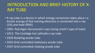
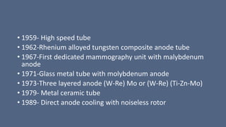
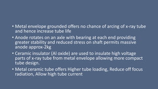
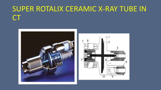
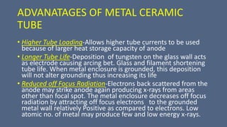
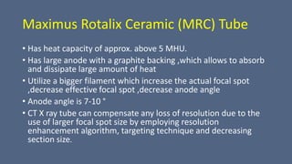
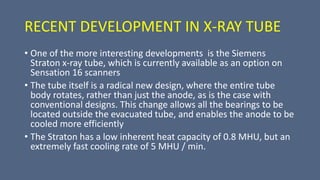
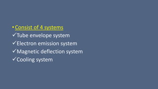
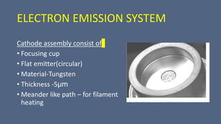
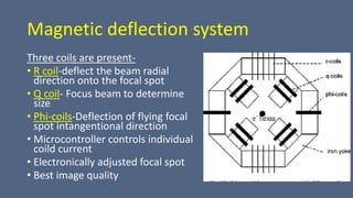
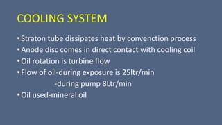
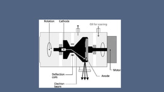
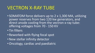
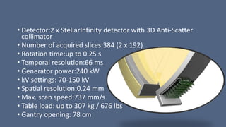
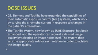
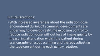
Kodo Millet PPT made by Ghanshyam bairwa college of Agriculture kumher bhara...
CT TUBES AND ITS TYPES.pptx...............
1. CT TUBES AND ITS TYPES
Presented by:Prativa khanal
BSc.MIT 2nd yr
NMCTH
2. INTRODUCTION TO CT
• CT Scanning is a non-invasive
diagnostic tool that uses X-rays to
create detailed images of the body
• Ct scanners obtain individual ,cross
sectional slices by utilizing thinly
collimated x-ray beams taken at
different angles around the
circumference of the same region
and reconstructing images through
the use of computer technology
3. CT HAS THREE MAJOR SYSTEMS
• Imaging system
A. Gantry B. Table
• Computer system
• The display, recording and storage system (operator console)
5. INTRODUCTION AND BRIEF HISTORY OF X-
RAY TUBE
• X-ray tube is a device in which energy conversion takes place i.e.
kinetic energy of fast moving electrons is converted into x-ray
(1%) and heat (99%)
• 1895- Roentgen discovered x-rays (using crock's type of tube)
• 1913- The Coolidge hot cathode x-ray tube
• 1929-Rotating anode tube
• 1932-Grid controlled stationary anode tube
• 1937-Grid controlled rotating anode tube
6. • 1959- High speed tube
• 1962-Rhenium alloyed tungsten composite anode tube
• 1967-First dedicated mammography unit with malybdenum
anode
• 1971-Glass metal tube with molybdenum anode
• 1973-Three layered anode (W-Re) Mo or (W-Re) (Ti-Zn-Mo)
• 1979- Metal ceramic tube
• 1989- Direct anode cooling with noiseless rotor
7. X-RAY TUBES USED IN CT
• Since CT require longer continuous exposure time at
higher KV and mA than needed for general radiography
hence; the general radiographic X-ray tubes cannot be
used for CT. It Should supply monochromatic X-ray beam
for accurate reconstruction.
• X-ray tubes for CT have been charged with heavy duty
rotating anode tube with higher thermal capacity and
smaller focal spot (up to 0.6mm). These tubes are air
cooled with current value up to 600mA.
8. The Manufacturing:
• The x-ray tube is made much like other types of
electrical diodes. The individual components,
including the cathode and anode, are placed
inside the tube envelope and vacuum sealed. The
tube is then situated into the protective housing,
which can then be attached to the rotating
portion of the scanner frame.
• To create the large amount of voltage needed to
produce x rays, an autotransformer is used. This
power supply device is made by winding wire
around a core.
• Electric tap connections are made at various
points along the coil and connected to the main
power source. With this device, output voltage
can be increased to approximately twice the
input voltage
9. X-RAY TUBE WITH FLYING FOCUS
• Has a magnet system for deflecting and focusing the electron
beam, whereby the magnet system including a carrier that is
constructed as an iron yoke and that has four pole projections
that are arranged around the axis of the electron beam offset
from one another by 90 °
• on which two pairs of coils (z-coils and .phi.-coils) are arranged
so as to be offset from one another 90°
• The individual coils of each pair supplied with a common high-
frequency alternating current that deflects the electron beam
in the .phi.-and z-directions, respectively, in a pulsed manner.
11. BRIEF HISTORY AND ADVANCEMENTS IN
CT X-RAY TUBE
• CT X-ray tubes which are located in the heart of the gantry,
Provide radiation source for CT
• First and second generation scanner used fixed anode
(relatively large (2x16mm)focal spot operated at 120 kVp &
30 mA & heavily filtered) , oil-cooled x-ray tube but with the
demand for increased output, gradually rotating anode tubes
become common in CT scanner
•
•
12. • The introduction of spiral/helical CT with the continuous rotation scanner
has placed new demands on CT tubes.
• Several technical advances in component design have been made to deal
with the problem of heat generation, heat storage and heat dissipation.
For example,
i)anode assembly including anode rotation- Anode is of larger diameter
with graphite backing, which allows the anode to absorb& dissipate large
amounts of heat
ii)target design- Anode target angle is made 7-10 ° to diminish heel effect
iii)cathode assembly- Bigger filament size, increased effective focal spot.
Focal spot size smaller (0.6 mm)
iv)the tube envelop-Although the borosilicate glass envelope in early CT
tube provides good thermal and electrical insulation, electrical arching
result from tungsten deposits on the glass caused by vaporization. To solve
above problem, tubes with metal envelop and ceramic insulator are now
common e.g. Metal ceramic x-ray tube
13. METAL CERAMIC X-RAY TUBE
• Glass envelope has been replaced by metal
casing and ceramic is used as insulation of high
voltage cable
• it’s construction includes alloy of chromium
and iron cylinder brazed to alumina ceramic
(aluminium oxide) insulators at each end.
These insulators carry the anode and cathode
assemblies
• smaller and more robust than their glass
equivalents. They have another advantage, in
that they enable more flexibility in the
electrical circuitary associated with the tube
14. • Metal envelope grounded offers no chance of arcing of x-ray tube
and hence increase tube life
• Anode rotates on an axle with bearing at each end providing
greater stability and reduced stress on shaft permits massive
anode approx-2kg
• Ceramic insulator (Al oxide) are used to insulate high voltage
parts of x-ray tube from metal envelope allowing more compact
tube design.
• Metal ceramic tube offers Higher tube loading, Reduce off focus
radiation, Allow high tube current
16. ADVANATAGES OF METAL CERAMIC
TUBE
• Higher Tube Loading-Allows higher tube currents to be used
because of larger heat storage capacity of anode
• Longer Tube Life-Deposition of tungsten on the glass wall acts
as electrode causing arcing bet. Glass and filament shortening
tube life. When metal enclosure is grounded, this deposition
will not alter grounding thus increasing its life
• Reduced off Focus Radiation-Electrons back scattered from the
anode may strike anode again producing x-rays from areas
other than focal spot. The metal enclosure decreases off focus
radiation by attracting off focus electrons to the grounded
metal wall relatively Positive as compared to electrons. Low
atomic no. of metal may produce few and low energy x-rays.
17. Maximus Rotalix Ceramic (MRC) Tube
• Has heat capacity of approx. above 5 MHU.
• Has large anode with a graphite backing ,which allows to absorb
and dissipate large amount of heat
• Utilize a bigger filament which increase the actual focal spot
,decrease effective focal spot ,decrease anode angle
• Anode angle is 7-10 °
• CT X ray tube can compensate any loss of resolution due to the
use of larger focal spot size by employing resolution
enhancement algorithm, targeting technique and decreasing
section size.
18. ADVANTAGES
• Higher tube loading
• Reduce off focus radiation
• Allow high tube current
• When metal enclosure is grounded this deposition do
not alter grounding thus increasing tube life
19. • The 200 mm graphite-backed anode disk used on the MRC
has a very high maximum heat loading capacity, direct heat
conduction
• a large surface area for rapid heat dissipation
• Additionally, the anode is directly cooled through the
patented spiral groove bearing, giving the MRC heat
management capability far superior to a conventional ball
bearing design. This capability is further supported by the
large heat capacity of the housing assembly and the
efficient heat exchanger
20. ADVANTAGES
• Noiselessly rotating anode that could be switched on
the morning and switched off in the evening
• Avoid waiting time during and between examination
• Liquid metal alloy as lubricant
• Possible to achieve dose saving filter technique in
angiography
21. • The second generation of MRC high performance CT tubes has
been updated to effectively manage gantry rotation times
shorter than 0.4 sec with motion-free focal spot performance.
• Motion-free focal spot guarantees optimized image quality in
ultra-high spatial resolution with Multislice CT.
• The Dynamic Focal Spot (DFS) doubles the data sampling
density from the detectors and allows for artifact-free ultra-
high spatial resolution in axial and spiral scanning. As a result,
the MRC is ideally suited to the specific requirements of Philips
new Brilliance CT 40-channel configuration
22. FEATURES
•Higher output and longer tube life
•Anode heat storage capacity – 8 MHU
•Tube voltage - 90 to 140 KV
•Tube current - 20 to 500 mA
•Anode angle - 7 °
•Directly cooled anode
USES
•Cardiovascular imaging
•MDCT
23. LIQUID METAL ANODE XRAY TUBE
(LIMAX)
• The electron beam would be
produced in a vacuum,
separated by a ‘‘thin’’electron
window from the liquid metal.
• Thus the high energy electron
beam penetrates the electron
window, losing as little energy
in the window as possible,
before entering the liquid metal
to produce X-rays.
24. AQUILLION X-RAY TUBE
• 320-row detector
• 160 mm wide Dynamic Volume CT
• Produces isotropic high resolution
• Maximum Load Capacity without Restrictions,
kg = 205 (+/-0.25)
• Tube Cooling = Oil/Air
• Heat Dissipation Rate, kHU/min = 1,386
Maximum
• Maximum mA for smallest Tube Spot = 450,
Optional: 522
• Tube Focal Spots, mm = 1.6 x 1.4, 0.9 x 0.8 (IEC
Standard)
25.
26. RECENT DEVELOPMENT IN X-RAY TUBE
• One of the more interesting developments is the Siemens
Straton x-ray tube, which is currently available as an option on
Sensation 16 scanners
• The tube itself is a radical new design, where the entire tube
body rotates, rather than just the anode, as is the case with
conventional designs. This change allows all the bearings to be
located outside the evacuated tube, and enables the anode to be
cooled more efficiently
• The Straton has a low inherent heat capacity of 0.8 MHU, but an
extremely fast cooling rate of 5 MHU / min.
27. • This compares with typical figures of 7-8 MHU and up to 1.4
MHU / min for existing tubes.
• The heat capacity and cooling rate combine to produce a
tube which Siemens claim is ‘0 MHU', implying that tube
cooling considerations are a thing of the past. Sensation 16
scanners fitted with the Straton tube now have a fastest scan
time of 0.37 seconds.
28. NEW STRATON TUBE
• New construction
• Focused and deflected beam of
thermal electron
• whole tube and anode assembly
rotates
• Bearing located out side
• Oil cooled
29. • Consist of 4 systems
Tube envelope system
Electron emission system
Magnetic deflection system
Cooling system
30. Tube envelope system
• Material used-non magnetic stainless steel
• Directly attached to anode disc
• annular/circular window(thickness-0.2 mm)
• Maximum rotation speed -9600rpm
31. Anode assembly
• Target track made up of 90%
Tungsten and 10% Rhenium
• Anode disc is made up of
Tungsten,Zirconium,Molybednum
body(TZM alloy)
• Boiling point -4612
• Melting point - 2600
32. ELECTRON EMISSION SYSTEM
Cathode assembly consist of
• Focusing cup
• Flat emitter(circular)
• Material-Tungsten
• Thickness -5μm
• Meander like path – for filament
heating
33. Magnetic deflection system
Three coils are present-
• R coil-deflect the beam radial
direction onto the focal spot
• Q coil- Focus beam to determine
size
• Phi-coils-Deflection of flying focal
spot intangentional direction
• Microcontroller controls individual
coild current
• Electronically adjusted focal spot
• Best image quality
34. COOLING SYSTEM
• Straton tube dissipates heat by convenction process
• Anode disc comes in direct contact with cooling coil
• Oil rotation is turbine flow
• Flow of oil-during exposure is 25ltr/min
-during pump 8Ltr/min
• Oil used-mineral oil
35. • When a power of 100KW is applied,the temperature
of focal spot reaches 2500 degree C and temperature
on focal tract reaches 2000 degree c.but the back of
anode has temperature of 200degree c
• This difference of temperature is due to direct cooling
36.
37. FEATURES
• Zero heat storage capacity
• Cooling rate 4.7 MHU/min
• Cooled down within 20 sec
• Enables gantry speed of 0.37 sec per rotation
• Tube current 500 mA
• Based on rotating envelope tube technology
• The electron beam in the tube is shaped and controlled by magnetic
deflection coil
• Focal spot – tungsten and rhenium
38. VECTRON X-RAY TUBE
• SOMATOM force delivers up to 2 x 1,300 MA, utilizing
power reserves from two 120 kw generators, and
direct anode cooling from the vectron x-ray tube:
offering voltages from 70 - 150 KV
• Tin filters
• Reworked with flying focal spot
• New stellar infinity detector
• Oncology, cardiac and paediatric
39. • Detector:2 x StellarInfinity detector with 3D Anti-Scatter
collimator
• Number of acquired slices:384 (2 x 192)
• Rotation time:up to 0.25 s
• Temporal resolution:66 ms
• Generator power:240 kW
• kV settings: 70-150 kV
• Spatial resolution:0.24 mm
• Max. scan speed:737 mm/s
• Table load: up to 307 kg / 676 lbs
• Gantry opening: 78 cm
40. DOSE ISSUES
• GE, Siemens and Toshiba have expanded the capabilities of
their automatic exposure control (AEC) systems, which work
by varying the x-ray tube current in response to changes in
the patient's attenuation
• The Toshiba system, now known as SURE Exposure, has been
expanded, and the operator can request a desired image
quality by selecting an image noise level. The system then
sets the appropriate mA for each rotation in order to achieve
this image quality
• .
41. • The Siemens CAREdose system is now capable of modulating
the tube current along the length of the patient, and also
accounts for overall patient size.
• CAREdose 4D takes a typical mAs value used for a standard
sized patient as the starting point for modulation, and adjusts
this based upon the patient size .This is in addition to the
rotational modulation of the mA that was available in existing
versions of CAREdose.
• GE's SmartScan system has been expanded on the LightSpeed
Pro 16 to allow rotational mA modulation, as well as the
existing z-axis and overall patient size adjustment that is
controlled by setting a desired image noise level
42. Future Directions:
• With increased awareness about the radiation dose
encountered during CT scanning, developments are
under way to develop real-time exposure control to
reduce radiation dose without loss of image quality by
measuring attenuation within the patient during
scanography or scout scanning and thereby adjusting
the tube current during each gantry rotation.
43. References
• Multislice CT: Current Technology and Future
Developments-Stefan Ulzheimer and Thomas Flohr
• Diagnostic Radiology Recent advances and applied
physics by Arun kumar,Veena Chaudhary, Niranjan
Khandelwal
A mounted framework that surrounds the patient in a vertical plane
Houses imaging component such as slip rings, x-ray tube, high voltage generator, collimators, detectors and data acquisition system
Two important feature of the gantry are the gantry aperture and tilting range..Gantry has a 50 to 90 cm aperture for the patient to pass through during the scan
There has been a series of advancement in the X-ray tube during the course of time
Titanium zinc molybednum
Alising artifact reduce,increses data sampling rate
METAL CERAMIC X-RAY TUBE USED IN SPIRAL CT
It is also the type of metal ceramic X-ray tube
Fig. Schematic diagram o f a Super Rotalix ceramic x-ray tube: 1.Metalcasing,2Anode, 3/6. Ball bearings, 4/8. Ceramic insulators,5.Cathode,7.Statorwindings,9.Anode shaft, 10. Beryllium window
In modern CT X ray tube….mega heat unit
Metal lubricated spiral groove bearings…direct conduction
Liquid metal selection Hg.GaLnSn,BiPbLnSn
International electrotechnical comission
The shorter scanning time also requires an increased X-ray dose per unit time, so a highoutput X-ray generation system is needed. Furthermore, faster scanning is expected to lead to an increase in the number of examinations, which means that the X-ray tube must have improved cooling efficiency. To satisfy these requirements, a completely new X-ray tube has been developed for Aquilion. This tube has a straddle bearing structure in which the anode is supported by bearings on both sides which provides a more precise and stable focus and a longer service life compared with the conventional cantilever anode type
In addition, the new tube is anode grounded, so the voltage is applied only to the cathode. This design eliminates the risk of electrical discharge between the anode and the housing, allowing them to be moved closer together, which resulted improved cooling. In an anode grounded tube, recoil electrons from the anode are prevented from re-entering the anode, minimizing heat buildup….. Elimination of recoil electrons is also effective in eliminating off-focus radiation, which will
improve accuracy of X-ray beam geometry
Strato cathode…inside the focusing head the emitter made of tungsten sheet is visible …the emitter has a diameter of 5mm
Scheme of magnet system with different coils
1.Unlike other rotating xray tube which dissipates heat by radiation,
ELECTRON BEAM IS SHAPED AND CINTROLLED BY MAGNETIC FIELD
COOLING SURFACE OF ANODE IN DIRECT CONTACT WITH COOLING FLUID whicb helps to dissipate heat