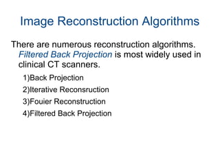
COMPUTED TOMOGRAPHY (CT)
Represented by
Kadriye Doğan
Introduction
Computed tomography (CT) is an imaging method
that uses special x-ray equipment to create
detailed scans of areas inside the body.
The term “tomography” comes from the Greek
words tomos (a slice) and graphein (to write or
record).
History

Invented by Godfrey Hounsfield in 1967,UK and
announced in 1972.

Allan McLeod Cormack in US, independently
invented similar process,and both Hounsfield
and Cormack shared the 1979 Nobel Prize in
Medicine.
Terms
Attenuation is a reduction of the beam intensity on
passing through the body section.
Scan is made up of multiple x-ray attenuation
measurements around a section.
Slice is the cross sectional portion of the body.
Voxel is a 3D version of the pixels.
CT number is a number which represents the x-
ray attenuation.
CT Scan
When X-rays are irradiated on the human body,
some of the rays are absorbed and some pass
through the body to produce an image.
X-ray detectors absorbs the penetrated X-rays,
measures the X-ray amount, and transmits the
data to a computer system.
Computer system calculates and analyzes data
from each detector in each level, and finally
reconstructs multiple,
two-dimensional, cross-sectional images.
Components of CT System

The scanning unit is consists of the gantry with
tube and detectors.

The patient table

The computer system for image reconstruction

Monitor and record system
Image Reconstruction-1

X-rays density is measured before and after
passing through the body section.

The difference between measurements are
recorded as attenuation of section which across
the detector.

The computer system converts digital data
which comes from detectors to CT numbers of
voxels for each section.
Image Reconstruction-2
The pixel value is colored
white,black or gray
according to the mean
attenuation of the section
that it corresponds to the
Hounsfield scale.
Image Reconstruction Algorithms
There are numerous reconstruction algorithms.
Filtered Back Projection is most widely used in
clinical CT scanners.
1)Back Projection
2)Iterative Reconsruction
3)Fouier Reconstruction
4)Filtered Back Projection
Back Projection
Back Projection
Iterative Reconstruction

Projection data are predicted based on an
assumption about the initial attenuation
coefficients of all voxels.

These predicted data are compared to
measured projection data.

The voxel attenuation values are modified until
an acceptable level of error between the
predicted and measured data is achieved.
Iterative Reconstruction
Fourier Reconstruction

Calculate the 1D Fourier transform of all
projections.[p(r) = P(k)]

Place P(k) on polar grid to get P(k,Θ)
 Resample in Cartesian space to get F(kx,ky)

Calculate the 2D inverse Fourier transform of
F(kx,ky) to get f(x,y) – image.
Fourier Reconstruction
Filtered Back Projection

A problem that is immediately apparent is the
blurring that occur in other parts of the
reconstructed image.

The combination of back projection and ramp
filtering is known as filtered back projection.
Filtered Back ProjectionFiltered Back Projection
Filtered Back Projection

Computed Tomography

  • 1.
  • 2.
    Introduction Computed tomography (CT)is an imaging method that uses special x-ray equipment to create detailed scans of areas inside the body. The term “tomography” comes from the Greek words tomos (a slice) and graphein (to write or record).
  • 3.
    History  Invented by GodfreyHounsfield in 1967,UK and announced in 1972.  Allan McLeod Cormack in US, independently invented similar process,and both Hounsfield and Cormack shared the 1979 Nobel Prize in Medicine.
  • 4.
    Terms Attenuation is areduction of the beam intensity on passing through the body section. Scan is made up of multiple x-ray attenuation measurements around a section. Slice is the cross sectional portion of the body. Voxel is a 3D version of the pixels. CT number is a number which represents the x- ray attenuation.
  • 6.
    CT Scan When X-raysare irradiated on the human body, some of the rays are absorbed and some pass through the body to produce an image. X-ray detectors absorbs the penetrated X-rays, measures the X-ray amount, and transmits the data to a computer system. Computer system calculates and analyzes data from each detector in each level, and finally reconstructs multiple, two-dimensional, cross-sectional images.
  • 7.
    Components of CTSystem  The scanning unit is consists of the gantry with tube and detectors.  The patient table  The computer system for image reconstruction  Monitor and record system
  • 8.
    Image Reconstruction-1  X-rays densityis measured before and after passing through the body section.  The difference between measurements are recorded as attenuation of section which across the detector.  The computer system converts digital data which comes from detectors to CT numbers of voxels for each section.
  • 9.
    Image Reconstruction-2 The pixelvalue is colored white,black or gray according to the mean attenuation of the section that it corresponds to the Hounsfield scale.
  • 10.
    Image Reconstruction Algorithms Thereare numerous reconstruction algorithms. Filtered Back Projection is most widely used in clinical CT scanners. 1)Back Projection 2)Iterative Reconsruction 3)Fouier Reconstruction 4)Filtered Back Projection
  • 11.
  • 12.
  • 13.
    Iterative Reconstruction  Projection dataare predicted based on an assumption about the initial attenuation coefficients of all voxels.  These predicted data are compared to measured projection data.  The voxel attenuation values are modified until an acceptable level of error between the predicted and measured data is achieved.
  • 14.
  • 15.
    Fourier Reconstruction  Calculate the1D Fourier transform of all projections.[p(r) = P(k)]  Place P(k) on polar grid to get P(k,Θ)  Resample in Cartesian space to get F(kx,ky)  Calculate the 2D inverse Fourier transform of F(kx,ky) to get f(x,y) – image.
  • 16.
  • 17.
  • 18.
    Filtered Back Projection  Aproblem that is immediately apparent is the blurring that occur in other parts of the reconstructed image.  The combination of back projection and ramp filtering is known as filtered back projection.
  • 19.
  • 20.