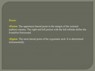
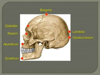
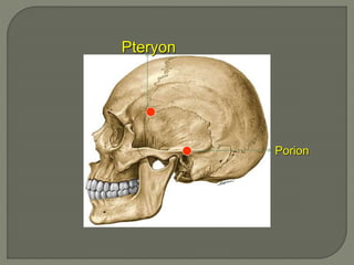
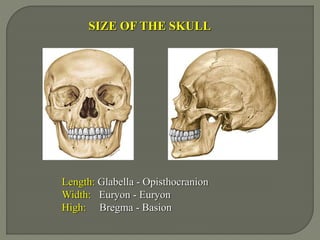
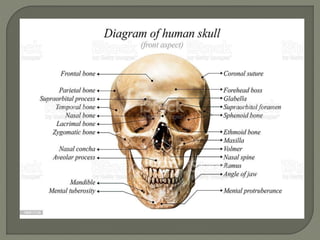
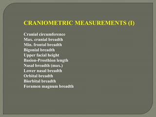
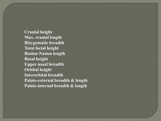
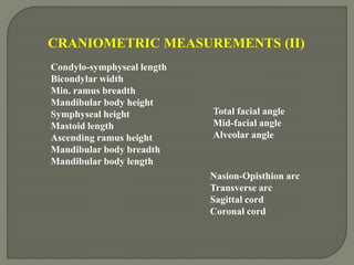
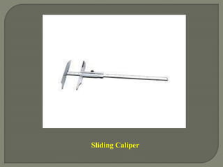
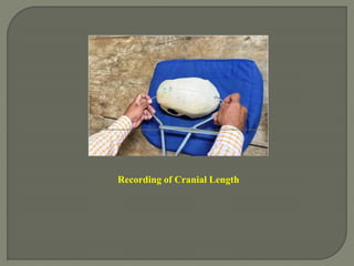
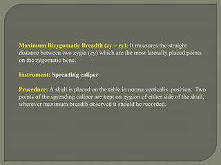
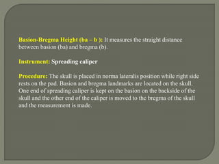
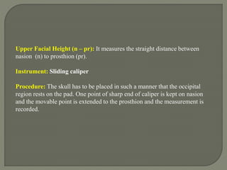
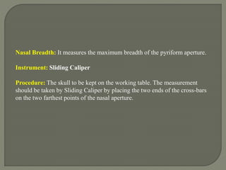
Craniometry is the technique used to measure the dry skull after removing soft tissues. Key landmarks are used as measurement points, including unpaired points like nasion, glabella, and bregma, as well as binate points like porion, zygion, and gonion. Standard craniometric measurements are taken using instruments like spreading calipers and sliding calipers to determine metrics of the entire skull as well as regions like the face, palate, and mandible. Length, width, and height are some of the metrics captured to characterize skull morphology.
Craniometry is the technique used to measure the dry skull after removing its soft parts using various craniometric points and landmarks as reference points. Standard craniometric measurements include maximum cranial length, breadth, bizygomatic breadth, basion-bregma height, and facial heights and breadths. Instruments used include spreading calipers, sliding calipers, and measuring tapes. Various indices can also be calculated from craniometric measurements to study relationships between measurements. Cephalometry is similarly used in dentistry to analyze tooth and jaw relationships and positions.
This document discusses craniometry and cephalometry, which are techniques used to measure the skull and head. Craniometry involves measuring the dry skull after removing soft tissues, while cephalometry measures the head with soft tissues intact. Both are branches of physical anthropology. Key craniometric and cephalometric points are identified which are landmarks used to take measurements of the skull, face, and palate. Various indices are also described, such as cephalic index which categorizes head shape, and facial and palatomaxillary indices relating to face and palate width. Cephalometry is also used in dentistry and orthodontics to analyze teeth, jaws, and skull relationships to inform treatment.
Sex determination from skull and mandibleRachit singh
This document discusses how to determine sex from the human skull. Several features of the skull are sexually dimorphic and can indicate if the skull belonged to a male or female. These features include the supraorbital ridges, orbits, glabella, zygomatic arches, mastoid process, and mandible. In general, male skulls tend to be larger and more robust than female skulls. Each of the sexually dimorphic features are described in terms of typical male and female characteristics. The document also discusses using metrics of the mandible alone to determine sex when the full skull is not available.
Somatometric measurements generally used for identification.pptxSuchita Rawat
Somatometric measurements refer to measurements taken on the living human body using well-defined anatomical landmarks. They are useful for identification, designing equipment, assessing nutrition, and studying populations. Measurements are generally taken with minimal clothing in a standardized position using instruments like calipers and tapes. Over 29 measurements are outlined covering dimensions of the head, face, nose, hands, feet, and various girths. Landmarks and techniques are provided to ensure accuracy. Ratios of measurements are also calculated as somatometric indices for identification and comparison across groups.
This document summarizes methods for determining sex from skull remains. It discusses both metrical and morphological characteristics that can be used. Metrical methods examine skull measurements and use statistics like means and standard deviations to differentiate male and female skulls. Morphological methods examine traits like robustness of features, shape of the orbits, palate shape, and mandible shape. The document indicates that sex determination from skulls can be 70-95% accurate using these methods, but accuracy depends on the population and may be lower for fragmented remains or those outside the 20-55 age range.
The document provides an overview of forensic facial reconstruction. It discusses how facial reconstruction can help identify unknown individuals when other techniques fail by recreating the face based on the skull. It outlines the history of the technique and some of the key pioneers. It also describes different reconstruction techniques, including 2D, 3D manual, and computerized 3D methods. The main techniques involve estimating soft tissue depths and positioning facial muscles and features on the skull.
this presentation deals with the forensic aspects of identification using various 2D & 3D techniques, including the facial approximation methods.....
The efforts of Dr. R V Singh and Ms. Alka Gupta in making of this presentation is appreciated..
whenever and wherever a Disaster takes place in the form of Tsunami, Earthquake, Terrorist attack or Bomb blast the bodies which we get at the crime scene are either damaged or sometimes face cannot be identified.
FACIAL RECONSTRUCTION by sant singh thakur.pptxSantSinghThakur
In these presentation we will discuss about what is facial reconstruction , how many types of methods in this technique and also we will discuss some facial landmarks through which this technique is done.
Radiology plays an important role in forensic investigations by using various imaging techniques like x-rays, CT scans, MRI and ultrasound. It helps identify individuals by comparing ante-mortem and post-mortem images of bones, teeth and implants. It can detect and document injuries, determine the cause of death by locating bullets, fractures and foreign objects. Imaging helps determine if injuries are accidental or non-accidental in cases of child and domestic abuse. It is also used to study firearm injuries by locating bullet paths and fragments. Forensic radiology thus provides crucial medical evidence for legal investigations and proceedings.
The National Integrated Ballistic Information Network (NIBIN) is a ballistics imaging system operated by the ATF that connects digital images of ballistic evidence from crimes involving firearms. It contains over 2.8 million images that can be rapidly searched to link unsolved shootings and identify repeat offenders. By automating the process of comparing ballistic marks on bullets and cartridge cases, NIBIN helps solve crimes faster than traditional microscopic examination. It has confirmed over 74,000 "hits" connecting different shootings since its creation in 1999.
The document discusses sutures of the human skull. It identifies the primary cranial sutures, including the coronal, sagittal, lambdoid, and metopic sutures. It also discusses the midpalatal suture and presents a classification system with 5 stages of maturation based on CBCT imaging. The classification aims to assess midpalatal suture morphology for determining candidacy for rapid maxillary expansion. The sutures most commonly affected by craniosynostosis are also identified.
Antropological Comparision Between Human and Non-human Skeleton RemainsG.S Shaktawat
The skeleton remains can be found at the scene of crime and to confirm the skeleton belongs to human or non human is very important. And for this one should able to do the camparision among them and should drive the investigation in right path. So, In this presentation you will see the some comparisions between human and non human skeleton remains,which is an important topic of the Forensic Anthropology.
The document discusses ear biometrics for human identification based on image analysis. It provides an introduction to traditional and biometric methods of human identification. Ear biometrics are proposed as a passive and accurate physiological method of identification. Existing ear biometric methods are described that use geometric distances, force field transformations, and principal component analysis. The document then outlines the author's approach using geometric feature extraction from ear contours and classification for identification. Current work involves improving algorithms using additional segmentation and contour-based geometric features.
This document provides information on the structure, characteristics, and analysis of human and animal hair. It describes the three main layers of hair - cuticle, cortex, and medulla. The key differences between human and animal hair are discussed, including the width and pigment distribution in the cortex and nature of the medulla. Methods for sampling, examining, and preparing hair samples microscopically are also outlined.
Forensic archaeology applies archaeological principles and techniques to legal investigations to locate, recover, and interpret evidence for past events within the constraints of the criminal justice system. It requires excavating crime scenes slowly and painstakingly, recording and preserving all finds and soil conditions as potential evidence. Forensic archaeologists combine knowledge of osteology and human remains with archaeological methods to recover vital evidence, provide field guidance on physical characteristics of remains, and accurately examine crime scenes.
Anatomy of neck spaces and levels of cervicalairwave12
The document discusses the anatomy of neck spaces and levels of cervical lymph nodes. It describes the various neck spaces such as the retropharyngeal space, prevertebral space, and carotid sheath space. It also details the levels of cervical lymph nodes including levels I-VII, specifying the location and boundaries of each level. A case example is then provided of a 27-year-old patient presenting with a sore throat and swollen lymph nodes on the left side of the neck, consistent with a cervical lymphadenitis infection.
This document discusses the importance of diagnostic imaging in detecting hidden injuries from trauma or abuse. It provides examples of different types of injuries that may be detected on imaging, including:
1) Soft tissue injuries like swelling, lacerations, and pneumothoraces visible on CT or MRI scans.
2) Fractures like hidden fractures in abused children, fractures of different ages, and metaphyseal fractures of long bones seen on x-rays.
3) Foreign bodies like glass, knives, bullets and wires that may be retained following assaults, visible on radiographs.
The document discusses how to determine sex from human skeletal remains. Key indicators of sex include the shape of the pelvis, skull, and long bones. The pelvis is the most accurate indicator, with features like the sciatic notch, subpubic angle, and pelvic inlet differing between males and females in ways related to reproduction and childbirth. The skull also displays sexual dimorphism through traits such as the supraorbital ridge, orbits, and mandible shape. Measurements of bones can provide clues as well.
The document summarizes the anatomy of the neck, including the boundaries and contents of the triangles of the neck. It describes the sternocleidomastoid muscle and its actions. It discusses the layers of the neck including the skin, fascia, muscles and vessels. It provides details of the contents of the anterior, posterior and muscular triangles, including the muscles, vessels, nerves and lymph nodes located in each triangle.
The document discusses the anatomy of the skull base and temporal bone. It describes how the skull base develops from cartilage precursors and separates the brain from facial structures. It details the development of various skull base structures including the parachordal cartilage, sclerotomal cartilage, hypophyseal cartilage and others. It also discusses the anatomy of the temporal bone, including its four parts - the squamous, mastoid, petrous and tympanic portions. Key anatomical structures and landmarks are described for surgical and pathological relevance.
This document provides a pictorial review of ultrasound images to illustrate benign and malignant features of thyroid nodules according to the U1-U5 classification system of the British Thyroid Association. It begins with an overview of normal thyroid ultrasound appearance and anatomy as a baseline for comparison. The majority of the document then features ultrasound images paired with descriptions of thyroid nodules demonstrating benign characteristics, such as a halo sign, microcystic/spongiform appearance, peripheral egg shell calcification, or peripheral vascularity, which correspond to a U2 classification. The aim is to help radiologists and clinicians recognize sonographic patterns to determine whether fine needle aspiration is necessary.
Cranial sutures connect the bones of the skull and allow flexibility during birth but become rigid in adults. There are several types of sutures located in different areas of the skull. Sutures close according to age-related timelines starting with fontanelles closing in infants and ending with some sutures closing in older adults. Three scoring systems are used to estimate age from cranial suture closure: Acsadi and Nemeskeri, Masset, and Meindl and Lovejoy. Each system assigns a score from 0-4 to describe the degree of suture closure.
The hyoid bone is a U-shaped bone located in the neck superior to the larynx. It has a body and paired greater and lesser horns. It develops from cartilage and ossifies from multiple centers between ages 16-30 years. Muscles of the oral cavity, pharynx, and larynx attach to the hyoid bone. Fractures can occur due to strangulation or blows to the neck. Inward or lateral fractures displace the fragments medially, while outward fractures displace them laterally. CT scan is the best way to diagnose hyoid fractures which are important in cases of hanging or strangulation.
DERMATOGLYPHICS.pptx by shree charan Nguestguest43
This document discusses dermatoglyphics, which is the study of epidermal ridge patterns on skin, particularly fingerprints. It notes that fingerprints form during fetal development and remain unchanged after the 21st week. Fingerprint patterns are classified into eight types including loops, whorls, arches. Specific fingerprint features are described like triradii, ATD angle, flexion creases, and simian creases. The document suggests dermatoglyphics can provide clues about genetic abnormalities and medical conditions.
Cephalometrics involves taking X-ray measurements of the head and skull to analyze facial structure and dental relationships. Key aspects include:
- Cephalometrics originated from measuring shadows of bony landmarks on X-ray images.
- Standardized head positions and planes like the Frankfort Horizontal are used for reproducible measurements.
- Analyses like Steiner and Downs involve measuring angles and distances between landmarks to assess skeletal and dental relationships compared to norms.
- Measurements are used for orthodontic diagnosis, treatment planning, and evaluating outcomes.
Cephalometrics involves taking radiographic measurements of head structures to analyze skeletal and dental relationships. Common cephalometric analyses include Steiner's analysis which evaluates skeletal, dental and soft tissue relationships and Tweed's diagnostic triangle which relates mandibular plane angle to lower incisor inclination. Cephalometric landmarks, reference planes and specific angular measurements are used to classify patients and guide orthodontic treatment planning.
This document provides an overview of cephalometrics and cephalometric analysis. It discusses the history and development of cephalometrics, types of cephalograms, cephalometric landmarks, planes, errors, and classifications of cephalometric analysis including Hard Tissue Analysis methods like Downs analysis and Steiner analysis. Downs analysis uses 10 parameters to evaluate skeletal and dental relationships while Steiner analysis divides analysis into skeletal, dental and soft tissue components to provide maximal information. Cephalometrics is important for orthodontic diagnosis and treatment planning.
Anthropometry is the measurement of the human body. It has various applications including identification of criminals, prevention of impersonation, and differentiation between ethnic groups. Modern anthropometry includes biometrics, nutrition/wellness assessments, ergonomics, and monitoring of growth. Key anthropometric measurements include length, breadth, circumference, and skinfolds of various body parts. Landmarks and standardized procedures are used to ensure accurate and reproducible measurements. Anthropometry provides information about human evolution, health, and identification.
Types of cephalogram, uses of cephalogram, technical aspects, cephaometric- soft tissue, hard tissue & PA landmark, lines and panes in cephalometrics, Analysis- Downs, Steiner,Tweed. Wits appraisal, computerized cephalometric system, errors in cephalometry, cephalometric superimposition.
This document provides information about postero-anterior cephalometric analysis, including its history, setup, landmarks, and purposes. Some key points:
- Postero-anterior cephalograms can provide important qualitative and quantitative skeletal and dentofacial data as a supplement to lateral cephalograms.
- Broadbent and Hofrath pioneered the methodology in 1931. Modern setup involves a headholder that can rotate 90 degrees from lateral to postero-anterior position.
- Analysis involves identifying landmarks like zygomatic arches, maxillary molars, and measuring widths, ratios, and angles to evaluate symmetry and proportions.
- Postero-anterior views have limitations due to superimposition
Cephalometrics involves the scientific measurement of the living head using lateral cephalometric radiographs. It allows orthodontists to analyze skeletal, dental, and soft tissue relationships and compare measurements to norms to diagnose orthodontic issues. Key aspects include standardized positioning of patients using a cephalostat to obtain reproducible lateral x-rays. Tracings of these x-rays are then made and landmarks identified to perform measurements of angles and distances. Common landmarks include nasion, sella, orbitale, and pogonion. Measurements are compared to norms for diagnosis, treatment planning, and evaluation.
Cephalometric analysis involves taking x-rays of the skull from different angles and measuring anatomical landmarks and relationships to assess skeletal patterns, dental patterns, and soft tissue profiles. Various instruments and techniques were developed over time to standardize cephalometric x-rays. Key developments included the cephalostat to orient the head in the same position, and advances like digital scans and 3D modeling to capture three-dimensional anatomy. Cephalometric analysis is used for diagnosis, treatment planning, evaluating treatment outcomes, and studying craniofacial growth and relapse.
This document describes various craniometric points and landmarks that are used as references in neurosurgery. It defines points such as the pterion, asterion, euryon, stephanion, vertex, nasion, inion, glabella, bregma, lambda, and others. It explains the location and anatomical relationships of each point. It also discusses how some craniometric points are used to localize structures like the ventricles, cortical areas such as the motor cortex, venous structures, and for strategically placing burr holes during craniotomies. Understanding the location of these points is important for surgical planning and navigation.
The Indian Dental Academy is the Leader in continuing dental education , training dentists in all aspects of dentistry and
offering a wide range of dental certified courses in different formats.
This document provides information about cephalometry and cephalometric analysis for general practitioners. It defines key terms like cephalometry, cephalogram, and standardization. It describes important cephalometric landmarks, lines, and planes used in analysis. Common cephalometric measurements are outlined that provide information about skeletal and dental relationships to assess malocclusions. Limitations of cephalometry including magnification, distortion, and blurring are also noted.
- Cephalometrics involves analyzing and measuring radiographic images of the head called cephalograms. Key landmarks are identified and linear and angular measurements are made between landmarks to assess craniofacial structures.
- There are several commonly used analyses in orthodontics including Downs analysis, Steiner analysis, Tweed analysis, and the Wits appraisal. These analyses establish norms for skeletal and dental relationships and angles that can be used to diagnose malocclusions.
- Landmarks, reference planes like the Frankfort horizontal and mandibular planes, and angular and linear measurements between them allow for evaluation of the positions of jaws, teeth, and soft tissues to develop treatment plans. Serial cephalograms also enable
The document provides an overview of cephalometric analysis. It defines cephalometry as the scientific measurement of the bones of the cranium and face using lateral radiographic images. It discusses the history and key figures in the development of cephalometry. The document outlines several commonly used cephalometric analyses including Downs analysis, Steiner analysis, and Tweed analysis. It describes the landmarks, planes, angles, and linear measurements used in cephalometric assessment. The principles, goals, types, and techniques of cephalometric analysis are summarized. Limitations and sources of error in cephalometrics are also mentioned.
Craniometry and functional craniology involve the measurement of the human skull and head to analyze differences between populations. This document outlines the history and objectives of craniometry, which include examining differences between species, investigating variations within species, and applying measurements in clinical and forensic applications. It then describes common anatomical landmarks, measuring devices and techniques, and analyses used to estimate characteristics like sex, age, and ethnicity from skull measurements.
This document discusses various morphometric measurements that can be taken when analyzing fish specimens. It lists 31 specific linear measurements that are commonly used, such as total length, fork length, standard length, head length, pre-dorsal length, and measurements of fins and other body parts. The measurements are useful for identification, comparisons between specimens, and studies of growth and maturity. References are provided for additional resources on fish morphology and measurement techniques.
Cephalometrics involves the analysis and interpretation of standardized radiographs of the skull and facial bones. It was introduced in the 1930s and modified from anthropological studies. Key aspects summarized include:
- Lateral and frontal cephalograms are used to assess skeletal, dental, airway, and soft tissue relationships.
- Landmarks, planes, and analyses like Downs, Steiner, and Tweed are used to evaluate relationships between cranial structures and compare to norms.
- Analyses evaluate skeletal, dental, and soft tissue parameters to diagnose abnormalities and plan orthodontic treatment.
- Cephalometrics is a valuable tool for orthodontic diagnosis, treatment planning, and evaluating treatment outcomes.
This document provides an overview of cephalometric analysis. It defines cephalometry as the scientific measurement of the bones of the cranium and face using lateral radiographs. Various cephalometric analyses are described, including landmarks, planes, and measurements used in Downs, Steiner, Tweed, and Ricketts analyses to evaluate the skeletal and dental relationships of the craniofacial structures. Limitations of cephalometric analysis are also discussed.
Cephalometrics as a diagnostic aid/orthodontic courses by indian dental academyIndian dental academy
Indian Dental Academy: will be one of the most relevant and exciting training center with best faculty and flexible training programs for dental professionals who wish to advance in their dental practice,Offers certified courses in Dental implants,Orthodontics,Endodontics,Cosmetic Dentistry, Prosthetic Dentistry, Periodontics and General Dentistry.
1. Cerebral edema can be cytotoxic, vasogenic, hydrostatic, osmotic, or hydrocephalic depending on the underlying cause and mechanism.
2. Management of cerebral edema focuses on controlling ICP, optimizing cerebral perfusion, and using specific therapies like hyperventilation, osmotherapy, and corticosteroids.
3. Mannitol is a commonly used osmotic agent that works by increasing plasma osmolality and creating an osmotic gradient to draw fluid from brain tissue, reducing edema. Its administration transiently improves edema but must be carefully monitored.
This document discusses spinal metastases. Key points include:
- Vertebral metastases are the first sign of malignancy in 12-20% of cases and commonly occur in the thoracolumbar region.
- Symptoms include spinal pain and neurologic deficit due to destruction of vertebral elements, instability, or compression/infiltration of spinal cord/nerves.
- Diagnosis involves imaging like MRI, CT, PET, or biopsy.
- Treatment includes medical options like chemotherapy, radiation, steroids, or bisphosphonates as well as surgical options depending on factors like instability, pain level, tumor type, and life expectancy.
- Scoring systems help evaluate patients for surgical vs palliative
This document provides information on the anatomy of the major lobes and structures of the human brain. It describes the key sulci (fissures) and gyri (convolutions) that make up the frontal, parietal, temporal, occipital and limbic lobes. For each lobe, it lists the sulci and gyri on the lateral surface, medial surface and basal surface. It also provides descriptions of other structures like the insular lobe and signs that can help identify sulci on MRI scans of the brain. Diagrams of brain sections in the sagittal, axial and coronal planes are included to illustrate the spatial relationships between lobes and sulci/gyri.
Bone tumors can be primary, originating in bone tissue, or secondary (metastatic) tumors that have spread from other sites. They are classified based on the normal cell type and include hematopoietic, chondrogenic, osteogenic, and others of unknown or various origins. Evaluation of bone tumors involves history, physical exam, labs/imaging, and biopsy. Radiography provides key information like the site and borders of the lesion, type of bone destruction, periosteal reaction, and matrix/soft tissue involvement to characterize the tumor and determine if it is benign or malignant.
The document provides an overview of spinal cord anatomy and spinal tumors. It describes the layers surrounding the spinal cord, blood supply, and classifications of spinal tumors as extradural, intradural extramedullary, or intramedullary. Common presentations include back pain, sensory and motor deficits, and sphincter disturbances. MRI is important for diagnosis. Extramedullary tumors are often metastases that compress the cord without exceeding disk spaces. Intradural tumors like meningiomas attach to dura and taper the CSF. Intramedullary gliomas are more common in thoracic regions and males.
The radial nerve is a continuation of the posterior cord of the brachial plexus. It supplies the posterior compartment of the upper limb. It courses through the axilla, arm, and spiral groove of the humerus before dividing into superficial and deep branches in the forearm. The radial nerve is susceptible to injury at several points along its course, which can result in weakness of wrist and finger extension as well as sensory loss on the back of the hand. Damage to specific branches can produce unique clinical presentations depending on the level and extent of injury.
The radial nerve provides motor innervation to the posterior forearm muscles and cutaneous sensation to the back of the arm and lateral forearm. It arises from the brachial plexus and travels through the spiral groove of the humerus. Damage to the radial nerve can result in wrist drop and sensory loss on the dorsal hand. Injuries commonly occur in the axilla, spiral groove, or at the radial tunnel at the elbow.
The spinal cord is a cylindrical structure running from the foramen magnum to the L1-L2 vertebrae. It contains white and gray matter and is divided into cervical, thoracic, lumbar, and sacral regions. The spinal cord transmits motor and sensory information between the brain and body via ascending and descending tracts. It is supplied by the anterior and posterior spinal arteries and drained by veins that communicate with the azygos system.
The document describes the anatomy of the brain including sulci and gyri. It discusses typical continuous fissures such as the interhemispheric fissure and sylvian fissure. It then describes the lobes of the brain including the frontal, parietal, temporal, occipital and limbic lobes. Key sulci and gyri are identified for each lobe on the lateral, medial and basal surfaces of the brain. Signs to identify sulci on MRI are also provided.
The thalamus is a large mass of gray matter located in the cerebrum that serves as a relay station for sensory and motor signals to and from the cerebral cortex. It has several nuclei that are involved in functions like sensory processing, motor control, arousal, memory, and cognition. The thalamus receives inputs from various subcortical structures and sends outputs to different regions of the cerebral cortex via fiber tracts called thalamic radiations. Damage to the thalamus can result in thalamic syndromes characterized by abnormal pain or sensory processing.
This document discusses various types of vascular malformations of the brain. It begins by describing the histopathology and classification of arteriovenous malformations (AVMs), venous angiomas, capillary telangiectasias, and cavernous malformations. It then covers the pathology, clinical features, diagnosis using angiography, and treatment options for AVMs including surgery, radiosurgery, and endovascular embolization. It also discusses dural arteriovenous fistulas, carotid cavernous fistulas, vein of Galen malformations, developmental venous anomalies, cavernous malformations, capillary telangiectasias, and sinus pericranii.
The document summarizes the embryology of the brain. It describes how the neural tube forms from the ectoderm and divides into subdivisions. It then discusses the development of specific brain structures like the medulla, pons, midbrain, cerebellum and cerebral hemispheres. Key events include formation of the neural plate and tube, development of brain vesicles and flexures, migration of neural crest cells, and growth and differentiation of structures derived from the prosencephalon, mesencephalon and rhombencephalon.
The optic nerve carries visual information from the retina to the brain. It has several parts:
1. The intraocular part passes through the eyeball.
2. The intraorbital part extends from the eyeball to the optic canal.
3. The intracanalicular part passes through the optic canal to the brain.
4. The intracranial part converges with the other optic nerve to form the optic chiasm in the brain.
This document provides an overview of smart and intelligent textiles. It defines smart textiles as textiles that can sense environmental stimuli and react or adapt in response through the integration of functionalities into the textile structure. Smart textiles are classified into three categories - passive, active, and ultra smart - based on their functional activity of sensing, reacting, and adapting. Examples of applications for smart textiles include military, healthcare, space exploration, and fashion. The document also discusses phase change materials and how they can be incorporated into textiles to provide thermoregulation properties for applications such as sportswear, bedding, and medical uses.
This document discusses solar cell textiles and flexible organic solar cells that can be incorporated into clothing and other fabrics. It describes how organic and inorganic photovoltaic technologies can be used to create flexible solar cells that are woven into fibers and fabrics. This allows for solar energy to be harvested from clothing and other textiles, providing a portable and lightweight source of renewable energy for various applications. Key benefits include lower costs compared to rigid solar panels and easier integration into apparel and other fabrics. However, more research is still needed to improve the efficiency and manufacturing of photovoltaic fibers and textile-based solar cells.
The document discusses technical textiles in India. It notes that India's specialty fabric industry is still developing compared to global players. The government is focusing on upgrading infrastructure using technical textiles like geosynthetics and automotive nonwovens. Other niche areas seeing growth are medical, agricultural, and protective textiles. The document also outlines 12 main categories of technical textiles and variables involved in their production like polymers, fibers, yarns, fabrics, and finishing techniques.
Sampling involves taking representative samples of raw materials or finished goods rather than testing entire populations due to constraints of time and cost. Samples are taken from test lots, consignments, packages, containers, and other groupings using random or unbiased sampling methods to ensure samples accurately represent properties of the whole. Common sampling methods include zoning, core sampling, numerical sampling, and random draw or cut square for fibers or yarns. Sample size and selection depends on material type, amount available, intended tests, and required accuracy.
This document discusses yarn hairiness, which refers to fibers protruding from the main body of the yarn. It is generally an undesirable property that can cause issues in fabric production and quality. The document examines various causes of hairiness, its effects, and methods for measuring hairiness, including the Shirley Yarn Hairiness Tester, Zweigle hairiness tester G565, and USTER TESTER 3 hairiness meter attachment. Measurement involves counting the number of hairs per unit length of yarn.
The document describes the Advanced Fiber Information System (AFIS), which provides quick and accurate fiber property measurements. It separates fibers individually using an aeromechanical fiber individualizer. Electro-optical sensors then measure fiber properties like length, diameter and neps. Data is analyzed to provide distributions of fiber characteristics. AFIS applications include analyzing card neps, fiber length for various processes, and trash content to evaluate material quality. It offers advantages over conventional testing like higher accuracy, speed, and avoiding human error.
Geotextiles are permeable textile structures made of synthetic polymers like polypropylene and polyethylene. They are used in civil engineering applications to provide separation, filtration, drainage, reinforcement, and erosion control. There are several types of geotextiles based on manufacturing methods, including woven, nonwoven (needle-punched and heat-bonded), and knitted fabrics. Key properties for geotextiles include mechanical strength, hydraulic conductivity, and chemical resistance to withstand stresses and soil/water interactions over time. Proper selection involves considering the geotextile material, structure, pore size, and strength properties matched to the soil conditions and design loads.
ISO/IEC 27001, ISO/IEC 42001, and GDPR: Best Practices for Implementation and...PECB
Denis is a dynamic and results-driven Chief Information Officer (CIO) with a distinguished career spanning information systems analysis and technical project management. With a proven track record of spearheading the design and delivery of cutting-edge Information Management solutions, he has consistently elevated business operations, streamlined reporting functions, and maximized process efficiency.
Certified as an ISO/IEC 27001: Information Security Management Systems (ISMS) Lead Implementer, Data Protection Officer, and Cyber Risks Analyst, Denis brings a heightened focus on data security, privacy, and cyber resilience to every endeavor.
His expertise extends across a diverse spectrum of reporting, database, and web development applications, underpinned by an exceptional grasp of data storage and virtualization technologies. His proficiency in application testing, database administration, and data cleansing ensures seamless execution of complex projects.
What sets Denis apart is his comprehensive understanding of Business and Systems Analysis technologies, honed through involvement in all phases of the Software Development Lifecycle (SDLC). From meticulous requirements gathering to precise analysis, innovative design, rigorous development, thorough testing, and successful implementation, he has consistently delivered exceptional results.
Throughout his career, he has taken on multifaceted roles, from leading technical project management teams to owning solutions that drive operational excellence. His conscientious and proactive approach is unwavering, whether he is working independently or collaboratively within a team. His ability to connect with colleagues on a personal level underscores his commitment to fostering a harmonious and productive workplace environment.
Date: May 29, 2024
Tags: Information Security, ISO/IEC 27001, ISO/IEC 42001, Artificial Intelligence, GDPR
-------------------------------------------------------------------------------
Find out more about ISO training and certification services
Training: ISO/IEC 27001 Information Security Management System - EN | PECB
ISO/IEC 42001 Artificial Intelligence Management System - EN | PECB
General Data Protection Regulation (GDPR) - Training Courses - EN | PECB
Webinars: https://pecb.com/webinars
Article: https://pecb.com/article
-------------------------------------------------------------------------------
For more information about PECB:
Website: https://pecb.com/
LinkedIn: https://www.linkedin.com/company/pecb/
Facebook: https://www.facebook.com/PECBInternational/
Slideshare: http://www.slideshare.net/PECBCERTIFICATION
Exploiting Artificial Intelligence for Empowering Researchers and Faculty, In...Dr. Vinod Kumar Kanvaria
Exploiting Artificial Intelligence for Empowering Researchers and Faculty,
International FDP on Fundamentals of Research in Social Sciences
at Integral University, Lucknow, 06.06.2024
By Dr. Vinod Kumar Kanvaria
Strategies for Effective Upskilling is a presentation by Chinwendu Peace in a Your Skill Boost Masterclass organisation by the Excellence Foundation for South Sudan on 08th and 09th June 2024 from 1 PM to 3 PM on each day.
Physiology and chemistry of skin and pigmentation, hairs, scalp, lips and nail, Cleansing cream, Lotions, Face powders, Face packs, Lipsticks, Bath products, soaps and baby product,
Preparation and standardization of the following : Tonic, Bleaches, Dentifrices and Mouth washes & Tooth Pastes, Cosmetics for Nails.
A review of the growth of the Israel Genealogy Research Association Database Collection for the last 12 months. Our collection is now passed the 3 million mark and still growing. See which archives have contributed the most. See the different types of records we have, and which years have had records added. You can also see what we have for the future.
How to Manage Your Lost Opportunities in Odoo 17 CRMCeline George
Odoo 17 CRM allows us to track why we lose sales opportunities with "Lost Reasons." This helps analyze our sales process and identify areas for improvement. Here's how to configure lost reasons in Odoo 17 CRM
বাংলাদেশের অর্থনৈতিক সমীক্ষা ২০২৪ [Bangladesh Economic Review 2024 Bangla.pdf] কম্পিউটার , ট্যাব ও স্মার্ট ফোন ভার্সন সহ সম্পূর্ণ বাংলা ই-বুক বা pdf বই " সুচিপত্র ...বুকমার্ক মেনু 🔖 ও হাইপার লিংক মেনু 📝👆 যুক্ত ..
আমাদের সবার জন্য খুব খুব গুরুত্বপূর্ণ একটি বই ..বিসিএস, ব্যাংক, ইউনিভার্সিটি ভর্তি ও যে কোন প্রতিযোগিতা মূলক পরীক্ষার জন্য এর খুব ইম্পরট্যান্ট একটি বিষয় ...তাছাড়া বাংলাদেশের সাম্প্রতিক যে কোন ডাটা বা তথ্য এই বইতে পাবেন ...
তাই একজন নাগরিক হিসাবে এই তথ্য গুলো আপনার জানা প্রয়োজন ...।
বিসিএস ও ব্যাংক এর লিখিত পরীক্ষা ...+এছাড়া মাধ্যমিক ও উচ্চমাধ্যমিকের স্টুডেন্টদের জন্য অনেক কাজে আসবে ...
How to Fix the Import Error in the Odoo 17Celine George
An import error occurs when a program fails to import a module or library, disrupting its execution. In languages like Python, this issue arises when the specified module cannot be found or accessed, hindering the program's functionality. Resolving import errors is crucial for maintaining smooth software operation and uninterrupted development processes.
2. Craniometry:
It is the technique used to measure dry skull after removal of its soft parts.
•A landmark on the skull from which craniometric measurements can be taken are
craniometric points.
•A landmark may be defined as a
definite anatomical point or an indefinite point located by geometrical relations
which form the termini of the direct measurement.
4. Unpaired:
•Nasion: The intersection of the nasofrontal suture with the midsagittal
plane. Nasion is the uppermost landmark for the measure of facial height.
•Glabella: The most forward projecting point in the midline of the forehead
at the level of the supra-orbital ridges and above the nasofrontal suture.
•Bregma: The intersection of the coronal and sagittal sutures in the midline.
5. •Lambda: The intersection of the sagittal and lambdoidal sutures in the
midline.
•Opisthocranion: The most posterior point on the skull not on the external
occipital protuberance. It is the posterior end point of maximum cranial
length measured from glabella. It is determined instrumentally.
•Basion: The midpoint of the anterior margin of the foramen magnum.
6. Binate:
•Porion: The uppermost lateral point in the margin of the external
auditory meatus. The right and left porion with the left orbitale define the
Frankfort Horizontal.
•Zygion: The most lateral point of the zygomatic arch. It is determined
instrumentally.
7. •Gonion: The midpoint of the angel of the mandible between body and ramus.
•Euryon: The two points on the opposite sides of the skull that form termini of the lines of
greatest breadth. The two points are determined instrumentally.
•Pterion: Estimated as 2 finger-breadths above the zygomatic arch and a thumb’s breadth
behind the frontal process of the zygomatic bone.
12. Length: Glabella - Opisthocranion
Width: Euryon - Euryon
High: Bregma - Basion
SIZE OF THE SKULL
13. Length: Nasion - Gnathion
Width: Zygion - Zygion
SIZE OF THE FACE
14. Width: Endomolara - Endomolare
Length: Orale - Staphylion
SIZE OF THE PALATUM
15. Skull is the upper most part of the human skeleton consisting of
head and face. The human skull consists of 22 bones. Except for the mandible
(lower jaw), all the bones of the skull are connected together by sutures. The
skeleton of a skull without mandible is called Cranium which is made up of 8
bones and thirteen bones form the facial skeleton. The mandible is a movable
bone held to the cranium by ligaments. The cranial bones can be divided into
two categories: the calvaria and the cranial base. The calvaria is the
domeshaped superior portion of the cranium. It is composed of the frontal,
occipital, and parietal bones, and the flat portion of the temporal bones. The
cranial base is composed of the two remaining cranial bones, the ethmoid and
the sphenoid bone. Fourteen facial bones form the other components of the
skull. The facial bones are composed of the inferior nasal conchae, lacrimal
bones, mandible, maxillary bones, nasal bones, palatine bones, vomer and
zygomatic bones.
SKULL
19. CRANIOMETRIC MEASUREMENTS (II)
Condylo-symphyseal length
Bicondylar width
Min. ramus breadth
Mandibular body height
Symphyseal height
Mastoid length
Ascending ramus height
Mandibular body breadth
Mandibular body length
Total facial angle
Mid-facial angle
Alveolar angle
Nasion-Opisthion arc
Transverse arc
Sagittal cord
Coronal cord
20. INSTRUMENTS USED FOR
CRANIOMETRY
Instruments like spreading caliper, sliding caliper, steel tape and mandibulometer
are used while recording craniometric measurements.
•Calipers
Calipers are the most important instruments available for measuring distances
between two definite points. There are two types of calipers-Spreading and
Sliding.
21. •Spreading Caliper
This instrument is available in two sizes i.e. 25 cm long for taking smaller
measurements and another one 60 cm long for taking the measurements on
Pelvis and hence it is called as Pelvimeter. The caliper comprises of two long
arms which are curved outwards and bounded at one end. A meter scale is
fixed to one of the arms and passes through the socket on the second arm.
The spreading calipers are made with blunt or rounded ends and sharp or
pointed ends. The spreading caliper with rounded or blunted ends is used to
make measurements on living human being, whereas, the spreading caliper
with sharp or pointed end is employed to record measurements on skeletons.
The calipers are graduated up to one millimeter
24. CRANIOMETRIC MEASUREMENTS
The procedures for various craniometric measurements are presented as
below:
Maximum Cranial Length (g - op): It measures the straight distance
between glabella (g) and opisthocranion (op).
Instrument: Spreading Caliper (Pointed end)
Procedure: A skull is placed on the working table or on the seed bag. One
point of spreading caliper is kept on the glabella and other point is moved to
the posterior region of the skull on the occipital bone in midsagittal plane and
wherever the maximum length is observed it should be measured.
26. Maximum Cranial Breadth (eu – eu): It is the maximum breadth
(width) measured on the skull on right angle (90º) to the mid-sagittal
plane.
Instrument: Spreading Caliper
Procedure: Place the skull on the working table or on the seed bag. Keep
the two points of the spreading caliper on two sides of the euryon with
the two hands in the same horizontal plane and record the measurement.
28. Maximum Bizygomatic Breadth (zy – zy): It measures the straight
distance between two zygia (zy) which are the most laterally placed points
on the zygomatic bone.
Instrument: Spreading caliper
Procedure: A skull is placed on the table in norma verticalis position. Two
points of the spreading caliper are kept on zygion of either side of the skull,
wherever maximum breadth observed it should be recorded.
29. Basion-Bregma Height (ba – b ): It measures the straight distance
between basion (ba) and bregma (b).
Instrument: Spreading caliper
Procedure: The skull is placed in norma lateralis position while right side
rests on the pad. Basion and bregma landmarks are located on the skull.
One end of spreading caliper is kept on the basion on the backside of the
skull and the other end of the caliper is moved to the bregma of the skull
and the measurement is made.
30. Upper Facial Height (n – pr): It measures the straight distance between
nasion (n) to prosthion (pr).
Instrument: Sliding caliper
Procedure: The skull has to be placed in such a manner that the occipital
region rests on the pad. One point of sharp end of caliper is kept on nasion
and the movable point is extended to the prosthion and the measurement is
recorded.
31. Minimum Frontal Breadth (ft): It measures the straight distance between
frontotemporale located on either side of the skull.
Instrument: Sliding caliper
Procedure: A skull is placed on the working table. Frontotemporale on either
side of the skull are identified. One sharp end of the caliper is placed on the
frontotemporale on the one side of the skull and the movable point is spread
up to the frontotemporal end on the other side of skull and wherever the least
distance is observed the measurement should be made.
32. Morphological Facial Height (n – gn): It measures the straight distance
between nasion and gnathion.
Instrument: Sliding Caliper
Procedure: The measuring skull should be placed towards the measurer.
Nasion and gnathion landmarks are identified on the skull. The sharp end of
the caliper is kept on the nasion and the movable part of the caliper touches
the gnathion and the measurement is made.
33. Nasal Height (n – ns): It measures the straight distance between nasion and
nasospinale.
Instrument: Sliding Caliper
Procedure: The skull is kept in such a manner that the basal region rests on
the pad. The fixed point of cross-bar is placed on nasion (n) and the movable
crossbar is adjusted against the nasospinale (ns).
34. Nasal Breadth: It measures the maximum breadth of the pyriform aperture.
Instrument: Sliding Caliper
Procedure: The skull to be kept on the working table. The measurement
should be taken by Sliding Caliper by placing the two ends of the cross-bars
on the two farthest points of the nasal aperture.
35. Facial length or Facial depth (ba-pr): It measures the straight distance
between basion and prosthian.
Instrument: Sliding Caliper
Procedure: The skull is placed in the position of norma basalis on the working
table facing the measurer. Identify the prosthion and basion landmarks on the
skull. One sharp point of Sliding Caliper is kept on the prosthion and the
movable point is spread to the level of basion and the measurement is made.
36. Upper Facial height (n-pr): It measures the straight distance between nasion
and prosthion.
Instrument: Sliding Caliper
Procedure: The skull is placed on the working table in frontal part facing the
measurer. Nasion and Prosthion landmarks are identified. One sharp end of the
sliding caliper is kept on nasion and the movable point is drawn to the prosthion.
37. Horizontal Circumference of skull (g-op-g): It is the maximum
circumference of the cranium in horizontal plane over glabella (g) –
opisthocranion (op)- glabella (g).
Instrument: Measuring tape
Procedure: The skull is placed facing frontal region facing the measurer. The
landmark opisthocranion is located which is found on the back of the skull.
Passing the tape through opisthocranion the maximum circumference between
glabella to glabella is measured.
38. Morphological Facial height (n-gn): It measures the straight distance
between nasion and gnathion.
Instruments: Sliding Caliper
Procedure: The skull is placed on the working table facing frontal region to
the measurer. Identify and mark the nasion and gnathion landmarks. Keep one
sharp point of the Sliding Caliper on gnathion and draw the movable sharp
point towards nasion and record the measurement.
39. Breadth of the Upper Jaw or Bimaxillary Breadth (zm-zm): It measures
the straight distance between two zygomaxillaria.
Instruments: Sliding Caliper
Procedure: Place the skull on the working table facing the measurer. Identify
Zygomaxillare on either side of the skull. Keep one sharp fix point on left
zygomaxillare and draw the movable sharp point towards the right
zygomaxillrea and record the measurement.
40. INDICES
An Index represents the relationship between two absolute measurements.
Various indices can be calculated based on craniometric measurements.
However, some of the indices are given below.
41. CEPHALOMETRY
•It is used in dentristy, especially in orthodontics, to gauge the size and
special relationships of the teeth, jaws and cranium.
• This analysis informs treatment planning, quantifies changes during
treatment, and provides data for clinical research.
43. CEPHALOMETRIC POINT, PLANE, LINE AND ANGLES
USED IN DENTISTRY
S (Sella):
Midpoint of sella turcica
N (Nasion):
Most anterior point on fronto-nasal suture
Or (Orbitale):
Most inferior anterior point on margin of orbit
Po (Porion):
Upper most point on bony external auditory meatus
ANS (Anterior Nasal Spine)
PNS (Posterior Nasal Spine)
44. Go (Gonion):
Most posterior inferior point on angle of mandible
Me (Menton):
Lower most point on the mandibular symphysis
A (A point):
Position of deepest concavity on anterior profile of maxilla
B (B point):
Position of deepest concavity on anterior profile of mandibular symphysis
45. Frankfort Plane: Po – Or
Equivalent to the true horizontal when patient is standing upright
Maxillary Plane: PNS - ANS
Gives inclination of maxilla relative to other lines/planes
Mandibular Plane: Go - Me
Gives inclination of mandible relative to other lines/planes