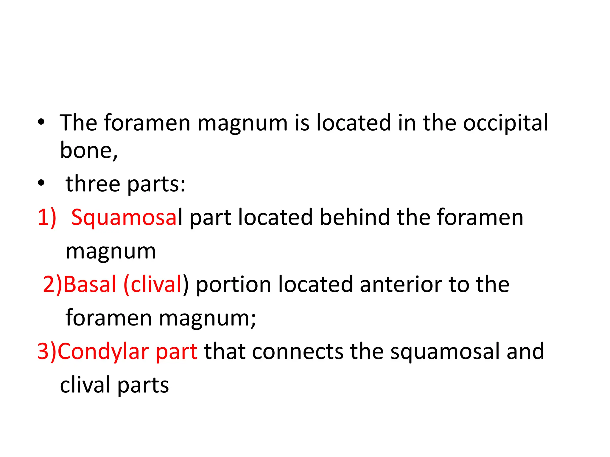
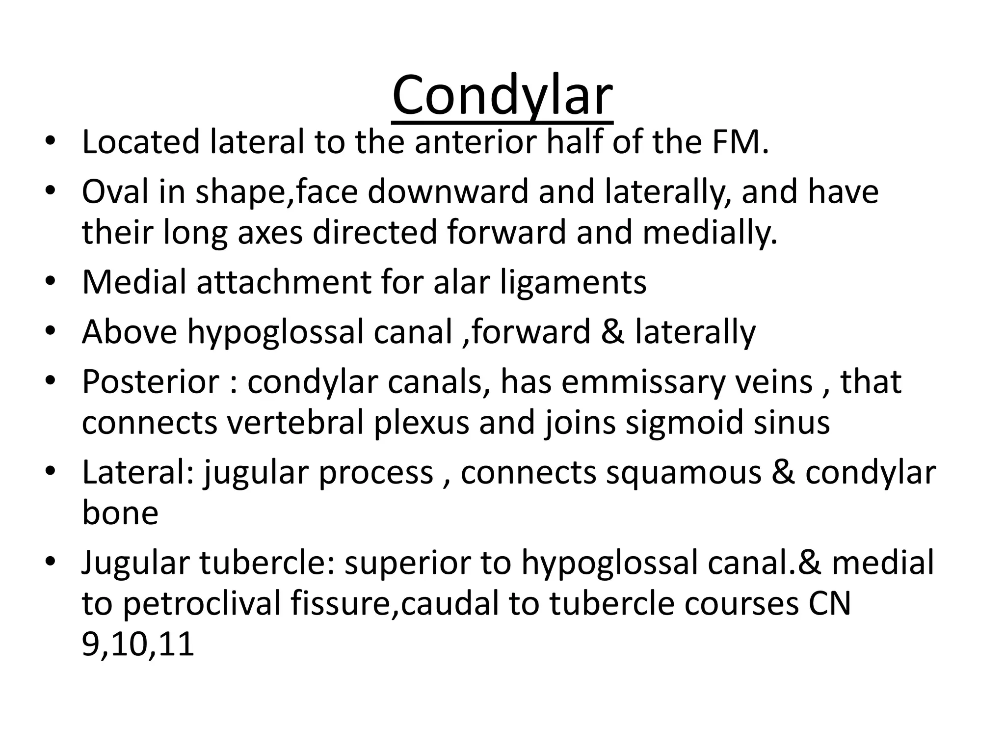
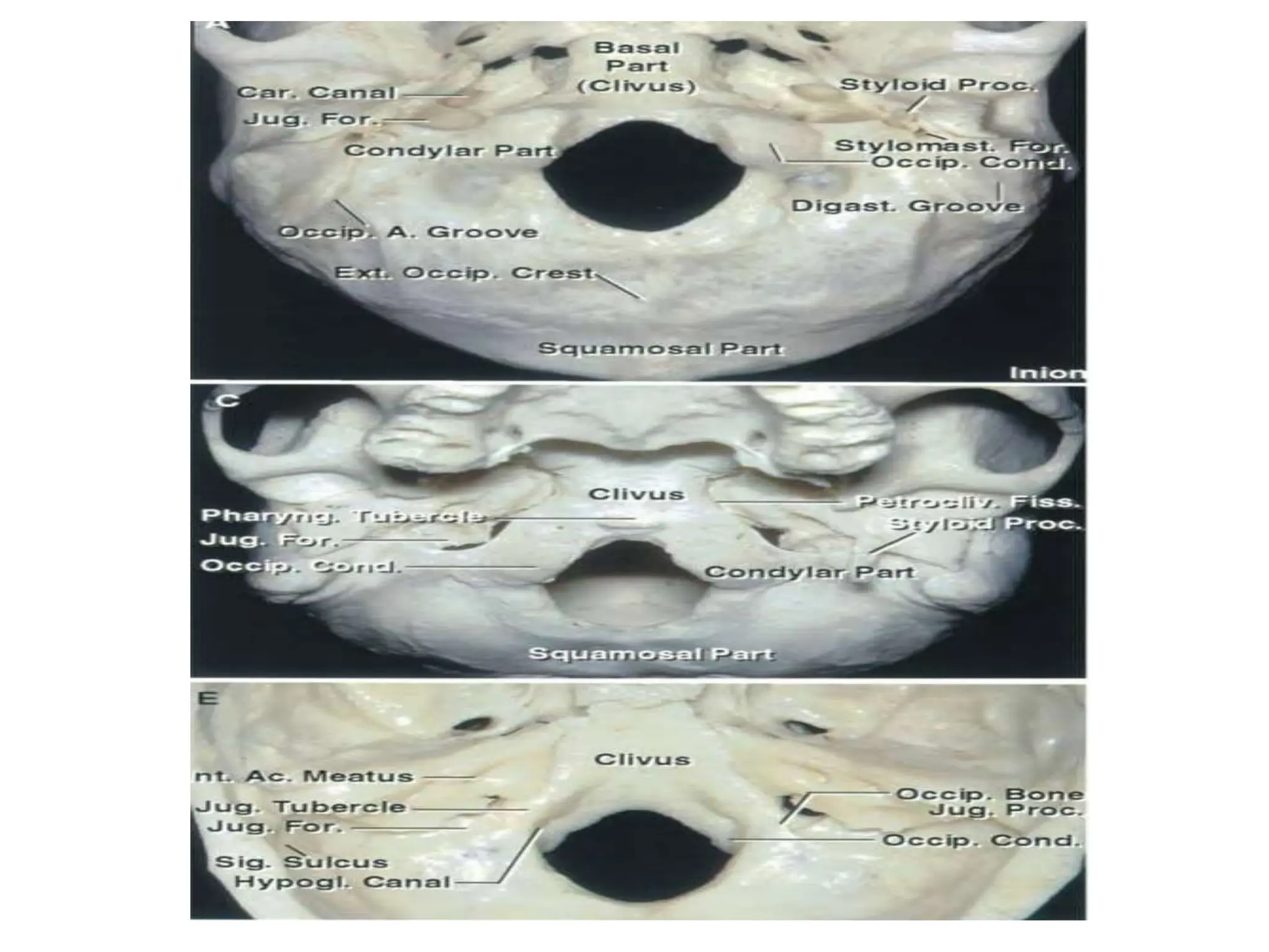
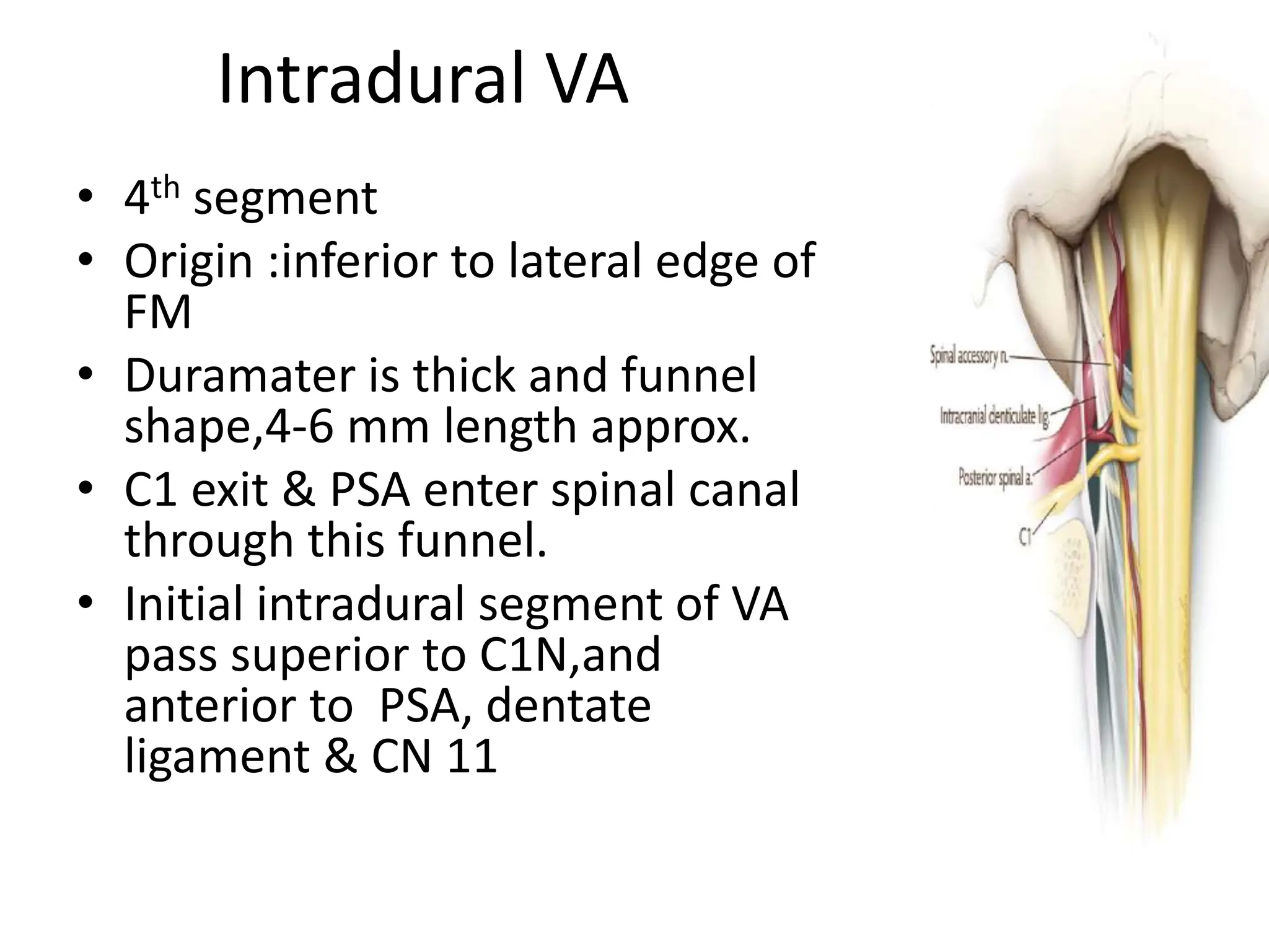
FORAMEN MAGNUM
ANATOMY
Dr.B Vinod
Mch Neurosurgery resident
Gandhi Medical college
Hyderabad.
• The foramen magnum is located in the occipital
bone,
• three parts:
1) Squamosal part located behind the foramen
magnum
2)Basal (clival) portion located anterior to the
foramen magnum;
3)Condylar part that connects the squamosal and
clival parts
RELATIONSHIPS
Osseous : 3 bony relations
• Occipital
• Atlas
• Axis
Occipital bone
• Anterior (Narrow): Clivus
• Posterior: Squamous
• Lateral:Condyles
• Clivus:
• Anterior to FM, thick quadrangular plate of bone
that extends forward and upward, at an angle of
about 45° from the foramen magnum.
• Superior surface concave side to side
• Separated from petrous bone of temporal by
petroclival fissure.
• Has inferior petrosal sinus on upper surface which
ends in jugular foramen .
• Inferior surface in front of the foramen magnum,
a small elevation, the pharyngeal tubercle, gives
attachment to the fibrous raphe of the pharynx.
Condylar
• Located lateral to the anterior half of the FM.
• Oval in shape,face downward and laterally, and have
their long axes directed forward and medially.
• Medial attachment for alar ligaments
• Above hypoglossal canal ,forward & laterally
• Posterior : condylar canals, has emmissary veins , that
connects vertebral plexus and joins sigmoid sinus
• Lateral: jugular process , connects squamous & condylar
bone
• Jugular tubercle: superior to hypoglossal canal.& medial
to petroclival fissure,caudal to tubercle courses CN
9,10,11
• Jugular foramen: anterior and superior to FM.
• 2 parts .
• Small anteromedial: petrous part,transmits IPS
• Large posterolateral: sigmoid part,transmits
sigmoid sinus.
• Intrajugular part. Transmits CN 9,10,11,
• Squamous part:
• External :
• External occipital protuberance(Inion)
convex
• 2 nucheal line
• External occipital crest, rough surface
for muscles
• Internal:
• concave & has internal protuberance .
• Superior fossa for occipital lobe
• Inferior fossa for cerebellar surface .
• Vermian fossa for vermis .
ATLAS
AXIS
Vertebral artery groove
LIGAMENTS
• Cruciform ligaments : transverse & vertical
• ALL
• PLL
• Atlanto-occipital
• Axis-occipital
LIGAMENTS
ALL
• Runs along anterior surface of VB
• From C1 to anterior sacrum
• Attached to VB and intervertebral discs along
its length
• PLL
• Along posterior surface of VB up c2
• Attaches along posterior surface of VB and
discs.
• Tectorial membrane :
• Upper part of PLL ,that connects C2 to clivus
MUSCULAR RELATIONS
• Posterior muscles
• Superficial
• Trapezius
• Sternocleidomastoid
• Deep
• Splenius capitis
• Semispinalis capitis
• Longissimus capitis
• Suboccipitalis muscle.
Suboccipitalis muscle
• 4 muscles in suboccipital area
Anterior muscles
• Longus colli (C1-T3)
• Longus capitis
(Clivus to C3 to C6 TP)
• Rectus capitus anterior
• Rectus capitus lateralis
NEURAL RELATIONS
• Spinal cord
• Dentate ligament
• Brainstem
• Cerebellum
• Cranial nerves
• Cervical nerve roots
Spinal cord
• Blends into medulla at level upper limit of
dorsal and ventral rootlets forming C1N
• Easy to identify level on ventral than dorsal
because of ventral rootlets are always present.
• Junction of SC and medulla present at rostral
end ventral C1.
• Medulla occupies FM not SC
• SC lies immediately below FM
Dentate ligament
• White sheeth attached to spinal cord
• Lateral projections of spinal piamater between dorsal
& ventral rootlets, forming triangle shape ligament
,that anchors the spinal cord along its length to
duramater on each side.
• At CVJ located
Anterior – between VA & C1N
ventral root
Posterior- PSA & CN 11
• Most attachments of ligament is at rostral end at level
of FM where VA pierces duramater.
BRAINSTEM
• Lower medulla present at C1n ventral
• Anterior medulla by medullary
pyramids faces
Anterior clivus
Anterior edge of FM
Rostral odontoid process
Lateral surface of MO by inferior olives.
Posterior MO
Superior : Midline inferior half of 4V
Lateral by inferior
cerebellar peduncles
Inferior :Gracious fasciculus and cuneatus.
Cerebellum
• Rests above the posterior and lateral edge of FM
• Tonsils- above and posterior edge
• Biventral and lobules-sits above and lateral
• Vermis lower part.
Cranial nerve
Spinal accessory nerve only nerve pass through FM
Cervical nerve roots
• C1N: suboccipital.N
• Dorsal and ventral root
• Dorsal root has medial and lateral
branches,supplies skin and muscles of posterior
region of neck.
• C1 leaves Vertebral canal between occipital bone
and atlas.
• Dorsal root courses between posterior arch of
atlas and VA ,reaches suboccipital traingle and
innervate muscles of this traingle and scalp .
• C1 ventral ramus courses between VA and
posterior arch of atlas , pass lateral to lateral
mass of C1 ,supplies Rectus capitis lateralis .
• C2N:
• Between posterior arch of
atlas and lamina of axis.
• Dorsal root has lateral and
medial branches
• Medial branch forms Greater
occipital nerve,supply
semispinalis capitis .
• Ascends with occipital artery
,supply scalp till vertex and
back ear.
• C2 ventral courses between
arch & TP of atlas ,axis .
ARTERIAL RELATIONS
• Vertebral artery
• PSA
• PICA
• ASA
• Meningeal artery.
VERTEBRAL ARTERY
• Starts at subclavian artery.
• Ascends through upper six cervical vertebra
• Enters duramater behind occipital condyles
• Into FM to front of MO
• Join Basilar artery at ponto medullary junction
.
• Has extradural & intradural
• Extradural VA:
• 1st segment ,origin to Transverse foramen of
C6
• 2nd : C6 to C1 infront of cervical roots
• 3rd : C1 to duramater entrance .
• VA after passing TP of C1 lies medial to rectus capitis
lateralis
• Pass medially below C1 lateral mass & A-O joint ,in
groove of posterior arch of C1 lies in floor of suboccipital
triangle.
• C1N pass through duramater of VA lower
surface,between VA and posterior arch of C1
• Terminal extradural gives branches
1. Posterior meningeal A
2. PSA
3. Br. To cervical muscles
4. PICA infrequently
Intradural VA
• 4th segment
• Origin :inferior to lateral edge of
FM
• Duramater is thick and funnel
shape,4-6 mm length approx.
• C1 exit & PSA enter spinal canal
through this funnel.
• Initial intradural segment of VA
pass superior to C1N,and
anterior to PSA, dentate
ligament & CN 11
Anterior spinal artery
• Origin anterior medial VA near basilar artery
• Descends through FM on anterior surface of
medulla & Spinal cord.
• Supplies :
1. Pryamids and dissucations
2. Medial lemniscus
3. Inter olivary bundles
4. Cn 12
5. Posterior longitudinal fasciculus
POSTERIOR SPINAL ARTERY
• Origin : outside duramater postero-medial of
VA
• Branches at medulla oblongata into ascending
and descending branches
• Ascending (Dura part):supplies,
1. Gracilis cuneatus
2. Cn11
3. Choroid plexus of foramen magendie
• Descending branch : supply
• Spinal cord posteriorly
• Anastomose with ascending radicular artery of
dorsal
PICA
• Largest
branch of VA,
• Usually fron
intradural
part
• Tonsomedulla
ry Pica which
forms caudal
loop in
relation to
FM
MENINGEAL ARTERY
1. Anterior meningeal
2. Posterior meningeal
3. Meningeal br.of pharyngeal & occipital
4. Dorsal meningeal br.of meningohypophyseal
of ICA
• Anterior meningeal artery:
• Origin immedialty above T.foramen of C3 medial
VA
• Ascends up C2-C1 region
• At dens apex joins opp side
• Supplies :
1. Clivus
2. Anterior part of FM
3. Sc
4. Axis, odontoid ,AA joint
Posterior meningeal artery
• Origin posterior superior of VA
• Course along lateral mass pf C1
• Enters FM
• Ascends near falx cerebelli
• Divides near torcula into branches
• Terminates posterior part of tentorium and
cerebral falx & posterior fossa dura ,
• Anastomose with ascending pharyngeal A.
Ascending pharyngeal artery
• Branch of ECA
• Two Branches that
1. Anastomose to dorsal meningeal A via
hypoglossal canal
2. With meningeal br of occipital & post
meningeal A via jugular foramen
Venous relations
Surgical approaches
Thank you

FORAMEN MAGNUM ANATOMY.pptx

  • 1.
    FORAMEN MAGNUM ANATOMY Dr.B Vinod MchNeurosurgery resident Gandhi Medical college Hyderabad.
  • 2.
    • The foramenmagnum is located in the occipital bone, • three parts: 1) Squamosal part located behind the foramen magnum 2)Basal (clival) portion located anterior to the foramen magnum; 3)Condylar part that connects the squamosal and clival parts
  • 3.
    RELATIONSHIPS Osseous : 3bony relations • Occipital • Atlas • Axis
  • 4.
    Occipital bone • Anterior(Narrow): Clivus • Posterior: Squamous • Lateral:Condyles
  • 5.
    • Clivus: • Anteriorto FM, thick quadrangular plate of bone that extends forward and upward, at an angle of about 45° from the foramen magnum. • Superior surface concave side to side • Separated from petrous bone of temporal by petroclival fissure. • Has inferior petrosal sinus on upper surface which ends in jugular foramen . • Inferior surface in front of the foramen magnum, a small elevation, the pharyngeal tubercle, gives attachment to the fibrous raphe of the pharynx.
  • 6.
    Condylar • Located lateralto the anterior half of the FM. • Oval in shape,face downward and laterally, and have their long axes directed forward and medially. • Medial attachment for alar ligaments • Above hypoglossal canal ,forward & laterally • Posterior : condylar canals, has emmissary veins , that connects vertebral plexus and joins sigmoid sinus • Lateral: jugular process , connects squamous & condylar bone • Jugular tubercle: superior to hypoglossal canal.& medial to petroclival fissure,caudal to tubercle courses CN 9,10,11
  • 8.
    • Jugular foramen:anterior and superior to FM. • 2 parts . • Small anteromedial: petrous part,transmits IPS • Large posterolateral: sigmoid part,transmits sigmoid sinus. • Intrajugular part. Transmits CN 9,10,11,
  • 9.
    • Squamous part: •External : • External occipital protuberance(Inion) convex • 2 nucheal line • External occipital crest, rough surface for muscles • Internal: • concave & has internal protuberance . • Superior fossa for occipital lobe • Inferior fossa for cerebellar surface . • Vermian fossa for vermis .
  • 10.
  • 11.
  • 12.
  • 13.
    LIGAMENTS • Cruciform ligaments: transverse & vertical • ALL • PLL • Atlanto-occipital • Axis-occipital
  • 14.
  • 15.
    ALL • Runs alonganterior surface of VB • From C1 to anterior sacrum • Attached to VB and intervertebral discs along its length • PLL • Along posterior surface of VB up c2 • Attaches along posterior surface of VB and discs.
  • 16.
    • Tectorial membrane: • Upper part of PLL ,that connects C2 to clivus
  • 17.
    MUSCULAR RELATIONS • Posteriormuscles • Superficial • Trapezius • Sternocleidomastoid • Deep • Splenius capitis • Semispinalis capitis • Longissimus capitis • Suboccipitalis muscle.
  • 18.
    Suboccipitalis muscle • 4muscles in suboccipital area
  • 19.
    Anterior muscles • Longuscolli (C1-T3) • Longus capitis (Clivus to C3 to C6 TP) • Rectus capitus anterior • Rectus capitus lateralis
  • 20.
    NEURAL RELATIONS • Spinalcord • Dentate ligament • Brainstem • Cerebellum • Cranial nerves • Cervical nerve roots
  • 21.
    Spinal cord • Blendsinto medulla at level upper limit of dorsal and ventral rootlets forming C1N • Easy to identify level on ventral than dorsal because of ventral rootlets are always present. • Junction of SC and medulla present at rostral end ventral C1. • Medulla occupies FM not SC • SC lies immediately below FM
  • 22.
    Dentate ligament • Whitesheeth attached to spinal cord • Lateral projections of spinal piamater between dorsal & ventral rootlets, forming triangle shape ligament ,that anchors the spinal cord along its length to duramater on each side. • At CVJ located Anterior – between VA & C1N ventral root Posterior- PSA & CN 11 • Most attachments of ligament is at rostral end at level of FM where VA pierces duramater.
  • 24.
    BRAINSTEM • Lower medullapresent at C1n ventral • Anterior medulla by medullary pyramids faces Anterior clivus Anterior edge of FM Rostral odontoid process Lateral surface of MO by inferior olives. Posterior MO Superior : Midline inferior half of 4V Lateral by inferior cerebellar peduncles Inferior :Gracious fasciculus and cuneatus.
  • 25.
    Cerebellum • Rests abovethe posterior and lateral edge of FM • Tonsils- above and posterior edge • Biventral and lobules-sits above and lateral • Vermis lower part. Cranial nerve Spinal accessory nerve only nerve pass through FM
  • 26.
    Cervical nerve roots •C1N: suboccipital.N • Dorsal and ventral root • Dorsal root has medial and lateral branches,supplies skin and muscles of posterior region of neck. • C1 leaves Vertebral canal between occipital bone and atlas. • Dorsal root courses between posterior arch of atlas and VA ,reaches suboccipital traingle and innervate muscles of this traingle and scalp .
  • 27.
    • C1 ventralramus courses between VA and posterior arch of atlas , pass lateral to lateral mass of C1 ,supplies Rectus capitis lateralis .
  • 28.
    • C2N: • Betweenposterior arch of atlas and lamina of axis. • Dorsal root has lateral and medial branches • Medial branch forms Greater occipital nerve,supply semispinalis capitis . • Ascends with occipital artery ,supply scalp till vertex and back ear. • C2 ventral courses between arch & TP of atlas ,axis .
  • 29.
    ARTERIAL RELATIONS • Vertebralartery • PSA • PICA • ASA • Meningeal artery.
  • 30.
    VERTEBRAL ARTERY • Startsat subclavian artery. • Ascends through upper six cervical vertebra • Enters duramater behind occipital condyles • Into FM to front of MO • Join Basilar artery at ponto medullary junction .
  • 31.
    • Has extradural& intradural • Extradural VA: • 1st segment ,origin to Transverse foramen of C6 • 2nd : C6 to C1 infront of cervical roots • 3rd : C1 to duramater entrance .
  • 32.
    • VA afterpassing TP of C1 lies medial to rectus capitis lateralis • Pass medially below C1 lateral mass & A-O joint ,in groove of posterior arch of C1 lies in floor of suboccipital triangle. • C1N pass through duramater of VA lower surface,between VA and posterior arch of C1 • Terminal extradural gives branches 1. Posterior meningeal A 2. PSA 3. Br. To cervical muscles 4. PICA infrequently
  • 33.
    Intradural VA • 4thsegment • Origin :inferior to lateral edge of FM • Duramater is thick and funnel shape,4-6 mm length approx. • C1 exit & PSA enter spinal canal through this funnel. • Initial intradural segment of VA pass superior to C1N,and anterior to PSA, dentate ligament & CN 11
  • 34.
    Anterior spinal artery •Origin anterior medial VA near basilar artery • Descends through FM on anterior surface of medulla & Spinal cord. • Supplies : 1. Pryamids and dissucations 2. Medial lemniscus 3. Inter olivary bundles 4. Cn 12 5. Posterior longitudinal fasciculus
  • 35.
    POSTERIOR SPINAL ARTERY •Origin : outside duramater postero-medial of VA • Branches at medulla oblongata into ascending and descending branches • Ascending (Dura part):supplies, 1. Gracilis cuneatus 2. Cn11 3. Choroid plexus of foramen magendie
  • 36.
    • Descending branch: supply • Spinal cord posteriorly • Anastomose with ascending radicular artery of dorsal
  • 37.
    PICA • Largest branch ofVA, • Usually fron intradural part • Tonsomedulla ry Pica which forms caudal loop in relation to FM
  • 38.
    MENINGEAL ARTERY 1. Anteriormeningeal 2. Posterior meningeal 3. Meningeal br.of pharyngeal & occipital 4. Dorsal meningeal br.of meningohypophyseal of ICA
  • 39.
    • Anterior meningealartery: • Origin immedialty above T.foramen of C3 medial VA • Ascends up C2-C1 region • At dens apex joins opp side • Supplies : 1. Clivus 2. Anterior part of FM 3. Sc 4. Axis, odontoid ,AA joint
  • 40.
    Posterior meningeal artery •Origin posterior superior of VA • Course along lateral mass pf C1 • Enters FM • Ascends near falx cerebelli • Divides near torcula into branches • Terminates posterior part of tentorium and cerebral falx & posterior fossa dura , • Anastomose with ascending pharyngeal A.
  • 41.
    Ascending pharyngeal artery •Branch of ECA • Two Branches that 1. Anastomose to dorsal meningeal A via hypoglossal canal 2. With meningeal br of occipital & post meningeal A via jugular foramen
  • 42.
  • 43.
  • 44.